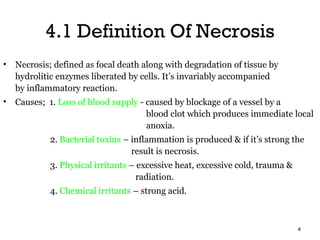
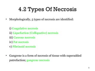
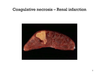
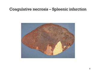
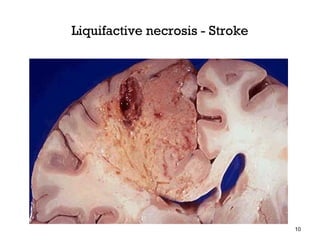
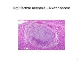
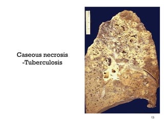
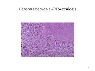
This document defines and describes different types of necrosis. It begins by defining necrosis as focal death and degradation of tissue by hydrolitic enzymes, accompanied by inflammation. It then describes several types of necrosis including coagulative, liquefactive, caseous, fat, and fibrinoid necrosis. Coagulative necrosis is the most common type caused by ischemia. Liquefactive necrosis occurs due to enzymatic degradation. Caseous necrosis is seen in tuberculosis. Fat necrosis occurs in the pancreas and breast. Fibrinoid necrosis involves fibrin deposition. The document also discusses pathologic changes seen in different types of necrosis both grossly and microscopically.
Difference between Apoptosis versus Necrosis and Types of Necrosis.pptxRukhshanda Ramzaan
Apoptosis Versus Nercosis
Apoptosis Necrosis
Predefined cell suicide or programmed cell death. Natural physiological Process. Involve one cell at a time. Cell shrinkage (Dense eosinophilic cytoplasm) Pyknosis (Condensation) and Karyorrhexis (fragmentation) of nuclear material Formation of membrane blebs and apoptotic bodies
Phagocytosis of apoptotic bodies by Macrophages
Caspase dependent pathway
No Inflammation (no immune response) Premature, unprogrammed cell death always pathological. Involve many cells Cell Swelling (Swelling of endoplasmic reticulum and mitochondria) and membrane blebs Pyknosis (condensation), Karyorrhexis (Fragmentation) and Karyolysis (lysis)of the nucleus. Breakdown of the plasma membrane, organelles (enzymatic digestion), leakage of cellular contents
Increased eosinophilia, Accumulation of Myelin figures (whorled precipitated Phospholipids)
Initiate Inflammation (Strong immune response)
- Coagulative Necrosis / summarized
- Description of the GROSS appearance of Coagulative necrosis.
- Description of the MICROSCOPIC appearance of Coagulative necrosis.
may start early after tissue damage
regeneration
by parenchymal cells of the same type
reparation
replacement by connective tissue (fibrosis)
result - scar
the Topics in JavaSE7 and some of Key features in JavaEE6.
the codes in this presentation are here. http://www.slideshare.net/akirakoyasu/java-up-to-date-sources
Hi..! This presentation series is about 'Cell Injury'.. So, here is the 1st part- 'OVERVIEW OF CELLULAR NECROSIS' which I tried to explain in simplest way.. i hope that it will be helpful.. Plz like n do share... Thank You..!!
CONTENTS,
Introduction
Necrosis
Fates of necrotic cells
Patterns of tissue necrosis
Causes of cell injury
The biomechanism of cell injury
Clinicopathological correlations; examples of cell injury and necrosis
Apoptosis
Causes of apoptosis
Apoptosis in physiologic conditions
Apoptosis in pathologic conditions
Mechanism of Apoptosis
The Mitochondrial pathway of Apoptosis
The Death receptor pathway of Apoptosis
Clearance of Apoptotic cells
Examples of Apoptosis
Summary
References
Instructions for Submissions thorugh G- Classroom.pptxJheel Barad
This presentation provides a briefing on how to upload submissions and documents in Google Classroom. It was prepared as part of an orientation for new Sainik School in-service teacher trainees. As a training officer, my goal is to ensure that you are comfortable and proficient with this essential tool for managing assignments and fostering student engagement.
Introduction to AI for Nonprofits with Tapp NetworkTechSoup
Dive into the world of AI! Experts Jon Hill and Tareq Monaur will guide you through AI's role in enhancing nonprofit websites and basic marketing strategies, making it easy to understand and apply.
How to Make a Field invisible in Odoo 17Celine George
It is possible to hide or invisible some fields in odoo. Commonly using “invisible” attribute in the field definition to invisible the fields. This slide will show how to make a field invisible in odoo 17.
Operation “Blue Star” is the only event in the history of Independent India where the state went into war with its own people. Even after about 40 years it is not clear if it was culmination of states anger over people of the region, a political game of power or start of dictatorial chapter in the democratic setup.
The people of Punjab felt alienated from main stream due to denial of their just demands during a long democratic struggle since independence. As it happen all over the word, it led to militant struggle with great loss of lives of military, police and civilian personnel. Killing of Indira Gandhi and massacre of innocent Sikhs in Delhi and other India cities was also associated with this movement.
June 3, 2024 Anti-Semitism Letter Sent to MIT President Kornbluth and MIT Cor...Levi Shapiro
Letter from the Congress of the United States regarding Anti-Semitism sent June 3rd to MIT President Sally Kornbluth, MIT Corp Chair, Mark Gorenberg
Dear Dr. Kornbluth and Mr. Gorenberg,
The US House of Representatives is deeply concerned by ongoing and pervasive acts of antisemitic
harassment and intimidation at the Massachusetts Institute of Technology (MIT). Failing to act decisively to ensure a safe learning environment for all students would be a grave dereliction of your responsibilities as President of MIT and Chair of the MIT Corporation.
This Congress will not stand idly by and allow an environment hostile to Jewish students to persist. The House believes that your institution is in violation of Title VI of the Civil Rights Act, and the inability or
unwillingness to rectify this violation through action requires accountability.
Postsecondary education is a unique opportunity for students to learn and have their ideas and beliefs challenged. However, universities receiving hundreds of millions of federal funds annually have denied
students that opportunity and have been hijacked to become venues for the promotion of terrorism, antisemitic harassment and intimidation, unlawful encampments, and in some cases, assaults and riots.
The House of Representatives will not countenance the use of federal funds to indoctrinate students into hateful, antisemitic, anti-American supporters of terrorism. Investigations into campus antisemitism by the Committee on Education and the Workforce and the Committee on Ways and Means have been expanded into a Congress-wide probe across all relevant jurisdictions to address this national crisis. The undersigned Committees will conduct oversight into the use of federal funds at MIT and its learning environment under authorities granted to each Committee.
• The Committee on Education and the Workforce has been investigating your institution since December 7, 2023. The Committee has broad jurisdiction over postsecondary education, including its compliance with Title VI of the Civil Rights Act, campus safety concerns over disruptions to the learning environment, and the awarding of federal student aid under the Higher Education Act.
• The Committee on Oversight and Accountability is investigating the sources of funding and other support flowing to groups espousing pro-Hamas propaganda and engaged in antisemitic harassment and intimidation of students. The Committee on Oversight and Accountability is the principal oversight committee of the US House of Representatives and has broad authority to investigate “any matter” at “any time” under House Rule X.
• The Committee on Ways and Means has been investigating several universities since November 15, 2023, when the Committee held a hearing entitled From Ivory Towers to Dark Corners: Investigating the Nexus Between Antisemitism, Tax-Exempt Universities, and Terror Financing. The Committee followed the hearing with letters to those institutions on January 10, 202
Synthetic Fiber Construction in lab .pptxPavel ( NSTU)
Synthetic fiber production is a fascinating and complex field that blends chemistry, engineering, and environmental science. By understanding these aspects, students can gain a comprehensive view of synthetic fiber production, its impact on society and the environment, and the potential for future innovations. Synthetic fibers play a crucial role in modern society, impacting various aspects of daily life, industry, and the environment. ynthetic fibers are integral to modern life, offering a range of benefits from cost-effectiveness and versatility to innovative applications and performance characteristics. While they pose environmental challenges, ongoing research and development aim to create more sustainable and eco-friendly alternatives. Understanding the importance of synthetic fibers helps in appreciating their role in the economy, industry, and daily life, while also emphasizing the need for sustainable practices and innovation.
Unit 8 - Information and Communication Technology (Paper I).pdfThiyagu K
This slides describes the basic concepts of ICT, basics of Email, Emerging Technology and Digital Initiatives in Education. This presentations aligns with the UGC Paper I syllabus.
Biological screening of herbal drugs: Introduction and Need for
Phyto-Pharmacological Screening, New Strategies for evaluating
Natural Products, In vitro evaluation techniques for Antioxidants, Antimicrobial and Anticancer drugs. In vivo evaluation techniques
for Anti-inflammatory, Antiulcer, Anticancer, Wound healing, Antidiabetic, Hepatoprotective, Cardio protective, Diuretics and
Antifertility, Toxicity studies as per OECD guidelines
2. OBJECTIVES
• Highlight the basic concepts and principles
of necrosis (cell death)
• Differentiate between different types of
necrosis
2
3. CONTENTS
4.1 Definition of necrosis
4.2 Different types of necrosis
4.3 Different types of necrosis base on pathologic
changes
3
4. 4.1 Definition Of Necrosis
• Necrosis; defined as focal death along with degradation of tissue by
hydrolitic enzymes liberated by cells. It’s invariably accompanied
by inflammatory reaction.
• Causes; 1. Loss of blood supply - caused by blockage of a vessel by a
blood clot which produces immediate local
anoxia.
2. Bacterial toxins – inflammation is produced & if it’s strong the
result is necrosis.
3. Physical irritants – excessive heat, excessive cold, trauma &
radiation.
4. Chemical irritants – strong acid.
4
5. 4.2 Types Of Necrosis
• Morphologically, 5 types of necrosis are identified:
i) Coagulative necrosis
ii) Liquefaction (Colliquative) necrosis
iii) Caseous necrosis
iv) Fat necrosis
v) Fibrinoid necrosis
• Gangrene is a form of necrosis of tissue with superadded
putrefaction; gangrene necrosis
5
6. i) Coagulative necrosis
Most common type of necrosis caused by irreversible
focal injury (mostly because ischaemia).
Less often bacterial & chemical agents.
The organs commonly affected are HEART, KIDNEY &
SPLEEN.
6
9. ii) Liquefaction (Colliquative) necrosis
Occurs commonly due to ischaemic injury & bacterial or
fungal infections.
Occurs due to degradation of tissue by the action of
powerful hydrolytic enzymes.
Examples are INFARCT BRAIN (stroke) & ABSCESS
CAVITY.
9
12. iii) Caseous necrosis
Found in the centre of foci of tuberculosis infections.
It combines features of both coagulative & liquefactive
necrosis.
12
15. iv) Fat necrosis
Special form of cell death occurring at two anatomically different
locations but morphologically similar lesions.
These are ACUTE PANCREATIC NECROSIS & TRAUMATIC FAT
NECROSIS (breast).
In the case of pancreas, there is liberation of pancreatic lipase from
injured or inflamed tissue that results in necrosis of the pancreas.
Fat necrosis in either of the two instances results in hydrolysis of
neutral fat present in adipose cells into glycerol & free fatty acid.
15
16. v) Fibrinoid necrosis
Characterized by deposition of fibrin-like material which has the
staining properties of fibrin.
It’s encountered in various examples of immunologic tissues injury,
arterioles in hypertension, peptic ulcer etc.
vi) Gangrene necrosis
The type of necrosis is usually coagulative due to ischaemia.
Is characterized by primarily inflammation provoked by virulent
bacteria.
3 types; dry, wet & gas gangrene.
16
17. 4.3 Types Of Necrosis Base On
Pathologic Changes
i) Coagulative necrosis
Grossly: foci of coagulative necrosis in the early stage are PALE, FIRM
& SLIGHTLY SWOLLEN.
: with progression, they become more YELLOWISH, SOFTER
& SHRUNKEN.
Micro: the pattern of microscopic change results from 2 processes
which are DENATURATION OF PROTEINS & ENZYMATIC
DIGESTION OF THE CELL.
17
18. ii) Liquefaction (Colliquative) necrosis
Grossly: the affected area is soft with liquefied centre containing necrotic
debris.
: later, a cyst wall is formed.
Micro: the cystic space contains necrotic cell debris & macrophages filled
with phagocytosed material.
: the cyst wall is formed by proliferating cappilaries,
inflammatory cells & gliosis (proliferating glial cells) in the
case of brain & proliferating fibroblasts in the case of
abscess cavity.
18
19. iii) Caseous necrosis
Grossly: resemble dry cheese and are soft, granular & yellowish.
Micro: the necrosed foci are structureless, eosinophilic & contain granular
debris.
iv) Fat necrosis
Grossly: appears as yellowish-white & firm deposits.
: formation of calcium soaps imparts the necrosed foci firmer &
chalky white appearance.
Micro: necrosed fat cells have cloudy appearance & are surrounded by an
inflammatory reaction.
19
20. v) Fibrinoid necrosis
Grossly: is identified by brightly eosinophilic, hyaline-like deposition in
the vessel wall or on the luminar surface of peptic ulcer.
: local haemorrhages may occur due to rupture of these blood
vessels.
vi) Dry gangrene
Macro: the affected part is dry, shrunken & dark black, resembling the foot
: it’s black due to liberation of haemoglobin from haemolysed red
blood cells.
Histo: the separation consists of inflammatory granulation tissue.
20
21. vii) Wet gangrene
Macro; the affected part is soft, swollen, putrid, rotten & dark.
: the example is gangrene of bowel.
Histo: there is ulceration of the mucosa & intense inflammatory infiltration.
viii) Gas gangrene
Grossly: the affected area is swollen, edematous, painful & crepitant due
to accumulation of gas bubbles within the tissues.
: the affected tissue becomes dark black & foul smelling.
Micro: at the periphery, a zone of leucocytic infiltration, edema &
congestion are found.
21