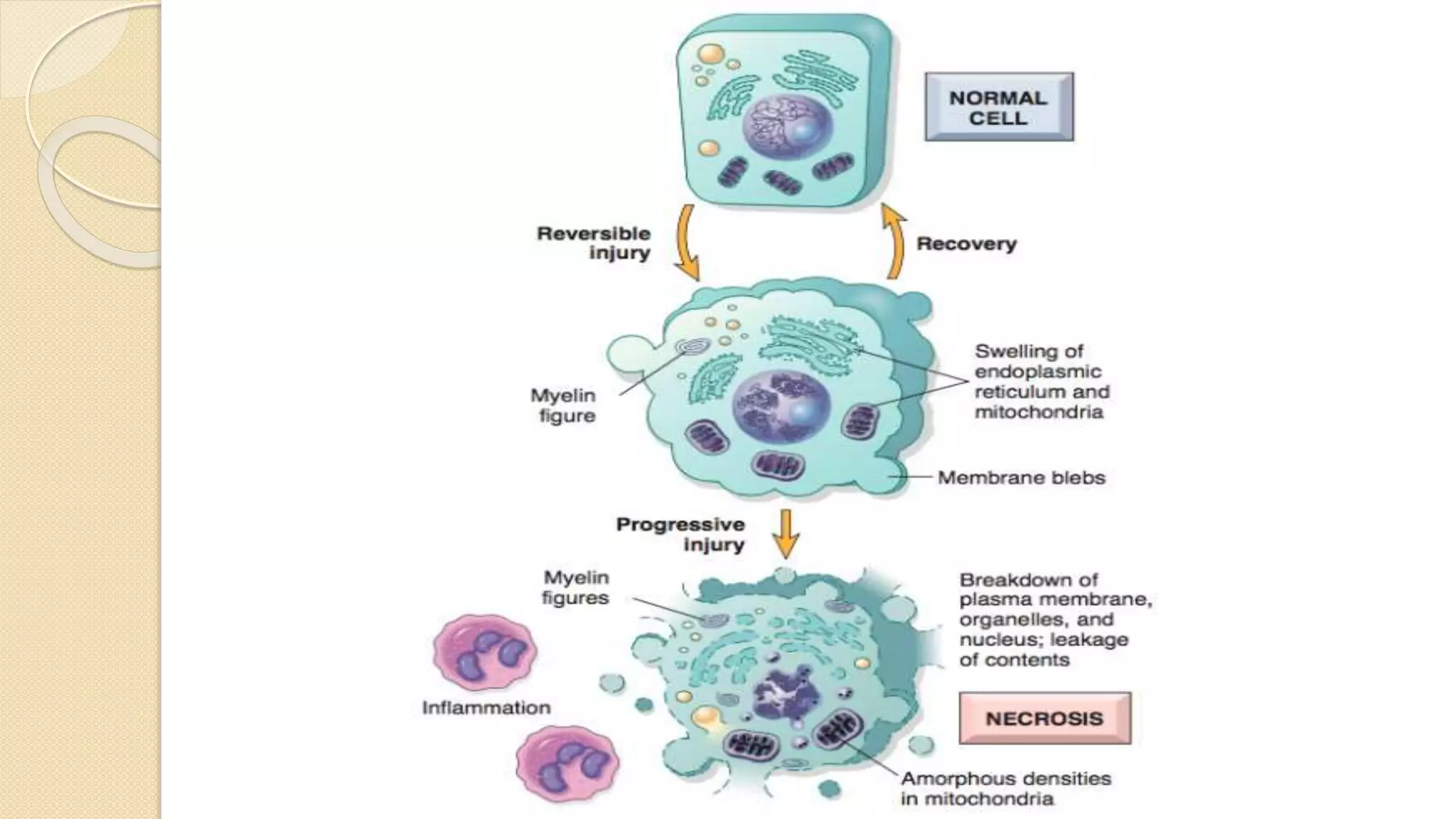
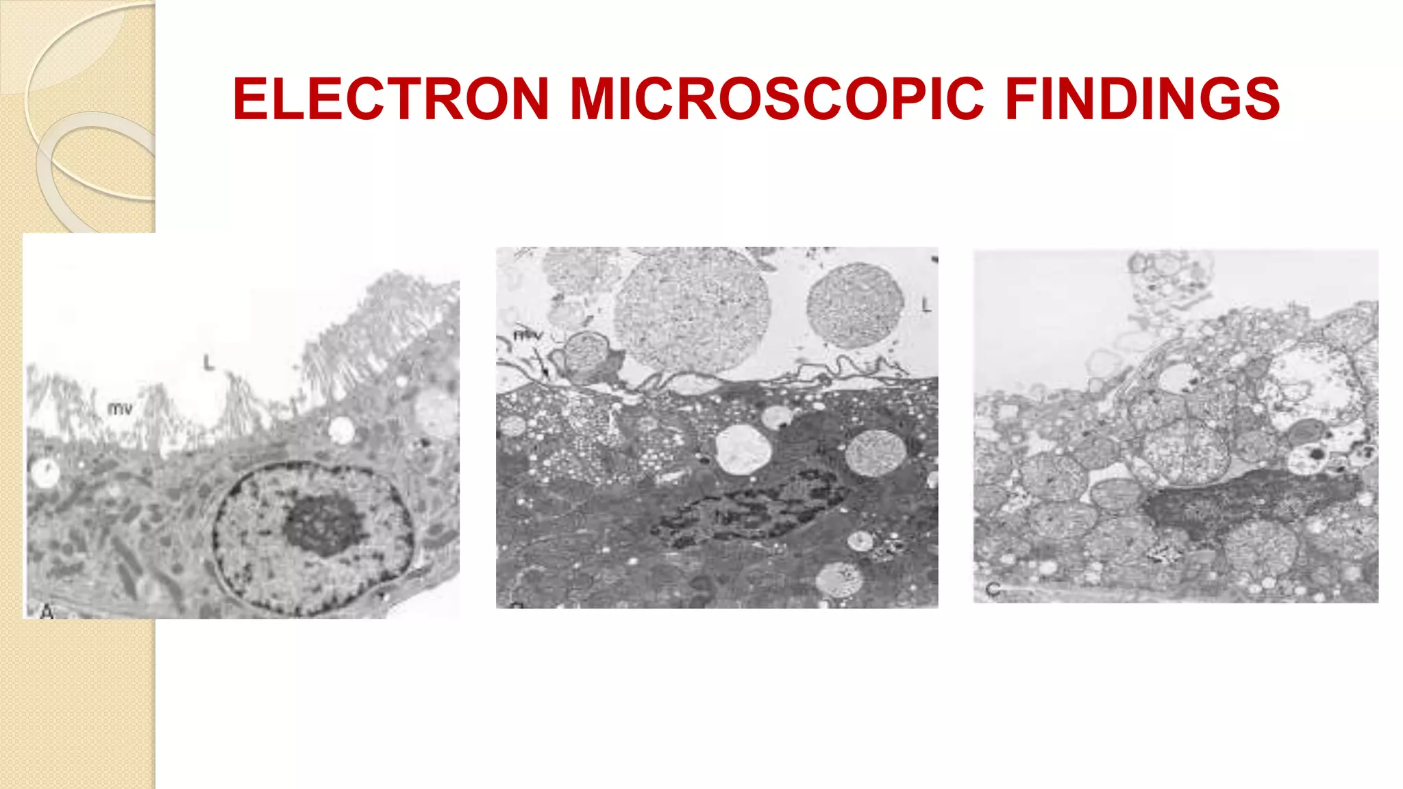
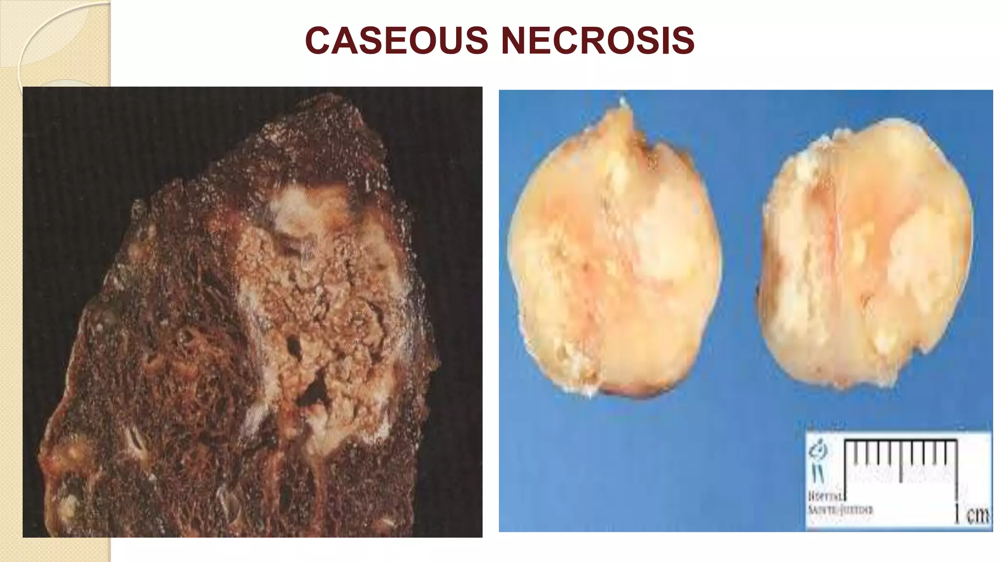
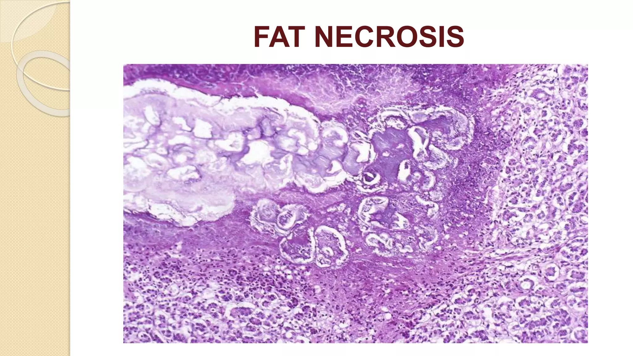
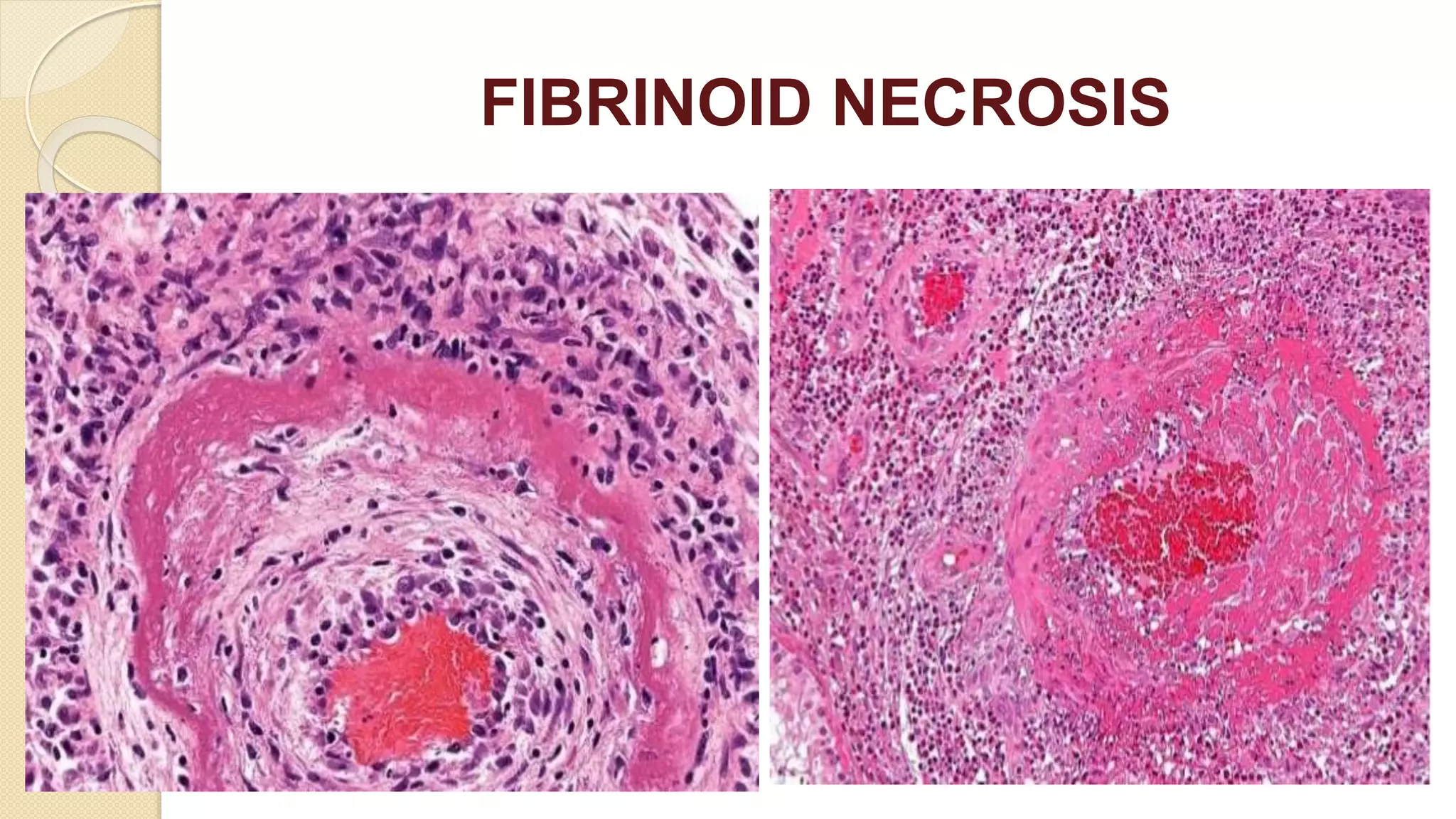
CELL INJURY
NECROSIS
Dr.V.Shanthi,
Associate Professor, Department of Pathology
Sri Venkateswara Institute of medical sciences
Tirupathi
CELL INJURY
Cellular responses to stress & injurious stimuli
Normal cell (homeostasis)
Altered physiologic stimuli and
non lethal injurious stimuli
Injurious stimuli like reduced oxygen
supply, chemical injury and microbial
infection
CELL ADAPTATIONS
CELL INJURY
Acute and transient Progressive and severe
REVERSIBLE CELL INJURY
IRREVERSIBLE CELL INJURY
CELL DEATH
• HYPERPLASIA
• HYPERTROPHY
• ATROPHY
• METAPLASIA
• DYSPLASIA
NECROSIS AND APOPTOSIS
NECROSIS
Definition
Necrosis refers to spectrum of morphologic changes that
follow cell death in living tissue, largely resulting from the
progressive degradative action of enzymes on lethally
injured cell
NECROSIS
• The morphologic appearance of necrosis is the result of
denaturation of intracellular proteins & enzymatic digestion of the
cell
• The enzymes are derived from either lysosomes of the dead cells
themselves (Autolysis) or from the lysosomes of the immigrant
leukocytes (heterolysis )
NECROSIS
Microscopic appearance
Cytoplasmic changes
• Increased eosinophilia – Due to denatured proteins and loss of cytoplasmic RNA
• Glassy homogenous appearance – loss of glycogen particles
• Moth eaten appearance of cytoplasm – enzymatic digestion of cytoplasmic
organelles
• Whorled phospholipid masses derived from damaged cell membranes – myelin
figures
• Calcifications – fatty acids derived from phospholipid masses
NECROSIS
NECROSIS
Electron microscopic findings
Necrotic cells are characterized by
 Discontinuities in plasma and organellar membrane
 Swollen mitochondria with large amorphous densities
 Intracytoplasmic myelin figures
 Aggregates of fluffy material representing denatured proteins
ELECTRON MICROSCOPIC FINDINGS
MYELIN FIGURES
NECROSIS
Microscopic appearance
Nuclear changes
Due to non-specific break down of DNA , 3 patterns are identified
 Karyolysis – fading of basophilia of chromatin due to enzymatic
degradation of DNA by endonucleases
 Pyknosis – nuclear shrinkage & increased basophilia
 Karyorrhexis – pyknotic nucleus undergoes fragmentation
NECROSIS
NECROSIS
Types of necrosis
• Coagulative necrosis
• Liquefactive necrosis
• Caseous necrosis
• Fat necrosis
• Fibrinoid necrosis
NECROSIS
COAGULATIVE NECROSIS
• There is preservation of basic outline of the cell
• The increasing intracellular acidosis denatures not only structural
proteins but also the enzymes & so blocks the proteolysis of the
cell
• E.g. Myocardial infarct
• This is characteristic of hypoxic death of cells in all tissues except
the brain
COAGULATIVE NECROSIS
NECROSIS
LIQUEFACTIVE NECROSIS
 Characterized by digestion of dead cells resulting in
transformation of the tissue into a liquid viscous mass
NECROSIS
LIQUEFACTIVE NECROSIS
• Is characteristic of focal bacterial, or occasional fungal infections
because microbes stimulate the accumulation of inflammatory
cells
• If the process is initiated by acute inflammation, the material is
creamy yellow because of the presence of dead white cells &
called pus
• E.g. hypoxic death in central nervous system
LIQUEFACTIVE NECROSIS
NECROSIS
GANGRENOUS NECROSIS
• The term is applied to a limb , usually lower leg that has lost its
blood supply & has undergone coagulation necrosis.
• When bacterial infection is superimposed , coagulative necrosis
is modified by the liquefactive action of the bacteria & the
attracted leukocytes (wet gangrene )
GANGRENE
Types of the gangrenes
 Dry gangrene – dead necrotised tissue remains uninfected.
 Wet gangrene -necrotic tissue becomes infected.
 Gas gangrene – Wet gangrene infected by one of the gas
forming Clostridia
DRY GANGRENE
 Gangrene begins in distal part of limb due to ischemia
 Causes-
◦ Atherosclerosis of anteries supplying limb
◦ Thromboangitis obliterans
◦ Raynauds disease
◦ Trauma
◦ Ergot poisoning
 Due to ischemia tissues undergoes infarction and line of separation is
formed between the necrotic tissue and healthy tissue
 At the margins of necrotic tissue, granulation tissue develops
DRY GANGRENE
Morphology
 Grossly the affected part is dry shrunken and dark in color resembling the
mummified foot.
 Black color is due to release of hemoglobin from the hemolysed RBC’s which are
acted upon by the hydrogen disulfide produced by bacteria which results in the
formation of black iron sulphide
 Gradually the gangrenous tissue falls from the healthy tissue at the line of
separation.
 Because of the risk of infection it should be surgically separated
WET GANGRENE
Wet gangrene typically occurs in the organs with venous blockage
or both venous and arterial blockage
 Diabetic foot –High glucose content in the necrosed tissue
favours bacterial growth
 Bed sores –In bed ridden patients at the pressure on the sites
like sacrum, buttocks and heel
 Also in organs like bowel (due to strangulated hernia, volvulus
or intusscesception ) lung, mouth, cervix and vulva
WET GANGRENE
Morphologic features:
 Grossly the affected part is black, rotten, and soft and pulpy
 The affected part is dark due to same reason as in dry gangrene
 Microscopically the tissue shows coagulative necrosis with
extensive areas of hemorrhage
 Line of demarcation between variable and necrosed tissue
is absent
GANGRENOUS NECROSIS
Dry gangrene Wet gangrene
Site Lower limbs More common in bowel
Mechanism Arterial occlusion More commonly venous
occlusion, less often
arterial occlusion
Macroscopy Organ is dry, shrunken
and black
Moist, soft swollen and
dark
Line of demarcation Present between
gangrenous and healthy
part
No line of demarcation
Presence of bacteria Bacteria fail to survive Numerous bacteria is
present
Prognosis General better due to
little septicemia
Generally poor due to
profound toxemia
GANGRENOUS NECROSIS
Gas gangrene
 It is wet gangrene infected by one of the gas forming
Clostridia (Gram positive anaerobic bacteria)
 Gas forming Clostridia enters the tissue through a
contaminated wound or as a complication of operation on
colon which normally contains Clostridia
 Clostridia rapidly spreads through tissues especially in
muscles and cause extensive necrosis and massive edema
Gas gangrene
Morphology
 Gross – the affected part is edematous, swollen and has
crepitations on palpation due to accumulation of gas in tissues
 Microscopically
◦ Muscle fibres undergo coagulative necrosis with liquefaction
◦ At the periphery, a zone of leukocytic infiltration, oedema and
congestion are found
NECROSIS
CASEOUS NECROSIS
• Distinctive form of coagulative necrosis most often
encountered in tuberculous infection
• The term caseous is derived from cheesy white gross
appearance of area of necrosis which is friable and white
CASEOUS NECROSIS
NECROSIS
CASEOUS NECROSIS
Microscopic appearance
• Necrotic focus – collection of fragmented or lysed cells and
amorphous granular debris enclosed with in distinctive
inflammatory border – granulomatous reaction
GRANULOMA FORMATION IN CASEOUS NECROSIS
GRANULOMA
GRANULOMA
NECROSIS
FAT NECROSIS
• It is descriptive term for focal areas of fat destruction , typically
occurring as a result of release of activated pancreatic lipases into
the substance of pancreas & the peritoneal cavity
• The fatty acids released combine with calcium to produce grossly
visible chalky white areas (fat saponification)
FAT NECROSIS
NECROSIS
FAT NECROSIS
Microscopic appearance
 Necrosis take the form of shadowy outlines of necrotic fat
cells, with basophilic calcium deposits surrounded by an
inflammatory reaction
FAT NECROSIS
FAT NECROSIS
NECROSIS
FIBRINOID NECROSIS
 Special form of necrosis seen in immune reactions involving blood
vessels
 Occurs when complexes of antigens and antibodies are deposited
in the walls of arteries
 Along with these complexes fibrin which has leaked out of vessels
give bright pink amorphous appearance called fibrinoid (Fibrin
like)
FIBRINOID NECROSIS
THANK YOU

necrosis.pptx

  • 1.
    CELL INJURY NECROSIS Dr.V.Shanthi, Associate Professor,Department of Pathology Sri Venkateswara Institute of medical sciences Tirupathi
  • 2.
    CELL INJURY Cellular responsesto stress & injurious stimuli Normal cell (homeostasis) Altered physiologic stimuli and non lethal injurious stimuli Injurious stimuli like reduced oxygen supply, chemical injury and microbial infection CELL ADAPTATIONS CELL INJURY Acute and transient Progressive and severe REVERSIBLE CELL INJURY IRREVERSIBLE CELL INJURY CELL DEATH • HYPERPLASIA • HYPERTROPHY • ATROPHY • METAPLASIA • DYSPLASIA NECROSIS AND APOPTOSIS
  • 3.
    NECROSIS Definition Necrosis refers tospectrum of morphologic changes that follow cell death in living tissue, largely resulting from the progressive degradative action of enzymes on lethally injured cell
  • 5.
    NECROSIS • The morphologicappearance of necrosis is the result of denaturation of intracellular proteins & enzymatic digestion of the cell • The enzymes are derived from either lysosomes of the dead cells themselves (Autolysis) or from the lysosomes of the immigrant leukocytes (heterolysis )
  • 6.
    NECROSIS Microscopic appearance Cytoplasmic changes •Increased eosinophilia – Due to denatured proteins and loss of cytoplasmic RNA • Glassy homogenous appearance – loss of glycogen particles • Moth eaten appearance of cytoplasm – enzymatic digestion of cytoplasmic organelles • Whorled phospholipid masses derived from damaged cell membranes – myelin figures • Calcifications – fatty acids derived from phospholipid masses
  • 7.
  • 8.
    NECROSIS Electron microscopic findings Necroticcells are characterized by  Discontinuities in plasma and organellar membrane  Swollen mitochondria with large amorphous densities  Intracytoplasmic myelin figures  Aggregates of fluffy material representing denatured proteins
  • 9.
  • 10.
  • 11.
    NECROSIS Microscopic appearance Nuclear changes Dueto non-specific break down of DNA , 3 patterns are identified  Karyolysis – fading of basophilia of chromatin due to enzymatic degradation of DNA by endonucleases  Pyknosis – nuclear shrinkage & increased basophilia  Karyorrhexis – pyknotic nucleus undergoes fragmentation
  • 12.
  • 13.
    NECROSIS Types of necrosis •Coagulative necrosis • Liquefactive necrosis • Caseous necrosis • Fat necrosis • Fibrinoid necrosis
  • 14.
    NECROSIS COAGULATIVE NECROSIS • Thereis preservation of basic outline of the cell • The increasing intracellular acidosis denatures not only structural proteins but also the enzymes & so blocks the proteolysis of the cell • E.g. Myocardial infarct • This is characteristic of hypoxic death of cells in all tissues except the brain
  • 15.
  • 16.
  • 17.
  • 18.
    NECROSIS LIQUEFACTIVE NECROSIS  Characterizedby digestion of dead cells resulting in transformation of the tissue into a liquid viscous mass
  • 19.
    NECROSIS LIQUEFACTIVE NECROSIS • Ischaracteristic of focal bacterial, or occasional fungal infections because microbes stimulate the accumulation of inflammatory cells • If the process is initiated by acute inflammation, the material is creamy yellow because of the presence of dead white cells & called pus • E.g. hypoxic death in central nervous system
  • 20.
  • 21.
    NECROSIS GANGRENOUS NECROSIS • Theterm is applied to a limb , usually lower leg that has lost its blood supply & has undergone coagulation necrosis. • When bacterial infection is superimposed , coagulative necrosis is modified by the liquefactive action of the bacteria & the attracted leukocytes (wet gangrene )
  • 22.
    GANGRENE Types of thegangrenes  Dry gangrene – dead necrotised tissue remains uninfected.  Wet gangrene -necrotic tissue becomes infected.  Gas gangrene – Wet gangrene infected by one of the gas forming Clostridia
  • 23.
    DRY GANGRENE  Gangrenebegins in distal part of limb due to ischemia  Causes- ◦ Atherosclerosis of anteries supplying limb ◦ Thromboangitis obliterans ◦ Raynauds disease ◦ Trauma ◦ Ergot poisoning  Due to ischemia tissues undergoes infarction and line of separation is formed between the necrotic tissue and healthy tissue  At the margins of necrotic tissue, granulation tissue develops
  • 24.
    DRY GANGRENE Morphology  Grosslythe affected part is dry shrunken and dark in color resembling the mummified foot.  Black color is due to release of hemoglobin from the hemolysed RBC’s which are acted upon by the hydrogen disulfide produced by bacteria which results in the formation of black iron sulphide  Gradually the gangrenous tissue falls from the healthy tissue at the line of separation.  Because of the risk of infection it should be surgically separated
  • 25.
    WET GANGRENE Wet gangrenetypically occurs in the organs with venous blockage or both venous and arterial blockage  Diabetic foot –High glucose content in the necrosed tissue favours bacterial growth  Bed sores –In bed ridden patients at the pressure on the sites like sacrum, buttocks and heel  Also in organs like bowel (due to strangulated hernia, volvulus or intusscesception ) lung, mouth, cervix and vulva
  • 26.
    WET GANGRENE Morphologic features: Grossly the affected part is black, rotten, and soft and pulpy  The affected part is dark due to same reason as in dry gangrene  Microscopically the tissue shows coagulative necrosis with extensive areas of hemorrhage  Line of demarcation between variable and necrosed tissue is absent
  • 27.
    GANGRENOUS NECROSIS Dry gangreneWet gangrene Site Lower limbs More common in bowel Mechanism Arterial occlusion More commonly venous occlusion, less often arterial occlusion Macroscopy Organ is dry, shrunken and black Moist, soft swollen and dark Line of demarcation Present between gangrenous and healthy part No line of demarcation Presence of bacteria Bacteria fail to survive Numerous bacteria is present Prognosis General better due to little septicemia Generally poor due to profound toxemia
  • 28.
  • 29.
    Gas gangrene  Itis wet gangrene infected by one of the gas forming Clostridia (Gram positive anaerobic bacteria)  Gas forming Clostridia enters the tissue through a contaminated wound or as a complication of operation on colon which normally contains Clostridia  Clostridia rapidly spreads through tissues especially in muscles and cause extensive necrosis and massive edema
  • 30.
    Gas gangrene Morphology  Gross– the affected part is edematous, swollen and has crepitations on palpation due to accumulation of gas in tissues  Microscopically ◦ Muscle fibres undergo coagulative necrosis with liquefaction ◦ At the periphery, a zone of leukocytic infiltration, oedema and congestion are found
  • 31.
    NECROSIS CASEOUS NECROSIS • Distinctiveform of coagulative necrosis most often encountered in tuberculous infection • The term caseous is derived from cheesy white gross appearance of area of necrosis which is friable and white
  • 32.
  • 33.
    NECROSIS CASEOUS NECROSIS Microscopic appearance •Necrotic focus – collection of fragmented or lysed cells and amorphous granular debris enclosed with in distinctive inflammatory border – granulomatous reaction
  • 34.
    GRANULOMA FORMATION INCASEOUS NECROSIS
  • 35.
  • 36.
  • 37.
    NECROSIS FAT NECROSIS • Itis descriptive term for focal areas of fat destruction , typically occurring as a result of release of activated pancreatic lipases into the substance of pancreas & the peritoneal cavity • The fatty acids released combine with calcium to produce grossly visible chalky white areas (fat saponification)
  • 38.
  • 39.
    NECROSIS FAT NECROSIS Microscopic appearance Necrosis take the form of shadowy outlines of necrotic fat cells, with basophilic calcium deposits surrounded by an inflammatory reaction
  • 40.
  • 41.
  • 42.
    NECROSIS FIBRINOID NECROSIS  Specialform of necrosis seen in immune reactions involving blood vessels  Occurs when complexes of antigens and antibodies are deposited in the walls of arteries  Along with these complexes fibrin which has leaked out of vessels give bright pink amorphous appearance called fibrinoid (Fibrin like)
  • 43.
  • 44.