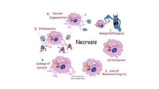
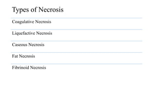
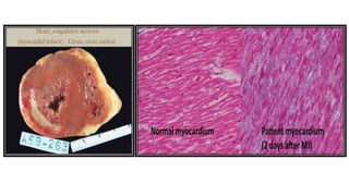
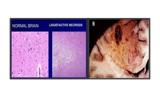
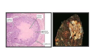
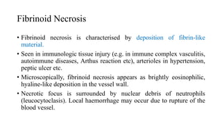
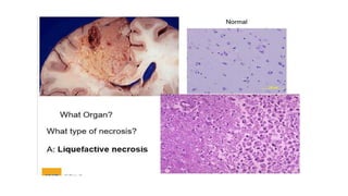
NECROSIS
Dr. Nishali Paladugu
Contents
•Definition and causes
•Pathogenesis
•Types of necrosis
Definition
• A localised area of death of tissue followed by degradation of tissue by
hydrolytic enzymes liberated from dead cells.
• Accompanied by inflammatory reaction.
Causes
• Hypoxia
• Chemical and physical agents
• Microbial agents
• Immunological injury, etc.
Cell digestion by lytic
enzymes
It is identified as homogeneous and intensely
eosinophilic cytoplasm
Denaturation of
proteins
Seen as characteristic nuclear changes in necrotic
cell which may include
• Condensation of nuclear chromatin (pyknosis)
• Which may either undergo dissolution
(karyolysis) OR
• Fragmentation into many granular clumps
(karyorrhexis)
Two essential changes cause irreversible cell injury in necrosis of all types
Pathogenesis
Types of Necrosis
Coagulative Necrosis
Liquefactive Necrosis
Caseous Necrosis
Fat Necrosis
Fibrinoid Necrosis
Coagulative Necrosis
• Most common type of necrosis.
• Caused by irreversible focal injury, mostly from sudden cessation of
blood flow (ischaemia).
• The organs commonly affected are the heart, kidney, and spleen.
• Grossly, foci of coagulative necrosis appears as
-Pale, firm, and slightly swollen in the early stage
-With progression, they become more yellowish, softer, and shrunken.
Coagulative Necrosis
• Microscopically, the hallmark of this is the conversion of normal cells
into their ‘tombstones’ i.e. outlines of the cells are retained so that the
cell type can still be recognized but their cytoplasmic and nuclear
details are lost.
• The necrosed cells are swollen and appear more eosinophilic than the
normal, along with nuclear changes.
• The necrosed focus is infiltrated by inflammatory cells, and the dead
cells are phagocytosed leaving granular debris and fragments of cells.
Liquefactive Necrosis
Liquefactive necrosis occurs due to ischaemic injury and bacterial or
fungal infections.
Characterized by digestion of the dead cells, resulting in transformation
of the tissue into a liquid viscous mass.
The common examples are infarct brain and abscess cavity.
• Grossly, it is soft with liquefied center containing necrotic debris.
Later, a cyst wall is formed.
• Microscopically, the cystic space contains necrotic cell debris and
macrophages filled with phagocytosed material.
Caseous Necrosis
Caseous necrosis is found in the centre of foci of tuberculous infections.
It combines features of both coagulative and liquefactive necrosis.
• Grossly, resemble dry cheese and are soft, granular and yellowish.
• Microscopically, the necrosed foci are structureless, eosinophilic, and
contain granular debris .The surrounding tissue shows characteristic
granulomatous inflammatory reaction consisting of epithelioid cells
with interspersed giant cells of Langhans’ or foreign body type and
peripheral mantle of lymphocytes
Fat Necrosis
Fat necrosis is a special form of cell death occurring at two anatomically
different locations but morphologically similar lesions.
• Acute pancreatic necrosis
• Traumatic fat necrosis commonly in breasts.
Fat necrosis hydrolyses neutral fat present in adipose cells into glycerol
and free fatty acids. The damaged adipose cells assume cloudy
appearance. The leaked out free fatty acids complex with calcium to
form calcium soaps (saponification).
Fat Necrosis
• Grossly, fat necrosis appears as yellowish-white and firm deposits.
Formation of calcium soaps imparts the necrosed foci firmer and
chalky white appearance.
• Microscopically, the necrosed fat cells have cloudy appearance and are
surrounded by an inflammatory reaction. Formation of calcium soaps
is identified in the tissue sections as amorphous, granular and
basophilic material
Fibrinoid Necrosis
• Fibrinoid necrosis is characterised by deposition of fibrin-like
material.
• Seen in immunologic tissue injury (e.g. in immune complex vasculitis,
autoimmune diseases, Arthus reaction etc), arterioles in hypertension,
peptic ulcer etc.
• Microscopically, fibrinoid necrosis appears as brightly eosinophilic,
hyaline-like deposition in the vessel wall.
• Necrotic focus is surrounded by nuclear debris of neutrophils
(leucocytoclasis). Local haemorrhage may occur due to rupture of the
blood vessel.
• Gangrenous necrosis is not a specific pattern of cell death, but the
term is commonly used in clinical practice. It is usually applied to a
limb, generally the lower leg, that has lost its blood supply
and has undergone necrosis (typically coagulative necrosis) involving
multiple tissue planes.
• When bacterial infection is superimposed there is more liquefactive
necrosis because of the actions of degradative enzymes in the bacteria
and the attracted leukocytes (giving rise to so-called wet gangrene).
THANK YOU!

Necrosis updated pathology powerpoint presentation .pptx

  • 1.
  • 2.
  • 3.
    Definition • A localisedarea of death of tissue followed by degradation of tissue by hydrolytic enzymes liberated from dead cells. • Accompanied by inflammatory reaction. Causes • Hypoxia • Chemical and physical agents • Microbial agents • Immunological injury, etc.
  • 4.
    Cell digestion bylytic enzymes It is identified as homogeneous and intensely eosinophilic cytoplasm Denaturation of proteins Seen as characteristic nuclear changes in necrotic cell which may include • Condensation of nuclear chromatin (pyknosis) • Which may either undergo dissolution (karyolysis) OR • Fragmentation into many granular clumps (karyorrhexis) Two essential changes cause irreversible cell injury in necrosis of all types Pathogenesis
  • 6.
    Types of Necrosis CoagulativeNecrosis Liquefactive Necrosis Caseous Necrosis Fat Necrosis Fibrinoid Necrosis
  • 7.
    Coagulative Necrosis • Mostcommon type of necrosis. • Caused by irreversible focal injury, mostly from sudden cessation of blood flow (ischaemia). • The organs commonly affected are the heart, kidney, and spleen. • Grossly, foci of coagulative necrosis appears as -Pale, firm, and slightly swollen in the early stage -With progression, they become more yellowish, softer, and shrunken.
  • 8.
    Coagulative Necrosis • Microscopically,the hallmark of this is the conversion of normal cells into their ‘tombstones’ i.e. outlines of the cells are retained so that the cell type can still be recognized but their cytoplasmic and nuclear details are lost. • The necrosed cells are swollen and appear more eosinophilic than the normal, along with nuclear changes. • The necrosed focus is infiltrated by inflammatory cells, and the dead cells are phagocytosed leaving granular debris and fragments of cells.
  • 10.
    Liquefactive Necrosis Liquefactive necrosisoccurs due to ischaemic injury and bacterial or fungal infections. Characterized by digestion of the dead cells, resulting in transformation of the tissue into a liquid viscous mass. The common examples are infarct brain and abscess cavity. • Grossly, it is soft with liquefied center containing necrotic debris. Later, a cyst wall is formed. • Microscopically, the cystic space contains necrotic cell debris and macrophages filled with phagocytosed material.
  • 12.
    Caseous Necrosis Caseous necrosisis found in the centre of foci of tuberculous infections. It combines features of both coagulative and liquefactive necrosis. • Grossly, resemble dry cheese and are soft, granular and yellowish. • Microscopically, the necrosed foci are structureless, eosinophilic, and contain granular debris .The surrounding tissue shows characteristic granulomatous inflammatory reaction consisting of epithelioid cells with interspersed giant cells of Langhans’ or foreign body type and peripheral mantle of lymphocytes
  • 14.
    Fat Necrosis Fat necrosisis a special form of cell death occurring at two anatomically different locations but morphologically similar lesions. • Acute pancreatic necrosis • Traumatic fat necrosis commonly in breasts. Fat necrosis hydrolyses neutral fat present in adipose cells into glycerol and free fatty acids. The damaged adipose cells assume cloudy appearance. The leaked out free fatty acids complex with calcium to form calcium soaps (saponification).
  • 15.
    Fat Necrosis • Grossly,fat necrosis appears as yellowish-white and firm deposits. Formation of calcium soaps imparts the necrosed foci firmer and chalky white appearance. • Microscopically, the necrosed fat cells have cloudy appearance and are surrounded by an inflammatory reaction. Formation of calcium soaps is identified in the tissue sections as amorphous, granular and basophilic material
  • 17.
    Fibrinoid Necrosis • Fibrinoidnecrosis is characterised by deposition of fibrin-like material. • Seen in immunologic tissue injury (e.g. in immune complex vasculitis, autoimmune diseases, Arthus reaction etc), arterioles in hypertension, peptic ulcer etc. • Microscopically, fibrinoid necrosis appears as brightly eosinophilic, hyaline-like deposition in the vessel wall. • Necrotic focus is surrounded by nuclear debris of neutrophils (leucocytoclasis). Local haemorrhage may occur due to rupture of the blood vessel.
  • 19.
    • Gangrenous necrosisis not a specific pattern of cell death, but the term is commonly used in clinical practice. It is usually applied to a limb, generally the lower leg, that has lost its blood supply and has undergone necrosis (typically coagulative necrosis) involving multiple tissue planes. • When bacterial infection is superimposed there is more liquefactive necrosis because of the actions of degradative enzymes in the bacteria and the attracted leukocytes (giving rise to so-called wet gangrene).
  • 21.