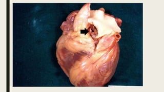
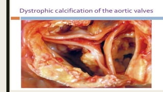
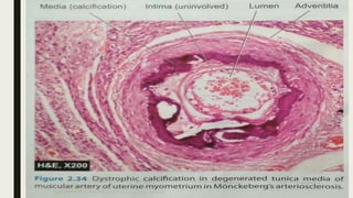
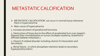
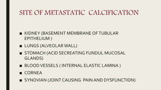
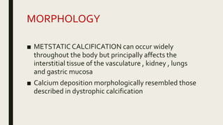
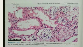
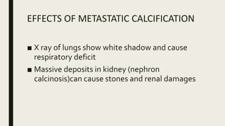
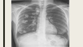
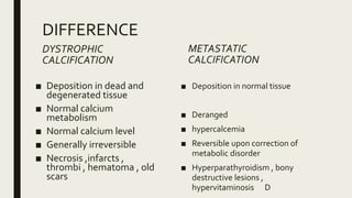
CALCIFICATION
By – AKHILESH KUMAR
PATHOLOGICAL CALCIFICATION
■ Pathological calcification is abnormal
deposition of CALCIUM salts with smaller
amounts of IRON, MAGNESIUM and other
minerals except teeth and bone`deposition
MECHANISM OF CALCIFICATION
■ Calcium deposits in the form of hydroxyapatite (natural
occurring mineral form of calcium) and involves two phases
■ INITIATION – initiation of calcium takes place in the matrix
vesicles where calcium and phosphates accumulate.The
matrix vesicles are extracellular membrane bound vesicles
that bud off from other cells.
■ PROPAGATION –The second phase involves further growth
of hydroxyapatite
TYPES OF PATHOLOGIC
CALCIFICATION
DYSTROPHIC CALCIFICATION
1. When deposition occurs in dead or dying tissue
2. It occurs with normal serum level of normal calcium
metabolism
METASTATIC CALCIFICATION
1. Deposition of calcium salts in normal tissue
2. It almost always reflects some derangement in calcium
metabolism and increase level of calcium(hypercalcemia)
DYSTROPHICCALCIFICATION
■ Dystrophic calcification is encountered in areas of
necrosis of any type
■ It is certain in atheromas of advanced atherosclerosis ,
associated with initial injury in the aorta and large
arteries
■ Although dystrophic calcification may be an incidental
finding indicating in significant past cell injury , it may
also be a cause of organ dysfunction
■ For example-dystrophic calcification of aortic valve is an
important cause of aortic stenosis in elderly
MORPHOLOGY OF DYSTROPHIC
CALCIFICATION
■ CALCIUM SALTS are grossly seen as fine white granules or
clumps , often felt as gritty deposition
■ Histologically calcification appears as intracellular and/or
extracellular basophilic deposits
■ In time heterotopic bone may be formed in the focus of
calcification
SITES OF DYSTROPHICCALCIFICATION
■ NECROTICTISSUE
a) Caseous necrosis
b) Fat necrosis
c) Liquifactive necrosis
■ Advanced atheromatous lesion
■ Hematoma
■ Thrombus
■ Infarct
■ Heart valve (example -sub acute infective endocarditis )
METASTATIC CALCIFICATION
 METASTATIC CALCIFICATION can occur in normal tissue whenever
there is hypercalcemia
 Main cause of hypercalcemia
1. increase secretion of parathyroid hormone
2. Destruction of bone due to the effect of accelerated turn over (paget’s
disease) then immobilisation or tumor (multiple myeloma, leukemia or
diffused skeletal metastasis )
3.Vitamin D related disorder including vitamin D intoxication and
sarcoidosis
4. Renal failure , in which phosphate retention leads to secondary
hyperparathyroidism
■
SITE OF METASTATIC CALCIFICATION
■ KIDNEY (BASEMENT MEMBRANE OFTUBULAR
EPITHELIUM )
■ LUNGS (ALVEOLAR WALL)
■ STOMACH (ACID SECREATING FUNDUL MUCOSAL
GLANDS)
■ BLOODVESSELS ( INTERNAL ELASTIC LAMINA )
■ CORNEA
■ SYNOVIAN (JOINT CAUSING PAIN AND DYSFUNCTION)
MORPHOLOGY
■ METSTATIC CALCIFICATION can occur widely
throughout the body but principally affects the
interstitial tissue of the vasculature , kidney , lungs
and gastric mucosa
■ Calcium deposition morphologically resembled those
described in dystrophic calcification
EFFECTS OF METASTATIC CALCIFICATION
■ X ray of lungs show white shadow and cause
respiratory deficit
■ Massive deposits in kidney (nephron
calcinosis)can cause stones and renal damages
DIFFERENCE
DYSTROPHIC
CALCIFICATION
■ Deposition in dead and
degenerated tissue
■ Normal calcium
metabolism
■ Normal calcium level
■ Generally irreversible
■ Necrosis ,infarcts ,
thrombi , hematoma , old
scars
METASTATIC
CALCIFICATION
■ Deposition in normal tissue
■ Deranged
■ hypercalcemia
■ Reversible upon correction of
metabolic disorder
■ Hyperparathyroidism , bony
destructive lesions ,
hypervitaminosis D
Pathologic Calcification

Pathologic Calcification

  • 1.
  • 2.
    PATHOLOGICAL CALCIFICATION ■ Pathologicalcalcification is abnormal deposition of CALCIUM salts with smaller amounts of IRON, MAGNESIUM and other minerals except teeth and bone`deposition
  • 3.
    MECHANISM OF CALCIFICATION ■Calcium deposits in the form of hydroxyapatite (natural occurring mineral form of calcium) and involves two phases ■ INITIATION – initiation of calcium takes place in the matrix vesicles where calcium and phosphates accumulate.The matrix vesicles are extracellular membrane bound vesicles that bud off from other cells. ■ PROPAGATION –The second phase involves further growth of hydroxyapatite
  • 4.
    TYPES OF PATHOLOGIC CALCIFICATION DYSTROPHICCALCIFICATION 1. When deposition occurs in dead or dying tissue 2. It occurs with normal serum level of normal calcium metabolism METASTATIC CALCIFICATION 1. Deposition of calcium salts in normal tissue 2. It almost always reflects some derangement in calcium metabolism and increase level of calcium(hypercalcemia)
  • 5.
    DYSTROPHICCALCIFICATION ■ Dystrophic calcificationis encountered in areas of necrosis of any type ■ It is certain in atheromas of advanced atherosclerosis , associated with initial injury in the aorta and large arteries ■ Although dystrophic calcification may be an incidental finding indicating in significant past cell injury , it may also be a cause of organ dysfunction ■ For example-dystrophic calcification of aortic valve is an important cause of aortic stenosis in elderly
  • 8.
    MORPHOLOGY OF DYSTROPHIC CALCIFICATION ■CALCIUM SALTS are grossly seen as fine white granules or clumps , often felt as gritty deposition ■ Histologically calcification appears as intracellular and/or extracellular basophilic deposits ■ In time heterotopic bone may be formed in the focus of calcification
  • 9.
    SITES OF DYSTROPHICCALCIFICATION ■NECROTICTISSUE a) Caseous necrosis b) Fat necrosis c) Liquifactive necrosis ■ Advanced atheromatous lesion ■ Hematoma ■ Thrombus ■ Infarct ■ Heart valve (example -sub acute infective endocarditis )
  • 11.
    METASTATIC CALCIFICATION  METASTATICCALCIFICATION can occur in normal tissue whenever there is hypercalcemia  Main cause of hypercalcemia 1. increase secretion of parathyroid hormone 2. Destruction of bone due to the effect of accelerated turn over (paget’s disease) then immobilisation or tumor (multiple myeloma, leukemia or diffused skeletal metastasis ) 3.Vitamin D related disorder including vitamin D intoxication and sarcoidosis 4. Renal failure , in which phosphate retention leads to secondary hyperparathyroidism ■
  • 12.
    SITE OF METASTATICCALCIFICATION ■ KIDNEY (BASEMENT MEMBRANE OFTUBULAR EPITHELIUM ) ■ LUNGS (ALVEOLAR WALL) ■ STOMACH (ACID SECREATING FUNDUL MUCOSAL GLANDS) ■ BLOODVESSELS ( INTERNAL ELASTIC LAMINA ) ■ CORNEA ■ SYNOVIAN (JOINT CAUSING PAIN AND DYSFUNCTION)
  • 13.
    MORPHOLOGY ■ METSTATIC CALCIFICATIONcan occur widely throughout the body but principally affects the interstitial tissue of the vasculature , kidney , lungs and gastric mucosa ■ Calcium deposition morphologically resembled those described in dystrophic calcification
  • 15.
    EFFECTS OF METASTATICCALCIFICATION ■ X ray of lungs show white shadow and cause respiratory deficit ■ Massive deposits in kidney (nephron calcinosis)can cause stones and renal damages
  • 17.
    DIFFERENCE DYSTROPHIC CALCIFICATION ■ Deposition indead and degenerated tissue ■ Normal calcium metabolism ■ Normal calcium level ■ Generally irreversible ■ Necrosis ,infarcts , thrombi , hematoma , old scars METASTATIC CALCIFICATION ■ Deposition in normal tissue ■ Deranged ■ hypercalcemia ■ Reversible upon correction of metabolic disorder ■ Hyperparathyroidism , bony destructive lesions , hypervitaminosis D