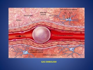
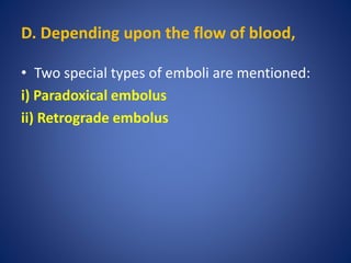
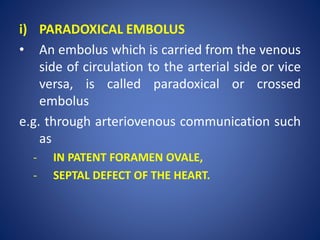
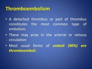
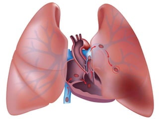
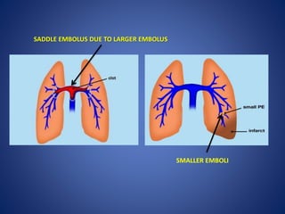
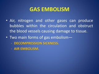
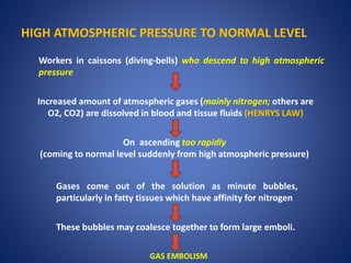
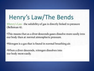
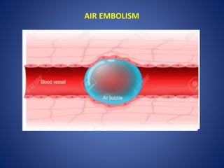
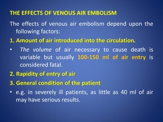
2. Definition
• Embolism is the process of partial or complete
obstruction of some part of the cardiovascular
system by any mass carried in the circulation;
the transported intravascular mass detached
from its site of origin is called an embolus.
• Most usual forms of emboli (90%) are
thromboemboli i.e. originating from thrombi
or their parts detached from the vessel wall.
3. Types
• Emboli may be of various types:
A. Depending upon the matter in the emboli
B. Depending upon whether infected or not
C. Depending upon the source of the emboli
D. Depending upon the flow of blood.
4. A. Depending upon the matter in the emboli
i) Solid
- e.g. detached thrombi (thromboemboli),
- atheromatous material,
- tumour cell clumps,
- tissue fragments,
- parasites,
- bacterial clumps,
ii) Liquid e.g. fat globules, amniotic fluid, bone
marrow.
iii) Gaseous e.g. air, other gases.
11. B. Depending upon whether infected or not
i) Bland, when sterile.
ii) Septic, when infected.
12. SEPTIC EMBOLI
JANEWAYS LESIONS: PAINLESS, NON-TENDER
SUBCUTANEOUS MACULOPAPULAR LESIONS
ON THE PULP OF THE FINGERS IN
ACUTE BACTERIAL ENDOCARDITIS
13. C. Depending upon the source of the emboli:
i) Cardiac emboli from left side of the heart
e.g. emboli originating in the atrium
ii) Arterial emboli
e.g. in systemic arteries in the brain
iii) Venous emboli
e.g. in pulmonary arteries.
iv) Lymphatic emboli can also sometimes occur.
14. D. Depending upon the flow of blood,
• Two special types of emboli are mentioned:
i) Paradoxical embolus
ii) Retrograde embolus
15. i) PARADOXICAL EMBOLUS
• An embolus which is carried from the venous
side of circulation to the arterial side or vice
versa, is called paradoxical or crossed
embolus
e.g. through arteriovenous communication such
as
- IN PATENT FORAMEN OVALE,
- SEPTAL DEFECT OF THE HEART.
17. ii) RETROGRADE EMBOLUS
• An embolus which travels against the flow of
blood is called retrograde embolus.
• For example, metastatic deposits in the spine
from carcinoma prostate in which case the
spread occurs by retrograde embolism.
• The prostatic venous plexus drains into the
internal iliac vein which connects with the
vertebral venous plexus, this is thought to be
the route of bone metastasis of prostate
cancer.
18.
19. • Due to low pressure in Intraspinal
veins/vertebral venous plexus, the retrograde
movement of venous blood into them from
prostrate through internal iliac vein can occur
in conditions when there is high pressure in
pelvis like during coughing/straining.
• In Prostrate cancer, the tumour cells move in
such direction due to the above reason
leading to bone metastasis.
20. Some of the important types of embolism are listed
below:
21. Thromboembolism
• A detached thrombus or part of thrombus
constitutes the most common type of
embolism.
• These may arise in the arterial or venous
circulation
• Most usual forms of emboli (90%) are
thromboemboli.
26. EFFECTS OF ARTERIAL EMBOLI
• The effects of arterial emboli depend upon their size, site of
lodgment, and adequacy of collateral circulation.
• If the vascular occlusion occurs, the following ill-effects may
result:
i) Infarction of the organ or its affected part e.g. ischaemic
necrosis in the lower limbs (70-75%), spleen, kidneys, brain,
intestine.
ii) Gangrene following infarction in the lower limbs if the
collateral circulation is inadequate.
iii) Myocardial infarction may occur following coronary
embolism.
iv) Sudden death may result from coronary embolism or
embolism in the middle cerebral artery.
27. EFFECT OF VENOUS EMBOLISM
• The most significant effect of venous
embolism is obstruction of pulmonary arterial
circulation leading to pulmonary embolism
28. PULMONARY THROMBOEMBOLISM
• Pulmonary embolism is the most common
and fatal form of venous thromboembolism
in which there is occlusion of pulmonary
arterial tree by thromboemboli.
29.
30. ETIOLOGY:
• Pulmonary emboli are more common in
hospitalized or bed-ridden patients, though
they can occur in ambulatory patients as
well.
- Late pregnancy,
- Puerperal state and
- Use of contraceptive pills.
31. SOURCES FOR EMBOLUS
More common sources:
- Originating from large veins of lower legs
(such as popliteal, femoral and iliac) are the
cause in 95% of pulmonary emboli.
Less common sources include
- Thrombi in varicosities of superficial veins of
the legs, and pelvic veins.
32. DRAINS INTO RIGHT SIDE OF THE HEART.
DETACHMENT OF THROMBI FROM ANY OF THE ABOVE-MENTIONED SITES
THROMBO-EMBOLUS FLOWS THROUGH VENOUS DRAINAGE INTO THE LARGER VEINS
IF THE THROMBUS IS LARGE, IT CAN GET
IMPACTED AT THE BIFURCATION OF
THE MAIN PULMONARY ARTERY
(SADDLE EMBOLUS)
MULTIPLE EMBOLI, OR A LARGE
EMBOLUS MAY BE FRAGMENTED INTO MANY
SMALLER EMBOLI WHICH ARE THEN IMPACTED
IN A NUMBER OF VESSELS, PARTICULARLY
AFFECTING THE LOWER LOBES OF LUNGS.
PATHOGENESIS:
34. CONSEQUENCES OF PULMONARY EMBOLISM
• Consequences of pulmonary embolism
depend mainly on
- the size of the occluded vessel,
- the number of emboli, and
- on the cardiovascular status of the patient.
35. • Following consequences can occur in pulmonary
embolism:
I) SUDDEN DEATH:
Massive pulmonary embolism results in
instantaneous death, without occurrence of
chest pain or dyspnoea.
However, if the death is somewhat delayed,
the clinical features resemble myocardial
infarction i.e. severe chest pain, dyspnoea and
shock.
36. II) ACUTE COR PULMONALE:
• Numerous small emboli may obstruct most of the
pulmonary circulation resulting in acute right heart
failure.
37. III) PULMONARY INFARCTION:
• Obstruction of relatively small sized pulmonary
arterial branches may result in pulmonary infarction.
• The clinical features include
- chest pain, Haemoptysis
- dyspnoea due to reduced functioning pulmonary
parenchyma
38. IV) PULMONARY HAEMORRHAGE:
• Obstruction of terminal branches (endarteries) leads
to central pulmonary haemorrhage.
• The clinical features are haemoptysis, dyspnoea, and
less commonly, chest pain due to central location of
pulmonary haemorrhage.
39. V) RESOLUTION:
• Vast majority of small pulmonary emboli (60-
80%) are resolved by fibrinolytic activity.
40. VI) PULMONARY HYPERTENSION, CHRONIC COR
PULMONALE AND PULMONARY ARTERIOSCLEROSIS
• These are the sequelae of multiple small
thromboemboli undergoing organisation
rather than resolution.
41.
42. Pulmonary Thrombus Vs Pulmonary Thromboembolism
• Pulmonary thrombosis is uncommon and may
occur in pulmonary atherosclerosis and
pulmonary hypertension.
• Differentiation of pulmonary thrombosis from
pulmonary thromboembolism is tabulated in
Table.
43.
44. GAS EMBOLISM
• Air, nitrogen and other gases can produce
bubbles within the circulation and obstruct
the blood vessels causing damage to tissue.
• Two main forms of gas embolism—
- DECOMPRESSION SICKNESS.
- AIR EMBOLISM.
45. Decompression Sickness
• This is a specialized form of gas embolism
known by various names such as
- CAISSON’S DISEASE,
- DIVERS’ PALSY OR
- AEROEMBOLISM.
47. PATHOGENESIS
• Decompression sickness is produced when the
individual decompresses suddenly, either from
- High atmospheric pressure to normal level,
or
- Low atmospheric pressure to normal levels
48. HIGH ATMOSPHERIC PRESSURE TO NORMAL LEVEL
Workers in caissons (diving-bells) who descend to high atmospheric
pressure
Increased amount of atmospheric gases (mainly nitrogen; others are
O2, CO2) are dissolved in blood and tissue fluids (HENRYS LAW)
On ascending too rapidly
(coming to normal level suddenly from high atmospheric pressure)
Gases come out of the solution as minute bubbles,
particularly in fatty tissues which have affinity for nitrogen
These bubbles may coalesce together to form large emboli.
GAS EMBOLISM
49.
50.
51. LOW ATMOSPHERIC PRESSURE TO NORMAL LEVELS
• Persons who ascend to high Altitudes or air
flight in unpressurised cabins, the individuals
are exposed to sudden decompression from
low atmospheric Pressure to normal levels.
• This results in similar effects as in Divers and
workers in caissons.
52. EFFECTS
The effects of decompression sickness depend
upon the following:
i) Depth or altitude reached
ii) Duration of exposure to altered pressure
iii) Rate of ascent or descent
iv) General condition of the individual
53. • Pathologic changes are more pronounced in
sudden decompression from high pressure to
normal levels than in those who decompress
from low pressure to normal levels.
• The changes are more serious in obese
persons as nitrogen gas is more soluble in fat
than in body fluids.
54. • Clinical effects of decompression sickness are
of 2 types—
1. Acute.
2. Chronic.
55. • Clinical effects of decompression sickness are
of 2 types—
1. Acute.
2. Chronic.
56. Acute form of DCS
• Occurs due to acute obstruction of small blood
vessels in the vicinity of joints and skeletal
muscles.
Clinically characterized by the following:
1. ‘The bends’, as the patient doubles up in bed
due to acute pain in joints, ligaments and
tendons.
2. ‘The chokes’ occur due to accumulation of
bubbles in the lungs, resulting in acute
respiratory distress.
3. Cerebral effects may manifest in the form of
vertigo, coma, and sometimes death.
57. Chronic form of DCS
The features of chronic form are as under:
1. Avascular necrosis of bones e.g. head of femur,
tibia, humerus.
2. Neurological symptoms include paraesthesia and
paraplegia.
3. Lung involvement in the form of haemorrhage,
oedema, emphysema and atelactasis may be seen.
4. Skin manifestations include itching, patchy
erythema, cyanosis and oedema.
58.
59.
60. AIR EMBOLISM
• Air embolism occurs when air is introduced
into venous or arterial circulation.
1. VENOUS AIR EMBOLISM:
- Occur when air enters systemic veins.
2. ARTERIAL AIR EMBOLISM:
- Occurs when air enters into pulmonary vein
or its tributaries.
62. VENOUS AIR EMBOLISM:
• Circumstances which causes entry of air into
systemic veins:
1. Operations on the head and neck, and trauma:
• The accidental opening of a major vein of the
neck like jugular, or neck wounds involving
the major neck veins, may allow air to be
drawn into venous circulation.
63. ii) Obstetrical operations and trauma:
• During childbirth by normal vaginal delivery,
caesarean section, abortions and other
procedures, fatal air embolism may result
from the entrance of air into the opened-up
uterine venous sinuses and endometrial veins.
64. iii) Intravenous infusion of blood and fluid:
• Air embolism may occur during intravenous
blood or fluid infusions if only positive
pressure is employed.
65. iv) Angiography:
• During venous angiographic procedures
(venogram) , air may be entrapped into a large
vein causing air embolism
66. The effects of venous air embolism depend upon the
following factors:
1. Amount of air introduced into the circulation.
• The volume of air necessary to cause death is
variable but usually 100-150 ml of air entry is
considered fatal.
2. Rapidity of entry of air
3. General condition of the patient
• e.g. in severely ill patients, as little as 40 ml of air
may have serious results.
THE EFFECTS OF VENOUS AIR EMBOLISM
67. 4. Position of the patient during or soon after entry of air:
• The air bubbles may ascend into the superior vena cava if the
position of head is higher than the trunk (e.g. in upright
position) and reach the brain.