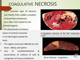
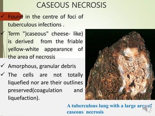
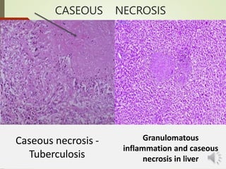
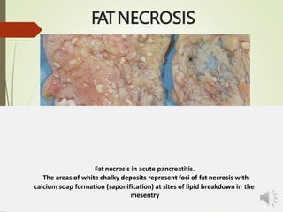
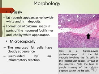
Necrosis
By
Assistant Professor
Dr. Jihad A. Ahmed
BVM,MSc,PhD Animal Pathology
NECROSIS
Defined as a localised area of death of tissue followed by
degradation of tissue by hydrolytic enzymes liberated from
dead cells .Or sequence of changes in a living tissue or
organism.
 It is invariably accompanied by inflammatory reaction.
 Necrosis occurs when cells are exposed to extreme variance
from physiological conditions (e.g., hypothermia, hypoxia)
which may result in damage to the plasma membrane.
 Two essential changes characterise irreversible cell injury
in necrosis of all types
–Cell digestion by lytic enzymes
–Denaturation ofproteins
Classification: necrosis & apoptosis
Causes of necrosis
1) Hypoxia (Oxygen deprivation)
2) Physical agents such as: Mechanical truma, temperature,
radiations, electric shock
3) Chemical agents and drugs such as: glucose or salt in
hypertonic concentration, high concentration of oxygen ,
poisons such as arsenic, cyanide or mercuric salt
4) Infectious agents such as: bacterial, fungal and parasite
5) Immunologic reactions
6) Genetic derangements
7) Nutritional imbalances
Morphology of necrosis :
Cytoplasmic changes:
1. Cellular swelling or rupture
2. Denaturation and coagulation of cytoplasmic proteins
3. Breakdown of cell organelles
4. Increased eosinophilia of cytoplasm
5. Glassy homogeneous appearance
6. Cytoplasm is vacuolated, moth eaten
7. Calcification of dead cells.
8. Appearance of myelin figures
9. Generation of calcium soaps
NUCLEAR CHANGES
 PYKNOSIS
Nuclear shrinkage and increased basophilia
 KARYORRHEXIS
Fragmentation of pyknotic nucleus
 KARYOLYSIS
Fading of basophilia of chromatin Due to DNAase activity.
Normal cell
Necrotic kidney
P: Pyknotic
nuclei K:
Karryorhexis
Types of Necrosis
Morphologically, there are five types of
necrosis
• Cogulative necrosis
• Liquefaction necrosis
• Caseous necrosis
• Fat necrosis
• Fibrinoid necrosis
COAGULATIVE NECROSIS
 Most common type of necrosis
and Mostly from sudden cessation
of blood flow (ischaemia)
 Less often from bacterial and
chemical agents.
 It’s characteristic of infarcts
(areas of ischemic necrosis)
in all solid organs except the
brain.
 The organs commonly affected are the
heart , kidney, and spleen
 Loss of nuclei(tombstone) &
preservation of outlines of cells &
recognition of tissue architecture
Coagulative necrosis of the left ventricular
wall
Renal Infarction - Coagulative
Morpholog
y
-With progression, they become more yellowish, softer,and
 Microscopically
 Gross
-Foci of coagulative necrosis in the early stage are pale, firm,
and slightly swollen.
shrunken.
-The hallmark of coagulative necrosis- the conversion of
normal cells into their ’tombstones’
• outlines of the cells are retained so that the cell type but
their cytoplasmic and nuclear details are lost.
-The necrosed cells are swollen and appear more eosinophilic
than the normal, along with nuclear changes described
above. But cell digestion and liquefaction fail to occur.
-Eventually, the necrosed focus is infiltrated by inflammatory
Cells And the dead cells are phagocytosed leaving granular
debris and fragments of cells
COAGULATIVE NECROSIS
Infarct Kidney
The affected area on right shows cells with intensely eosinophilic cytoplasm of tubular cells
but the outlines of tubules are still maintained. The nuclei show granulardebris.
The interface between viable and non-viable area shows nonspecific chronic inflammation
and proliferating vessels
Skeletal muscle - Necrosis in a
male F344/N rat from a chronic
skeletal muscle exhibits
study. This cross section of
an
extensive, highly fragmented and
vacuolated
fibers;
collection of muscle
some are
hypereosinophilic, and others are
pale.
CASEOUS NECROSIS
 Found in the centre of foci of
tuberculous infections .
 Term ")caseous" cheese- like)
is derived from the friable
yellow-white appearance of
the area of necrosis
 Amorphous, granular debris
 The cells are not totally
liquefied nor are their outlines
preserved(coagulation and
liquefaction).
A tuberculous lung with a large area of
caseous necrosis
Morphology
Grossly
 Foci of caseous necrosis, as the name implies, resemble dry cheese
granular and yellowish.
 This appearance is partly attributed to the histotoxic effect of
lipopolysaccharides present in the capsule of the tubercle bacilli,
mycrobacterium tuberculosis.
 Microscopically
 The necrosed foci are structureless, eosinophilic, and contain
granular debris
 The surrounding tissue shows characteristic granulomatous
inflammatory reaction
 Consisting of epithelioid cells with interspersed giant cells of
and also peripheral mantle of
Langhans’ or foreign body type
lymphocytes.
CASEOUS NECROSIS
Caseous necrosis -
Tuberculosis
Granulomatous
inflammation and caseous
necrosis in liver
FAT NECROSIS
 Refers to focal areas of fat destruction
–Acute pancreatic necrosis,
–traumatic fat necrosis commonly in breasts
 Pancreatic enzymes that have leaked out acinar of cells and
ducts
 liquefy the membranes of fat cells in theperitoneum.
 lipases split the triglyceride esters contained within fat cellsto
fatty acid.
 These combines calcium to produce grossly visible chalky
white areas (fat saponification )
 Shadowy outlines of necrotic fat cells
 Inflammatory reaction
FATNECROSIS
Fat necrosis in acute pancreatitis.
The areas of white chalky deposits represent foci of fat necrosis with
calcium soap formation (saponification) at sites of lipid breakdown in the
mesentry
Morphology
• Grossly
– fat necrosis appears as yellowish-
white and firm deposits.
– Formation of calcium soaps in
parts of the necrosed foci firmer
and chalky white appearance.
• Microscopically
– The necrosed fat cells have
cloudy appearance
– Surrounded by an
inflammatory reaction.
This is a higher-power
photomicrograph of the fat
necrosis involving the fat cells in
the interlobular spaces (arrow) of
the pancreas. Note the blue to
purple staining of the calcium
deposits within the fat cells.
Fig. 1B —Stages of fat necrosis as
seen on photomicrographs. Early
fat necrosis shows
adipocytes (arrows)
by areas
stage of
individual
surrounded
hemorrhage.
of
(Papanicolaou, ×20)
Fig. 1F —Stages of fat necrosis as
seen on photomicrographs. Late
stage of fat necrosis shows
calcifications, which are common
in late stage of fat necrosis.
(Papanicolaou, ×40)
LIQUEFACTION (COLLIQUATIVE)NECROSIS
of brain tissue and bacterial or
 Due to ischemic destruction
fungal infections
 degradation of tissue by the action of powerful hydrolytic
enzyme.
 The common examples are infarct brain and abscess
cavity.
 Whatever the pathogenesis, liquefaction completely digests the dead
cells, resulting in transformation of the tissue into a liquid viscous
mass.
 If the process was initiated by acute inflammation, the material is
frequently creamy yellow and is called pus
 Cystic structure filled with debris and fluid.
 The dead area softens, liquefies.
LIQUEFACTION NECROSIS
Gross
Morphology
 The affected area is soft with liquefied centre containing
necrotic debris.
 Later, a cyst wall is formed.
Microscopically,
 The cystic space contains necrotic cell debris and
macrophages filled with phagocytosed material.
 The cyst wall is formed by proliferating capillaries, inflammatory
cells, and gliosis (proliferating glial cells) in the case ofbrain
 proliferating fibroblasts in the case of abscesscavity.
LIQUEFACTION NECROSIS
Liquefactive necrosis brain The necrosed area on right
side of the field shows a cystic space containing cell
debris, while the surrounding zone shows granulation
tissue and gliosis.
FIBRINOID NECROSIS
• Characterised by deposition of fibrin-like material which
has the staining properties of fibrin.
Location: interstitial collagen and blood vessels (small
artery and arteriole)
• It is encountered in various examples of immunologic tissue
ulcer, active
injury, Arterioles in hypertension, peptic
rheumatism, polyarteritis nodose. etc.
– immune complex vasculitis,
– autoimmune diseases,
 Does not have any distinctive gross appearance.
o Grossly
o Microscopically
– Identified by brightly
eosinophilic, hyaline-like
deposition in the vessel
wall.
– Necrotic focus is surrounded by
nuclear debris of neutrophils
(leucocytoclasis)
Local haemorrhage may occur due
to rupture of the blood vessel.
–
Fibrinoid necrosis in autoimmune
vasculitis.
The vessel wall shows brightly
pink amorphous material and
nuclear fragments of necrosed
neutrophils
 Is identified by brightly eosinophilic,
hyaline-like deposition in the vessel
wall or on the luminar surface of
peptic ulcer. : local haemorrhages
may occur due to rupture of these
blood vessels.
FIBRINOID NECROSIS
Fibrinoid necrosis in an artery in a patient with polyarteritis
nodosa.
The wall of the artery shows a circumferential bright pink area of necrosis with
protein and inflammation(dark nuclei ofneutrophils).
Fate of necrosis:
1) Acute or chronic inflammation
2) Immunological reaction
3) Fibrosis & scar formation
4) Encapsulation & calcification of dead
cells
5) Cyst formation
6) Isolation and organization
7) lysis and absorption

1590180412.pptx

  • 1.
    Necrosis By Assistant Professor Dr. JihadA. Ahmed BVM,MSc,PhD Animal Pathology
  • 2.
    NECROSIS Defined as alocalised area of death of tissue followed by degradation of tissue by hydrolytic enzymes liberated from dead cells .Or sequence of changes in a living tissue or organism.  It is invariably accompanied by inflammatory reaction.  Necrosis occurs when cells are exposed to extreme variance from physiological conditions (e.g., hypothermia, hypoxia) which may result in damage to the plasma membrane.  Two essential changes characterise irreversible cell injury in necrosis of all types –Cell digestion by lytic enzymes –Denaturation ofproteins Classification: necrosis & apoptosis
  • 3.
    Causes of necrosis 1)Hypoxia (Oxygen deprivation) 2) Physical agents such as: Mechanical truma, temperature, radiations, electric shock 3) Chemical agents and drugs such as: glucose or salt in hypertonic concentration, high concentration of oxygen , poisons such as arsenic, cyanide or mercuric salt 4) Infectious agents such as: bacterial, fungal and parasite 5) Immunologic reactions 6) Genetic derangements 7) Nutritional imbalances
  • 4.
    Morphology of necrosis: Cytoplasmic changes: 1. Cellular swelling or rupture 2. Denaturation and coagulation of cytoplasmic proteins 3. Breakdown of cell organelles 4. Increased eosinophilia of cytoplasm 5. Glassy homogeneous appearance 6. Cytoplasm is vacuolated, moth eaten 7. Calcification of dead cells. 8. Appearance of myelin figures 9. Generation of calcium soaps
  • 5.
    NUCLEAR CHANGES  PYKNOSIS Nuclearshrinkage and increased basophilia  KARYORRHEXIS Fragmentation of pyknotic nucleus  KARYOLYSIS Fading of basophilia of chromatin Due to DNAase activity. Normal cell
  • 6.
  • 7.
    Types of Necrosis Morphologically,there are five types of necrosis • Cogulative necrosis • Liquefaction necrosis • Caseous necrosis • Fat necrosis • Fibrinoid necrosis
  • 8.
    COAGULATIVE NECROSIS  Mostcommon type of necrosis and Mostly from sudden cessation of blood flow (ischaemia)  Less often from bacterial and chemical agents.  It’s characteristic of infarcts (areas of ischemic necrosis) in all solid organs except the brain.  The organs commonly affected are the heart , kidney, and spleen  Loss of nuclei(tombstone) & preservation of outlines of cells & recognition of tissue architecture Coagulative necrosis of the left ventricular wall Renal Infarction - Coagulative
  • 9.
    Morpholog y -With progression, theybecome more yellowish, softer,and  Microscopically  Gross -Foci of coagulative necrosis in the early stage are pale, firm, and slightly swollen. shrunken. -The hallmark of coagulative necrosis- the conversion of normal cells into their ’tombstones’ • outlines of the cells are retained so that the cell type but their cytoplasmic and nuclear details are lost. -The necrosed cells are swollen and appear more eosinophilic than the normal, along with nuclear changes described above. But cell digestion and liquefaction fail to occur. -Eventually, the necrosed focus is infiltrated by inflammatory Cells And the dead cells are phagocytosed leaving granular debris and fragments of cells
  • 10.
    COAGULATIVE NECROSIS Infarct Kidney Theaffected area on right shows cells with intensely eosinophilic cytoplasm of tubular cells but the outlines of tubules are still maintained. The nuclei show granulardebris. The interface between viable and non-viable area shows nonspecific chronic inflammation and proliferating vessels
  • 11.
    Skeletal muscle -Necrosis in a male F344/N rat from a chronic skeletal muscle exhibits study. This cross section of an extensive, highly fragmented and vacuolated fibers; collection of muscle some are hypereosinophilic, and others are pale.
  • 12.
    CASEOUS NECROSIS  Foundin the centre of foci of tuberculous infections .  Term ")caseous" cheese- like) is derived from the friable yellow-white appearance of the area of necrosis  Amorphous, granular debris  The cells are not totally liquefied nor are their outlines preserved(coagulation and liquefaction). A tuberculous lung with a large area of caseous necrosis
  • 13.
    Morphology Grossly  Foci ofcaseous necrosis, as the name implies, resemble dry cheese granular and yellowish.  This appearance is partly attributed to the histotoxic effect of lipopolysaccharides present in the capsule of the tubercle bacilli, mycrobacterium tuberculosis.  Microscopically  The necrosed foci are structureless, eosinophilic, and contain granular debris  The surrounding tissue shows characteristic granulomatous inflammatory reaction  Consisting of epithelioid cells with interspersed giant cells of and also peripheral mantle of Langhans’ or foreign body type lymphocytes.
  • 14.
    CASEOUS NECROSIS Caseous necrosis- Tuberculosis Granulomatous inflammation and caseous necrosis in liver
  • 15.
    FAT NECROSIS  Refersto focal areas of fat destruction –Acute pancreatic necrosis, –traumatic fat necrosis commonly in breasts  Pancreatic enzymes that have leaked out acinar of cells and ducts  liquefy the membranes of fat cells in theperitoneum.  lipases split the triglyceride esters contained within fat cellsto fatty acid.  These combines calcium to produce grossly visible chalky white areas (fat saponification )  Shadowy outlines of necrotic fat cells  Inflammatory reaction
  • 16.
    FATNECROSIS Fat necrosis inacute pancreatitis. The areas of white chalky deposits represent foci of fat necrosis with calcium soap formation (saponification) at sites of lipid breakdown in the mesentry
  • 17.
    Morphology • Grossly – fatnecrosis appears as yellowish- white and firm deposits. – Formation of calcium soaps in parts of the necrosed foci firmer and chalky white appearance. • Microscopically – The necrosed fat cells have cloudy appearance – Surrounded by an inflammatory reaction. This is a higher-power photomicrograph of the fat necrosis involving the fat cells in the interlobular spaces (arrow) of the pancreas. Note the blue to purple staining of the calcium deposits within the fat cells.
  • 18.
    Fig. 1B —Stagesof fat necrosis as seen on photomicrographs. Early fat necrosis shows adipocytes (arrows) by areas stage of individual surrounded hemorrhage. of (Papanicolaou, ×20) Fig. 1F —Stages of fat necrosis as seen on photomicrographs. Late stage of fat necrosis shows calcifications, which are common in late stage of fat necrosis. (Papanicolaou, ×40)
  • 19.
    LIQUEFACTION (COLLIQUATIVE)NECROSIS of braintissue and bacterial or  Due to ischemic destruction fungal infections  degradation of tissue by the action of powerful hydrolytic enzyme.  The common examples are infarct brain and abscess cavity.  Whatever the pathogenesis, liquefaction completely digests the dead cells, resulting in transformation of the tissue into a liquid viscous mass.  If the process was initiated by acute inflammation, the material is frequently creamy yellow and is called pus  Cystic structure filled with debris and fluid.  The dead area softens, liquefies.
  • 20.
  • 21.
    Gross Morphology  The affectedarea is soft with liquefied centre containing necrotic debris.  Later, a cyst wall is formed. Microscopically,  The cystic space contains necrotic cell debris and macrophages filled with phagocytosed material.  The cyst wall is formed by proliferating capillaries, inflammatory cells, and gliosis (proliferating glial cells) in the case ofbrain  proliferating fibroblasts in the case of abscesscavity.
  • 22.
    LIQUEFACTION NECROSIS Liquefactive necrosisbrain The necrosed area on right side of the field shows a cystic space containing cell debris, while the surrounding zone shows granulation tissue and gliosis.
  • 23.
    FIBRINOID NECROSIS • Characterisedby deposition of fibrin-like material which has the staining properties of fibrin. Location: interstitial collagen and blood vessels (small artery and arteriole) • It is encountered in various examples of immunologic tissue ulcer, active injury, Arterioles in hypertension, peptic rheumatism, polyarteritis nodose. etc. – immune complex vasculitis, – autoimmune diseases,  Does not have any distinctive gross appearance.
  • 24.
    o Grossly o Microscopically –Identified by brightly eosinophilic, hyaline-like deposition in the vessel wall. – Necrotic focus is surrounded by nuclear debris of neutrophils (leucocytoclasis) Local haemorrhage may occur due to rupture of the blood vessel. – Fibrinoid necrosis in autoimmune vasculitis. The vessel wall shows brightly pink amorphous material and nuclear fragments of necrosed neutrophils  Is identified by brightly eosinophilic, hyaline-like deposition in the vessel wall or on the luminar surface of peptic ulcer. : local haemorrhages may occur due to rupture of these blood vessels.
  • 25.
    FIBRINOID NECROSIS Fibrinoid necrosisin an artery in a patient with polyarteritis nodosa. The wall of the artery shows a circumferential bright pink area of necrosis with protein and inflammation(dark nuclei ofneutrophils).
  • 26.
    Fate of necrosis: 1)Acute or chronic inflammation 2) Immunological reaction 3) Fibrosis & scar formation 4) Encapsulation & calcification of dead cells 5) Cyst formation 6) Isolation and organization 7) lysis and absorption