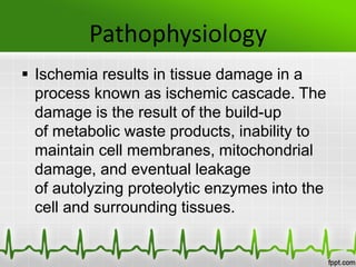
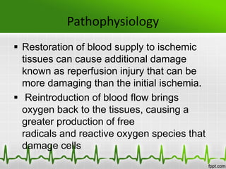
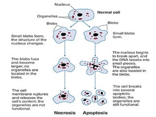
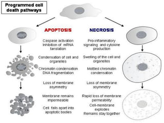
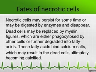
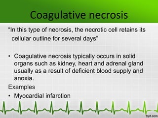
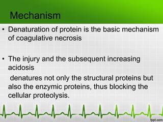
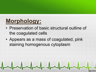
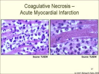
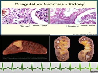
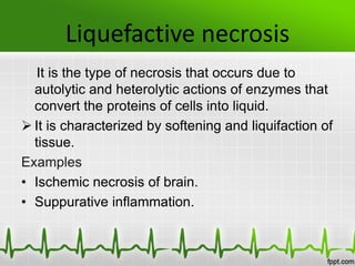
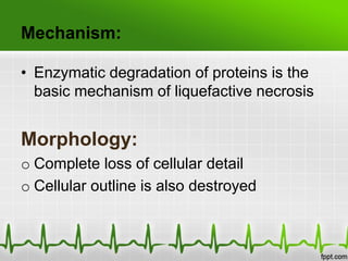
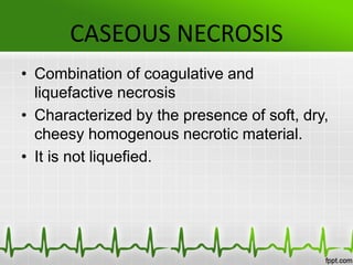
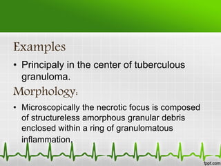
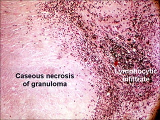
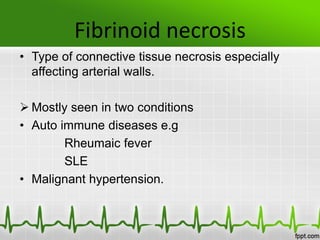
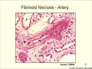
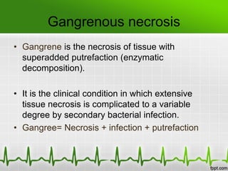
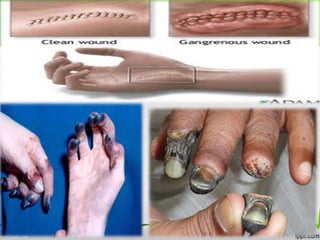
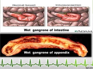
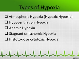
Topics
Ischemia
Necrosis
Hypoxia
Ischemia
Introduction
Causes of ischemia
Signs and Symptoms
Types of Ischemia
Presented By:
Muhammad Ammar
Roll No. 01
Ischemia
Ischemia is a restriction in blood supply
to tissues, causing a shortage of oxygen
and glucose needed for cellular
metabolism (to keep tissue alive).
Ischemia is generally caused by problems
with blood vessels, with resultant damage
to or dysfunction of tissue.
Ischemia
Ischemia comprises not only insufficiency
of oxygen, but also reduced availability
of nutrients and inadequate removal
of metabolites.
Causes of Ischemia
 It can be caused by:
Embolism, thrombosis of
an atherosclerosis artery, or trauma.
 Venous problems like venous outflow
obstruction and low-flow states can
cause acute arterial ischemia.
 An aneurysm is one of the most frequent
causes of acute arterial ischemia.
Causes of Ischemia
Other causes are heart conditions
including myocardial infarction, mitral
valve disease, chronic atrial
fibrillation, cardiomyopathies,
and prosthesis, in all of which thrombi are
prone to develop.
Signs and symptoms
Since oxygen is carried to tissues in
the blood, insufficient blood supply causes
tissue to become starved of oxygen.
 In the highly aerobic tissues of
the heart and brain, irreversible damage to
tissues can occur in as little as 3–4
minutes at body temperature.
Signs and symptoms
The kidneys are also quickly damaged by
loss of blood flow. Tissues with slower
metabolic rates may undergo irreversible
damage after 20 minutes.
Without immediate intervention, ischemia
may progress quickly to
tissue necrosis and gangrene within a few
hours.
Signs and symptoms
 Paralysis is a very late sign of acute
arterial ischemia and signals the death
of nerves supplying the extremity. Foot
drop may occur as a result of nerve
damage.
Types of Ischemia
 Cardiac ischemia
 Bowel ischemia
 Brain ischemia
 Limb ischemia
 Cutaneous ischemia
Cardiac Ischemia
o Cardiac ischemia may be asymptomatic or
may cause chest pain, known as angina
pectoris.
o It occurs when the heart muscle,
or myocardium, receives insufficient blood
flow. This most frequently results
from atherosclerosis.
Cardiac Ischemia
o Ischemic heart disease is the most
common cause of death in most Western
countries and a major cause of hospital
admissions.
Bowel Ischemia
o Both large and small intestine can be
affected by ischemia.
o Ischemia of the large intestine may result
in an inflammatory process known
as ischemic colitis.
o Ischemia of the small bowel is
called mesenteric ischemia.
Brain Ischemia
o Brain ischemia is insufficient blood flow to
the brain, and can be acute or chronic.
o Acute ischemic stroke is a neurologic
emergency that may be reversible if
treated rapidly.
o Chronic ischemia of the brain may result
in a form of dementia called vascular
dementia. ".
Brain Ischemia
o A brief episode of ischemia affecting the
brain is called a transient ischemic
attack (TIA), often referred to as a "mini-
stroke”.
Limb Ischemia
o Lack of blood flow to a limb results in
acute limb ischemia.
Cutaneous Ischemia
o Reduced blood flow to the skin layers may
result in mottling or uneven, patchy
discoloration of the skin.
Pathophysiology of Ischemia
Treatment of Ischemia
Introduction of Necrosis
Causes of Necrosis
Changes in necrotic cell
Fate of necrotic cell
Presented by:
Hamayoun Ashraf
Roll No. 06
Pathophysiology
 Ischemia results in tissue damage in a
process known as ischemic cascade. The
damage is the result of the build-up
of metabolic waste products, inability to
maintain cell membranes, mitochondrial
damage, and eventual leakage
of autolyzing proteolytic enzymes into the
cell and surrounding tissues.
Pathophysiology
 Restoration of blood supply to ischemic
tissues can cause additional damage
known as reperfusion injury that can be
more damaging than the initial ischemia.
 Reintroduction of blood flow brings
oxygen back to the tissues, causing a
greater production of free
radicals and reactive oxygen species that
damage cells
 It also brings more calcium ions to the
tissues causing further calcium
overloading and can result in potentially
fatal cardiac arrhythmias and also
accelerates cellular self-destruction.
 The restored blood flow also exaggerates
the inflammation response of damaged
tissues, causing white blood cells to
destroy damaged cells that may otherwise
still be viable.
Treatment
Early treatment is essential to keep the
affected limb viable.
The treatment options include injection of
an anticoagulant, thrombolysis,
embolectomy, surgical revascularisation,
or amputation.
Treatment
Anticoagulant therapy is initiated to
prevent further enlargement of
the thrombus. Continuous
IV unfractionated heparin has been the
traditional agent of choice.
Necrosis
Necrosis is the type of cell death that is
associated with loss of membrane integrity
and leakage of cellular contents culminating
in dissolution of cells, largely resulting from
the degradative action of enzymes on
lethally injured cells.
Causes of Necrosis
• Anoxia
• Ischemia
• Physical agents
• Chemical agents
• Biological agents
• Hypersenstivity
Changes in necrotic cell
Necrosis is characterized by changes in the
cytoplasm and nuclei of the injured cells.
Cytoplasmic changes: Necrotic cells show
increased eosinophilia. The cell may have a
more glassy, homogeneous appearance,
mostly because of the loss of glycogen
particles. Myelin figures are more prominent
in necrotic cells than during reversible injury.
Changes in necrotic cell
Nuclear changes. Nuclear changes
assume one of three patterns, all due to
breakdown of DNA and chromatin.
• The basophilia of the chromatin may fade
(karyolysis), presumably secondary to
deoxyribonuclease (DNase) activity.
Changes in necrotic cell
A second pattern is
• pyknosis, characterized by nuclear
shrinkage and increased basophilia; the
DNA condenses into a solid shrunken
mass.
In the third pattern,
• karyorrhexis, the pyknotic nucleus
undergoes fragmentation
Fates of necrotic cells
Necrotic cells may persist for some time or
may be digested by enzymes and disappear.
Dead cells may be replaced by myelin
figures, which are either phagocytosed by
other cells or further degraded into fatty
acids. These fatty acids bind calcium salts,
which may result in the dead cells ultimately
becoming calcified.
Types of Necrosis
Presented by:
Amjid Afridi
Roll No. 55
Types of necrosis
Basic types:
• Coagulative necrosis
• Liquefactive necrosis
• Caseous necrosis
In special sites
• Fat necrosis
• Fibrinoid necrosis
• Gangrenous necrosis
Coagulative necrosis
“In this type of necrosis, the necrotic cell retains its
cellular outline for several days”
• Coagulative necrosis typically occurs in solid
organs such as kidney, heart and adrenal gland
usually as a result of deficient blood supply and
anoxia.
Examples
• Myocardial infarction
Mechanism
• Denaturation of protein is the basic mechanism
of coagulative necrosis
• The injury and the subsequent increasing
acidosis
denatures not only the structural proteins but
also the enzymic proteins, thus blocking the
cellular proteolysis.
Morphology:
• Preservation of basic structural outline of
the coagulated cells
• Appears as a mass of coagulated, pink
staining homogenous cytoplasm
Coaglative necrosis:a wedge shaped kidney
infarct
Liquefactive necrosis
It is the type of necrosis that occurs due to
autolytic and heterolytic actions of enzymes that
convert the proteins of cells into liquid.
 It is characterized by softening and liquifaction of
tissue.
Examples
• Ischemic necrosis of brain.
• Suppurative inflammation.
Mechanism:
• Enzymatic degradation of proteins is the
basic mechanism of liquefactive necrosis
Morphology:
o Complete loss of cellular detail
o Cellular outline is also destroyed
Liquificative necrosis an infract in brain
CASEOUS NECROSIS
• Combination of coagulative and
liquefactive necrosis
• Characterized by the presence of soft, dry,
cheesy homogenous necrotic material.
• It is not liquefied.
Examples
• Principaly in the center of tuberculous
granuloma.
Morphology:
• Microscopically the necrotic focus is composed
of structureless amorphous granular debris
enclosed within a ring of granulomatous
inflammation.
Caseous necrosis. Tuberculosis of the lung
Necrosis in special sites
Fat Necrosis
It occurs in two forms:
• Enzymatic fat necrosis
• Traumatic fat necrosis
Enzymetic Fat Necrosis
 Most commenly seen in acute pancreatitis.
“Refers to the necrosis in adipose tissue, induced
by the action of pancreatic enzymes which are
lead due to trauma to the pancreas”
Morphology :
• Chalky white opaque spots surrounded by
inflammatory margins are seen
• Necrotic area shows acute inflammatory
changes with dissolved fat cells
Traumatic Fat necrosis
It occur following severe injury to the tissues with
high fat content such as the breast
subcutaneous tissue and abdomen.
Morphology
• Foam cells and gaint cells are seen.
 necrotic foci contain a lot of phagocytes
containing fat known as foam cells
Fibrinoid necrosis
• Type of connective tissue necrosis especially
affecting arterial walls.
 Mostly seen in two conditions
• Auto immune diseases e.g
Rheumaic fever
SLE
• Malignant hypertension.
Fibrinoid necrosis in an artery
Gangrenous necrosis
• Gangrene is the necrosis of tissue with
superadded putrefaction (enzymatic
decomposition).
• It is the clinical condition in which extensive
tissue necrosis is complicated to a variable
degree by secondary bacterial infection.
• Gangree= Necrosis + infection + putrefaction
Causes of Gangrene
 Arterial obstructon due to:
• Thrombosis of atherosclerotic artery
• Embolus
• Diabetes:- atherosclerotic artery , loss of sensation
results reapeted trauma & increase chances of
infection
 Infection
• Gas gangrene
• Gangrene of scrotum
Trauma
• Crush injuries
Physical agents
• Burns
• Chemicals
Types of gangrene
• Dry gangrene
• Wet gangrene
• Gas gangrene
Dry Gangrene
It is usually secondary to slow occlusive vascular
disease
Etiology
Gradual loss of arterial supply to an organ or tissue as
happens in
Arteriosclerosis
Atherosclerosis
Trauma
Ergot poisoning
Common sites
 limbs; especially foot
Pathogenesis:
• It is a traditional term used to describe the
infarction of the limbs.
• It is not true gangrene because the infection in
necrotic tissue is insignificant and putrefaction is
absent or minimal.
• The necrotic area becomes black due to
breakdown of hemoglobin and formation of iron
sulfide
Dry Gangrene of foot
Wet Gangrene
• It is a type of gangrene in which tissue appears moist.
• It results from severe bacterial infection superimposed
on necrosis
Common sites
 Intestine
 Appendix
 Limbs
Pathogenesis
• It is a true gangrene because it shows the
severe infection and putrefaction of tissue with
edema and foul smell.
• Arterial obstruction present.
• blackening of the tissue is due to formation of
iron sulphide
It is not clearly demarcated from adjacent
healthy tissues.
Gas Gangrene
“In this type of gangrene bacterial infection causes necrosis
and then gangrene with abundant gas formation in the tissue”
• Gas gangrene=wet gangrene + gas formation
Predisposing factors:
• Foreign bodies in wound cause tissue ischemia
• Foreign bodies favour infection
• Contamination of wound by soil is dangerous bqz its ionisable
calcium salts and silicic acid may lead to tissue necrosis.
• Infection by aerobic organisms at the same time serve to
produce anaerobic environment that is favourable for
anaerobic clostridia.
Etiology:
Two groups of clostridia cause gas gangrene
• Saccharolytic:
Clostridia profringes
• Proteolytic:
Clostridia isolyticum
Pathogenesis:
• Deep wound----anerobic condition---caused by
spores of clostridia
• Necrosis of muscle fiber occur
• Fermentation of muscle carbohydrate occur with
formation of lactic acid and gas.
• Arterial supply of the area is cut down
• Muscles become greenish- black due to iron
sulphide & foul smell
Common Sites:
• Muscles
• Liver
Complicatons:
• Rapidly spreading gangrene
• Shock and hemolytic anemia
Treatment of gangrene:
• Treatment of predisposing factor:
• Amputation:
Surgical removal of gangrene tissue to prevent spreading of
the infection to the healthy tissue.
Gas gangrene of muscles Gas gangrene of Liver
Introduction of Hypoxia
Causes of hypoxia
Effects of hypoxia on body
Types of hypoxia
Presented by:
Naeemullah
Roll No. 54
Hypoxia
An abnormally reduced O2 supply to
tissue
 A pathological condition in which the body
as a whole (generalized hypoxia) or a
region of the body (regional hypoxia) is
deprived of adequate oxygen supply.
Causes of Hypoxia
Inadequate oxygenation
Deficiency of oxygen in atmosphere
Hypoventilation (neuromuscular disorders)
Pulmonary disease
 Hypoventilation due to increased airway
Resistance.
Causes of Hypoxia
 Diminished respiratory membrane
diffusion
Inadequate oxygen transport to tissues
 Anaemia or abnormal Hb
 General circulatory deficiency
 Localized circulatory deficiency
 Tissue oedema
Causes of Hypoxia
 Inadequate tissue capability of using
oxygen
 Poisoning of cellular oxidation enzymes
 Diminished cellular metabolic capacity for
using oxygen, because of toxicity, vitamin
deficiency or other factors
Effects of Hypoxia on body
Hypoxia, if severe
• can cause death of cells throughout the
body
In less severe degrees
• Depressed mental activity, sometimes
results in coma
• Reduced work capacity of muscles
Types of Hypoxia
 Atmospheric Hypoxia (Hypoxic Hypoxia)
 Hypoventilation Hypoxia
 Anemic Hypoxia
 Stagnant or ischemic Hypoxia
 Histotoxic or cytotoxic Hypoxia
Atmospheric Hypoxia (Hypoxic Hypoxia)
An insufficient O2 supply reaches the
blood due to
Decreased atmospheric PO2 at high
altitudes
 Reduced alveolar ventilation
 Impaired alveolar gas exchange
Hypoventilation Hypoxia
A reduced amount of air enters the alveoli
in your lungs, resulting in hypoxia and
hypercapnia
 COPD
 Scoliosis, nasal septum deformation
 Weakened respiratory muscles - motor
neurone disease
Anemic Hypoxia
Reduced O2-carrying capacity of blood
 Due to decreased total Hb or RBC
Stagnant or ischemic Hypoxia
Insufficient O2 reaches the tissue due to
reduced blood flow
 Systemic or local
Histotoxic or cytotoxic Hypoxia
Impaired utilization of O2 by the tissues
despite a sufficient supply of O2 in the
mitochondria
 Cyanide poisoning
 Cyanide (HCN) blocks oxidative cellular
metabolism by inhibiting cytochromoxidase
Ischemia,necrosis,hypoxia

Ischemia,necrosis,hypoxia

  • 2.
  • 3.
    Ischemia Introduction Causes of ischemia Signsand Symptoms Types of Ischemia Presented By: Muhammad Ammar Roll No. 01
  • 4.
    Ischemia Ischemia is arestriction in blood supply to tissues, causing a shortage of oxygen and glucose needed for cellular metabolism (to keep tissue alive). Ischemia is generally caused by problems with blood vessels, with resultant damage to or dysfunction of tissue.
  • 5.
    Ischemia Ischemia comprises notonly insufficiency of oxygen, but also reduced availability of nutrients and inadequate removal of metabolites.
  • 6.
    Causes of Ischemia It can be caused by: Embolism, thrombosis of an atherosclerosis artery, or trauma.  Venous problems like venous outflow obstruction and low-flow states can cause acute arterial ischemia.  An aneurysm is one of the most frequent causes of acute arterial ischemia.
  • 7.
    Causes of Ischemia Othercauses are heart conditions including myocardial infarction, mitral valve disease, chronic atrial fibrillation, cardiomyopathies, and prosthesis, in all of which thrombi are prone to develop.
  • 8.
    Signs and symptoms Sinceoxygen is carried to tissues in the blood, insufficient blood supply causes tissue to become starved of oxygen.  In the highly aerobic tissues of the heart and brain, irreversible damage to tissues can occur in as little as 3–4 minutes at body temperature.
  • 9.
    Signs and symptoms Thekidneys are also quickly damaged by loss of blood flow. Tissues with slower metabolic rates may undergo irreversible damage after 20 minutes. Without immediate intervention, ischemia may progress quickly to tissue necrosis and gangrene within a few hours.
  • 10.
    Signs and symptoms Paralysis is a very late sign of acute arterial ischemia and signals the death of nerves supplying the extremity. Foot drop may occur as a result of nerve damage.
  • 11.
    Types of Ischemia Cardiac ischemia  Bowel ischemia  Brain ischemia  Limb ischemia  Cutaneous ischemia
  • 12.
    Cardiac Ischemia o Cardiacischemia may be asymptomatic or may cause chest pain, known as angina pectoris. o It occurs when the heart muscle, or myocardium, receives insufficient blood flow. This most frequently results from atherosclerosis.
  • 13.
    Cardiac Ischemia o Ischemicheart disease is the most common cause of death in most Western countries and a major cause of hospital admissions.
  • 14.
    Bowel Ischemia o Bothlarge and small intestine can be affected by ischemia. o Ischemia of the large intestine may result in an inflammatory process known as ischemic colitis. o Ischemia of the small bowel is called mesenteric ischemia.
  • 15.
    Brain Ischemia o Brainischemia is insufficient blood flow to the brain, and can be acute or chronic. o Acute ischemic stroke is a neurologic emergency that may be reversible if treated rapidly. o Chronic ischemia of the brain may result in a form of dementia called vascular dementia. ".
  • 16.
    Brain Ischemia o Abrief episode of ischemia affecting the brain is called a transient ischemic attack (TIA), often referred to as a "mini- stroke”.
  • 17.
    Limb Ischemia o Lackof blood flow to a limb results in acute limb ischemia.
  • 18.
    Cutaneous Ischemia o Reducedblood flow to the skin layers may result in mottling or uneven, patchy discoloration of the skin.
  • 19.
    Pathophysiology of Ischemia Treatmentof Ischemia Introduction of Necrosis Causes of Necrosis Changes in necrotic cell Fate of necrotic cell Presented by: Hamayoun Ashraf Roll No. 06
  • 20.
    Pathophysiology  Ischemia resultsin tissue damage in a process known as ischemic cascade. The damage is the result of the build-up of metabolic waste products, inability to maintain cell membranes, mitochondrial damage, and eventual leakage of autolyzing proteolytic enzymes into the cell and surrounding tissues.
  • 21.
    Pathophysiology  Restoration ofblood supply to ischemic tissues can cause additional damage known as reperfusion injury that can be more damaging than the initial ischemia.  Reintroduction of blood flow brings oxygen back to the tissues, causing a greater production of free radicals and reactive oxygen species that damage cells
  • 22.
     It alsobrings more calcium ions to the tissues causing further calcium overloading and can result in potentially fatal cardiac arrhythmias and also accelerates cellular self-destruction.  The restored blood flow also exaggerates the inflammation response of damaged tissues, causing white blood cells to destroy damaged cells that may otherwise still be viable.
  • 23.
    Treatment Early treatment isessential to keep the affected limb viable. The treatment options include injection of an anticoagulant, thrombolysis, embolectomy, surgical revascularisation, or amputation.
  • 24.
    Treatment Anticoagulant therapy isinitiated to prevent further enlargement of the thrombus. Continuous IV unfractionated heparin has been the traditional agent of choice.
  • 25.
    Necrosis Necrosis is thetype of cell death that is associated with loss of membrane integrity and leakage of cellular contents culminating in dissolution of cells, largely resulting from the degradative action of enzymes on lethally injured cells.
  • 26.
    Causes of Necrosis •Anoxia • Ischemia • Physical agents • Chemical agents • Biological agents • Hypersenstivity
  • 27.
    Changes in necroticcell Necrosis is characterized by changes in the cytoplasm and nuclei of the injured cells. Cytoplasmic changes: Necrotic cells show increased eosinophilia. The cell may have a more glassy, homogeneous appearance, mostly because of the loss of glycogen particles. Myelin figures are more prominent in necrotic cells than during reversible injury.
  • 28.
    Changes in necroticcell Nuclear changes. Nuclear changes assume one of three patterns, all due to breakdown of DNA and chromatin. • The basophilia of the chromatin may fade (karyolysis), presumably secondary to deoxyribonuclease (DNase) activity.
  • 29.
    Changes in necroticcell A second pattern is • pyknosis, characterized by nuclear shrinkage and increased basophilia; the DNA condenses into a solid shrunken mass. In the third pattern, • karyorrhexis, the pyknotic nucleus undergoes fragmentation
  • 32.
    Fates of necroticcells Necrotic cells may persist for some time or may be digested by enzymes and disappear. Dead cells may be replaced by myelin figures, which are either phagocytosed by other cells or further degraded into fatty acids. These fatty acids bind calcium salts, which may result in the dead cells ultimately becoming calcified.
  • 33.
    Types of Necrosis Presentedby: Amjid Afridi Roll No. 55
  • 34.
    Types of necrosis Basictypes: • Coagulative necrosis • Liquefactive necrosis • Caseous necrosis In special sites • Fat necrosis • Fibrinoid necrosis • Gangrenous necrosis
  • 35.
    Coagulative necrosis “In thistype of necrosis, the necrotic cell retains its cellular outline for several days” • Coagulative necrosis typically occurs in solid organs such as kidney, heart and adrenal gland usually as a result of deficient blood supply and anoxia. Examples • Myocardial infarction
  • 36.
    Mechanism • Denaturation ofprotein is the basic mechanism of coagulative necrosis • The injury and the subsequent increasing acidosis denatures not only the structural proteins but also the enzymic proteins, thus blocking the cellular proteolysis.
  • 37.
    Morphology: • Preservation ofbasic structural outline of the coagulated cells • Appears as a mass of coagulated, pink staining homogenous cytoplasm
  • 38.
    Coaglative necrosis:a wedgeshaped kidney infarct
  • 40.
    Liquefactive necrosis It isthe type of necrosis that occurs due to autolytic and heterolytic actions of enzymes that convert the proteins of cells into liquid.  It is characterized by softening and liquifaction of tissue. Examples • Ischemic necrosis of brain. • Suppurative inflammation.
  • 41.
    Mechanism: • Enzymatic degradationof proteins is the basic mechanism of liquefactive necrosis Morphology: o Complete loss of cellular detail o Cellular outline is also destroyed
  • 43.
    Liquificative necrosis aninfract in brain
  • 44.
    CASEOUS NECROSIS • Combinationof coagulative and liquefactive necrosis • Characterized by the presence of soft, dry, cheesy homogenous necrotic material. • It is not liquefied.
  • 45.
    Examples • Principaly inthe center of tuberculous granuloma. Morphology: • Microscopically the necrotic focus is composed of structureless amorphous granular debris enclosed within a ring of granulomatous inflammation.
  • 46.
  • 47.
    Necrosis in specialsites Fat Necrosis It occurs in two forms: • Enzymatic fat necrosis • Traumatic fat necrosis
  • 48.
    Enzymetic Fat Necrosis Most commenly seen in acute pancreatitis. “Refers to the necrosis in adipose tissue, induced by the action of pancreatic enzymes which are lead due to trauma to the pancreas” Morphology : • Chalky white opaque spots surrounded by inflammatory margins are seen • Necrotic area shows acute inflammatory changes with dissolved fat cells
  • 49.
    Traumatic Fat necrosis Itoccur following severe injury to the tissues with high fat content such as the breast subcutaneous tissue and abdomen. Morphology • Foam cells and gaint cells are seen.  necrotic foci contain a lot of phagocytes containing fat known as foam cells
  • 51.
    Fibrinoid necrosis • Typeof connective tissue necrosis especially affecting arterial walls.  Mostly seen in two conditions • Auto immune diseases e.g Rheumaic fever SLE • Malignant hypertension.
  • 52.
  • 53.
    Gangrenous necrosis • Gangreneis the necrosis of tissue with superadded putrefaction (enzymatic decomposition). • It is the clinical condition in which extensive tissue necrosis is complicated to a variable degree by secondary bacterial infection. • Gangree= Necrosis + infection + putrefaction
  • 55.
    Causes of Gangrene Arterial obstructon due to: • Thrombosis of atherosclerotic artery • Embolus • Diabetes:- atherosclerotic artery , loss of sensation results reapeted trauma & increase chances of infection  Infection • Gas gangrene • Gangrene of scrotum
  • 56.
    Trauma • Crush injuries Physicalagents • Burns • Chemicals
  • 57.
    Types of gangrene •Dry gangrene • Wet gangrene • Gas gangrene
  • 58.
    Dry Gangrene It isusually secondary to slow occlusive vascular disease Etiology Gradual loss of arterial supply to an organ or tissue as happens in Arteriosclerosis Atherosclerosis Trauma Ergot poisoning
  • 59.
    Common sites  limbs;especially foot Pathogenesis: • It is a traditional term used to describe the infarction of the limbs. • It is not true gangrene because the infection in necrotic tissue is insignificant and putrefaction is absent or minimal. • The necrotic area becomes black due to breakdown of hemoglobin and formation of iron sulfide
  • 60.
  • 61.
    Wet Gangrene • Itis a type of gangrene in which tissue appears moist. • It results from severe bacterial infection superimposed on necrosis Common sites  Intestine  Appendix  Limbs
  • 62.
    Pathogenesis • It isa true gangrene because it shows the severe infection and putrefaction of tissue with edema and foul smell. • Arterial obstruction present. • blackening of the tissue is due to formation of iron sulphide It is not clearly demarcated from adjacent healthy tissues.
  • 64.
    Gas Gangrene “In thistype of gangrene bacterial infection causes necrosis and then gangrene with abundant gas formation in the tissue” • Gas gangrene=wet gangrene + gas formation Predisposing factors: • Foreign bodies in wound cause tissue ischemia • Foreign bodies favour infection • Contamination of wound by soil is dangerous bqz its ionisable calcium salts and silicic acid may lead to tissue necrosis. • Infection by aerobic organisms at the same time serve to produce anaerobic environment that is favourable for anaerobic clostridia.
  • 65.
    Etiology: Two groups ofclostridia cause gas gangrene • Saccharolytic: Clostridia profringes • Proteolytic: Clostridia isolyticum
  • 66.
    Pathogenesis: • Deep wound----anerobiccondition---caused by spores of clostridia • Necrosis of muscle fiber occur • Fermentation of muscle carbohydrate occur with formation of lactic acid and gas. • Arterial supply of the area is cut down • Muscles become greenish- black due to iron sulphide & foul smell
  • 67.
    Common Sites: • Muscles •Liver Complicatons: • Rapidly spreading gangrene • Shock and hemolytic anemia Treatment of gangrene: • Treatment of predisposing factor: • Amputation: Surgical removal of gangrene tissue to prevent spreading of the infection to the healthy tissue.
  • 68.
    Gas gangrene ofmuscles Gas gangrene of Liver
  • 69.
    Introduction of Hypoxia Causesof hypoxia Effects of hypoxia on body Types of hypoxia Presented by: Naeemullah Roll No. 54
  • 70.
    Hypoxia An abnormally reducedO2 supply to tissue  A pathological condition in which the body as a whole (generalized hypoxia) or a region of the body (regional hypoxia) is deprived of adequate oxygen supply.
  • 71.
    Causes of Hypoxia Inadequateoxygenation Deficiency of oxygen in atmosphere Hypoventilation (neuromuscular disorders) Pulmonary disease  Hypoventilation due to increased airway Resistance.
  • 72.
    Causes of Hypoxia Diminished respiratory membrane diffusion Inadequate oxygen transport to tissues  Anaemia or abnormal Hb  General circulatory deficiency  Localized circulatory deficiency  Tissue oedema
  • 73.
    Causes of Hypoxia Inadequate tissue capability of using oxygen  Poisoning of cellular oxidation enzymes  Diminished cellular metabolic capacity for using oxygen, because of toxicity, vitamin deficiency or other factors
  • 74.
    Effects of Hypoxiaon body Hypoxia, if severe • can cause death of cells throughout the body In less severe degrees • Depressed mental activity, sometimes results in coma • Reduced work capacity of muscles
  • 75.
    Types of Hypoxia Atmospheric Hypoxia (Hypoxic Hypoxia)  Hypoventilation Hypoxia  Anemic Hypoxia  Stagnant or ischemic Hypoxia  Histotoxic or cytotoxic Hypoxia
  • 76.
    Atmospheric Hypoxia (HypoxicHypoxia) An insufficient O2 supply reaches the blood due to Decreased atmospheric PO2 at high altitudes  Reduced alveolar ventilation  Impaired alveolar gas exchange
  • 77.
    Hypoventilation Hypoxia A reducedamount of air enters the alveoli in your lungs, resulting in hypoxia and hypercapnia  COPD  Scoliosis, nasal septum deformation  Weakened respiratory muscles - motor neurone disease
  • 78.
    Anemic Hypoxia Reduced O2-carryingcapacity of blood  Due to decreased total Hb or RBC
  • 79.
    Stagnant or ischemicHypoxia Insufficient O2 reaches the tissue due to reduced blood flow  Systemic or local
  • 80.
    Histotoxic or cytotoxicHypoxia Impaired utilization of O2 by the tissues despite a sufficient supply of O2 in the mitochondria  Cyanide poisoning  Cyanide (HCN) blocks oxidative cellular metabolism by inhibiting cytochromoxidase