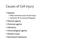
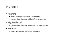
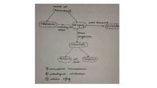
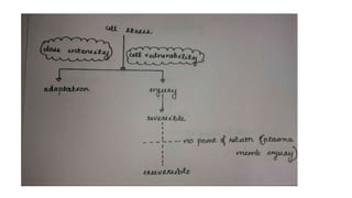
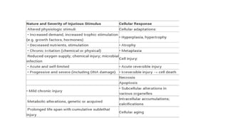
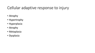
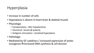
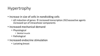
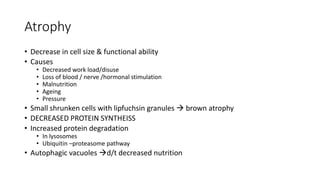
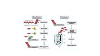
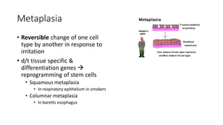
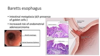
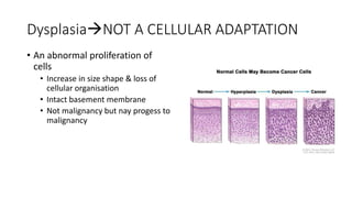
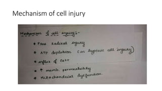
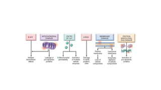
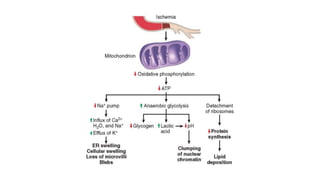
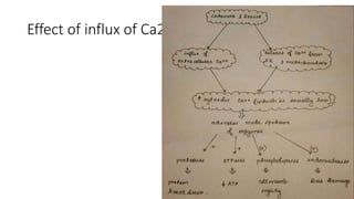
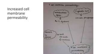
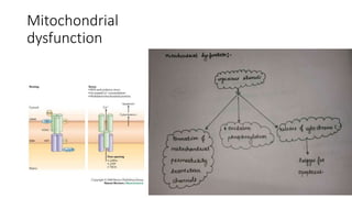
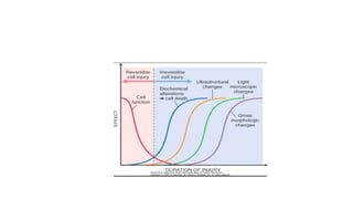
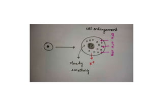
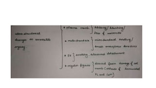
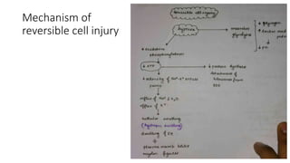
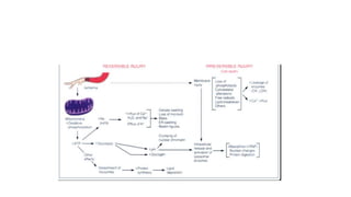
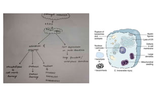
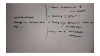
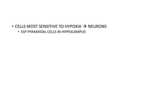
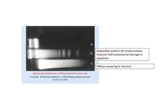
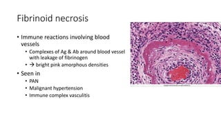
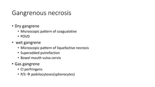
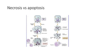
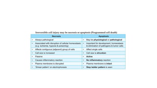
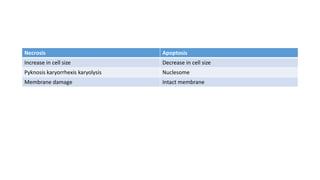
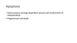
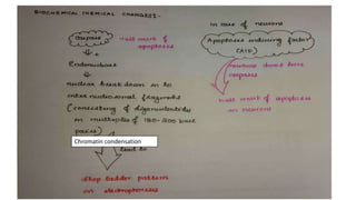
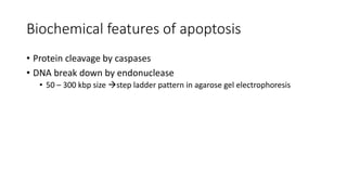
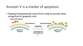
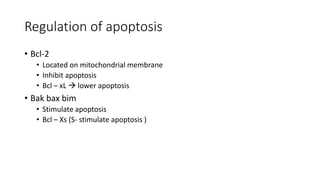
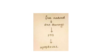
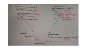
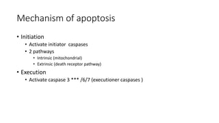
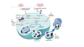
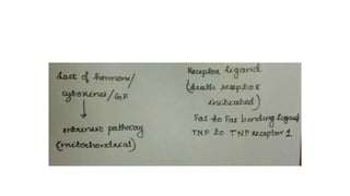
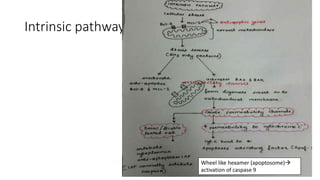
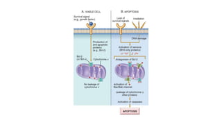
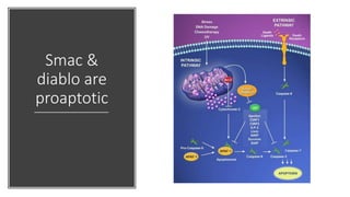
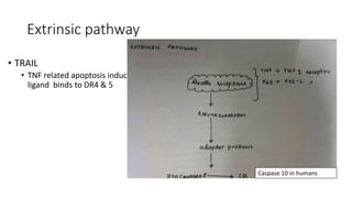
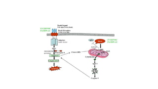
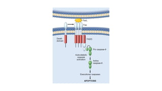
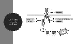
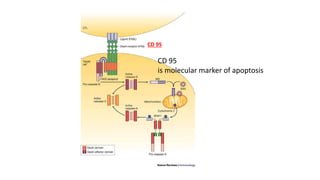
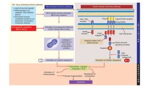
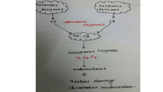
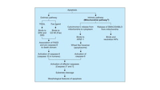
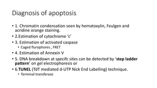
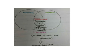
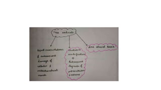
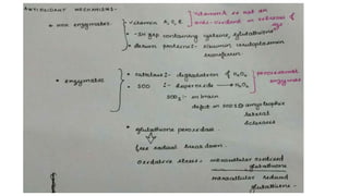
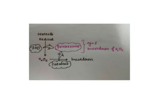
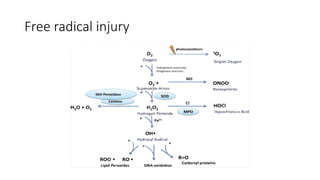
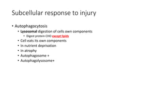
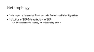
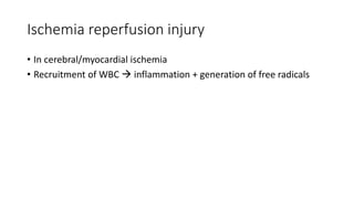
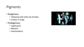
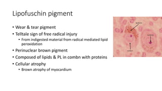
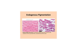
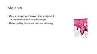
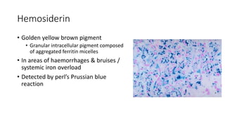
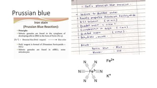
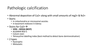
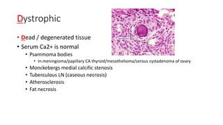
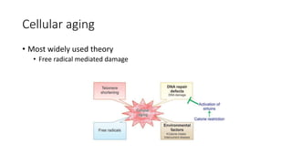
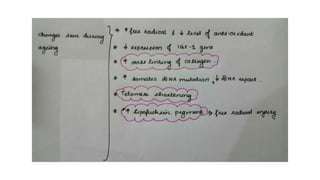
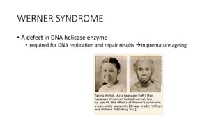
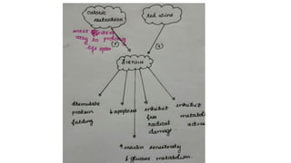
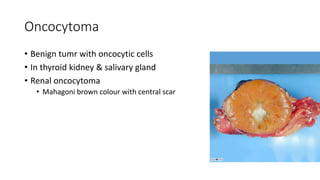
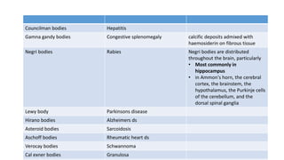
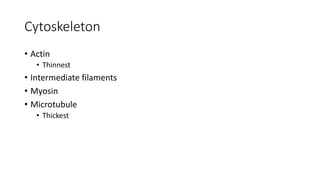
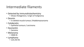
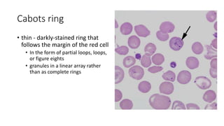
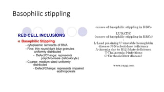
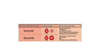
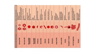
2. Causes of Cell injury
• Hypoxia
• Most common cause of cell injury
• Ischemia mc cause of hypoxia
• Physical agents
• Chemical agents
• Infections
• Immunological agents
• Genetic cause
• Nutritional imbalance
3. Hypoxia
• Neurons
• Most susceptible tissue to ischemia
• Irreversible damage with in 3 to 4 minutes
• Myocardial cells
• Irreversible damage with in 20 to 30 minutes
• Fibroblast
• Most resistant to ischemic damage
9. Hyperplasia
• Increase in number of cells
• Hyperplasia is absent in heart brain & skeletal muscle
• Physiologic
• Compensatory : after hepatectomy
• Hormonal : breast @ puberty
• Antigenic stimulation : lymphoid hyperplasia
• Pathologic
• Mediated by GF cytokines / increased expression of proto-
oncogenesincreased DNA synthesis & cell division
10. Hypertrophy
• Increase in size of cells in nondividing cells
• d/t induction of genes increased transcription /GF/vasoactive agents
increased syn of intracellular components
• Increased mechanical demand
• Physiological
• Skeletal muscle
• Pathological
• Increased endocrine stimulation
• Lactating breast
11. • Hyperplasia & hypertrophy occur together
• in gravid uterus
• Mainly hypertrophy + only some hyperplasia
• In breast during puberta period & pregnancy
12. Atrophy
• Decrease in cell size & functional ability
• Causes
• Decreased work load/disuse
• Loss of blood / nerve /hormonal stimulation
• Malnutrition
• Ageing
• Pressure
• Small shrunken cells with lipfuchsin granules brown atrophy
• DECREASED PROTEIN SYNTHEISS
• Increased protein degradation
• In lysosomes
• Ubiquitin –proteasome pathway
• Autophagic vacuoles d/t decreased nutrition
13.
14. Metaplasia
• Reversible change of one cell
type by another in response to
irritation
• d/t tissue specific &
differentiation genes
reprogramming of stem cells
• Squamous metaplasia
• In respiratory epithelium in smokers
• Columnar metaplasia
• In baretts esophagus
15. • Vitamin A deficiency or excess metaplasia
• Deficiency squamous metaplasia in respiratory epithelium
16. Baretts esophagus
• Intestinal metaplasia (d/t presence
of goblet cells )
• Increased risk of endometrial
adenocarcinoma
17. DysplasiaNOT A CELLULAR ADAPTATION
• An abnormal proliferation of
cells
• Increase in size shape & loss of
cellular organisation
• Intact basement membrane
• Not malignancy but nay progess to
malignancy
26. Features of reversible injury
• Cellular swelling
• First manifestation of all cell injury
• Except for apoptosis in which cell shrinkage is seen
• Hydropic degeration /cloudy swelling
• Intacellular water accumulation
32. Myelin figures are seen in cell inury
• Whorls of laminates PL & Ca2+
• More promineny in necrosis than in
reversible cell injury
• Break down of membrane organelles
composed of phospholipids
33. Light microscopic changes in reversible injury
• Irregular swelling /cloudy
swelling/hydropic change
• Prominent in kidney
• Earliest
• Fatty changes
• In cells involved in fat metabolism
• In hepatocytes & myocardial cells
34. Irreversible cell injury
• Severe membrane damage
• Masive influx of ca2+
• Efflux of intracellular enzymes in to circulation
• Marked mitochondrial dysfunction
35.
36.
37.
38. • CELLS MOST SENSITIVE TO HYPOXIA NEURONS
• ESP PYRAMIDAL CELLS IN HIPPOCAMPUS
39. Stepladder pattern d/t endonuclease
induced internucleosomal damage in
apoptosis
Diffuse smearing in necrosis
40. Cytoplasmic changes in necrosis
• Increased eosinophilia
• d/t protein denaturation
• Glassy appearance d/t loss of glycogen
• Cytoplasm is vacuolated moth eaten
• D/T ENZYMATIC DIGESTION VACOULE APPEAR
• Finally calcification
44. Coagulative necrosis
• Most common type
• Architecture of tissue preserved
• Ghost cell outline /tomb stone
• Cellular outline is maintained
• Loss of nucleus
• a/w ischemia (hypoxia)
• d/t denaturation of protein
• Seen in all tissues (heart***(mc) liver kidney)
• except brain LIQUEFACTIVE
• Zenkers degeration necrosis
• Waxy hyaline degenerstion of skeletal muscle in a/c infectious ds
45. Liquefactive necrosis
• Seen in
• focal bacterial
• Most common cause
• ischemia in brain
• d/t local stromal support & abundant liquefactive necrosis
• occasionally fungal infections
• d/t enzymatic degradation
• Inflammatory cells –> hydrolytic enzymes tissue degradation into liquid
viscuous mass no cellular details
46. Caseous necrosis
• Combination of liquefactive & coagulative
necrosis
• d/t mycolic acid of TB
• Friable cottage cheese like reaction
• Characteristic feature of TB
• Unlike coagulative necrosis
• Tissue architecture is completely obliterated
47.
48. • If caseation is absent & lymphoid infiltrate is absent
• In naked granuloma sarcoidosis
49. Fat necrosis
• d/t lipases on fatty tissue
• Seen in breast omentum pancreatitis
• Chalky white in appearance
50. Fibrinoid necrosis
• Immune reactions involving blood
vessels
• Complexes of Ag & Ab around blood vessel
with leakage of fibrinogen
• bright pink amorphous densities
• Seen in
• PAN
• Malignant hypertension
• Immune complex vasculitis
60. Biochemical features of apoptosis
• Protein cleavage by caspases
• DNA break down by endonuclease
• 50 – 300 kbp size step ladder pattern in agarose gel electrophoresis
84. Diagnosis of apoptosis
• 1. Chromatin condensation seen by hematoxylin, Feulgen and
acridine orange staining.
• 2.Estimation of cytochrome ‘c’
• 3. Estimation of activated caspase
• Caged flurophores , FRET
• 4. Estimation of Annexin V
• 5. DNA breakdown at specifc sites can be detected by ‘step ladder
pattern’ on gel electrophoresis or
• 6.TUNEL (TdT mediated d-UTP Nick End Labelling) technique.
• Terminal transferase
85.
86. Necroptosis
• Hybrid of necrosis + apoptosis
• Programmed cell death with out caspase activation
• Cell swelling
• Increased lysosomal permeability
• Cell membrane damage
• Presence of inflammation
88. Free radical
• Single unpaired electron in outer orbit
• Reactive oxygen species
• Normally produced as an unavoidable by product of mitochondrial respiration
96. Subcellular response to injury
• Autophagocytosis
• Lysosomal digestion of cells own components
• Digest protein CHO except lipids
• Cell eats its own components
• In nutrient deprivation
• In atrophy
• Autophagosome +
• Autophagolysosome+
97. Deletion of Atg5 increased susceptibility to
tuberculosis
• Atg5 autophagy related genes
98.
99. Heterophagy
• Cells ingest substances from outside for intracellular digestion
• Induction of SERhypertrophy of SER
• On phenobarbitone therapy hypertrophy of SER
100. Ischemia reperfusion injury
• In cerebral/myocardial ischemia
• Recruitment of WBC inflammation + generation of free radicals
103. Lipids
• Intracellular lipid accumulation steatosis
• Mainly in liver
• Triglyceride ***(mc)
• Cholestrol &CE
• PL
• Tigeroid / tabby cat myocardial cells
• Alternating bands of dark red normal myocardium with yellow streaks
• d/t lipid accumaltion
• Special stains
• Oil red O stain
• Sudan black B
• Sudan IV
• Osmium tetroxide
104. Proteins
• In proteinuria Protein accumalates in proximal renal tubule
• Russel bodies
• Intracytoplasmic accumaltion of Ig
• Defective protein folding accumulation
• Alzheimers d/s /parkinsons ds/huntingtons ds
Russel bodies
109. Lipofuschin pigment
• Wear & tear pigment
• Telltale sign of free radical injury
• From indigested material from radical mediated lipid
peroxidation
• Perinuclear brown pigment
• Composed of lipids & PL in combn with proteins
• Cellular atrophy
• Brown atrophy of myocardium
110.
111. Melanin
• Only endogenous brown black pigment
• In melanocytes & substantia nigra
• Detected by fontanna masson staining
112. Hemosiderin
• Golden yellow brown pigment
• Granular intracellular pigment composed
of aggregated ferritin micelles
• In areas of haemorrhages & bruises /
systemic iron overload
• Detected by perl’s Prussian blue
reaction
114. Pathologic calcification
• Abnormal deposition of Ca2+ along with small amounts of mg2+ & fe2+
• Starts
• In mitochondria or microsomal vesicles
• In basement mebrane in kidney
• Stains for Ca2+
• VON – KOSSA (BEST)
• ALIZARIN RED-S
• Calcein stain
• Tetracycline labelling index (best method to detect bone demineralisation)
• 2 types
• Dystrophic
• Metastatic
115. Dystrophic
• Dead / degenerated tissue
• Serum Ca2+ is normal
• Psammoma bodies
• In meningioma/papillary CA thyroid/mesothelioma/serous cystadenoma of ovary
• Monckebergs medial calcific stenosis
• Tuberculous LN (caseous necrosis)
• Atherosclerosis
• Fat necrosis
116. Metastatic
• Raised Ca2+ calcification in normal tissues
• Causes
• Hyperparathyroidism ***
• **Parathyroid adenoma ***
• Vitamin A toxicity
• Vitamin A activates osteoclast increased bone resorption & hypercalcemia
• Milk alkali syndrome
• Renal failure
• Vitamin D related disorders
• Vitamin D intoxication
• Williams syndrome (idiopathic hypercalcemia of infancy)
• Sarcoidosis
• Saracoid gramuloma synthesise excess 1,25 OH vitamin D3
117. Metastatic calcification
• Most coomon site
• Lung alveoli
• Systemic arteries & pulmonary V
• Gastric mucosa
• Cornea & conjunctiva
118. Glycogen storage ds
• Best carmine or PAS with diastase sensitivity
• Best fixative is alcohol
121. Telomeres
• Short stretches of DNA @ end of chromosomes
• Important in ensuring complete replication of chromosomes & protects
chromosomal ends from fusion & degradation
123. Telomerase
• Aare reverse transcriptases or RNA dependent polymerase
• Normally present in germ cells >> stem cells
• Presence in normal somatic cells neoplastic transformation
124.
125.
126. WERNER SYNDROME
• A defect in DNA helicase enzyme
• required for DNA replication and repair results in premature ageing
127. sirtuin
• Plays an important role in aging / DM & various cancers
• NAD dependant deacetylase
• Stimulates protein folding
• Decrease apoptosis
• Inhibit free radical damage
• Increase insulin sensitivity
128.
129.
130. • Commonest fixative
• In light microscopy 10 % bufferd formalin
• In e microscopy glutaraldehyde
135. Oncocytoma
• Benign tumr with oncocytic cells
• In thyroid kidney & salivary gland
• Renal oncocytoma
• Mahagoni brown colour with central scar
136. Councilman bodies Hepatitis
Gamna gandy bodies Congestive splenomegaly calcific deposits admixed with
haemosiderin on fibrous tissue
Negri bodies Rabies Negri bodies are distributed
throughout the brain, particularly
• Most commonly in
hippocampus
• in Ammon's horn, the cerebral
cortex, the brainstem, the
hypothalamus, the Purkinje cells
of the cerebellum, and the
dorsal spinal ganglia
Lewy body Parkinsons disease
Hirano bodies Alzheimers ds
Asteroid bodies Sarcoidosis
Aschoff bodies Rheumatic heart ds
Verocay bodies Schwannoma
Cal exner bodies Granulosa
145. Cabots ring
• thin - darkly-stained ring that
follows the margin of the red cell
• In the form of partial loops, loops,
or figure eights
• granules in a linear array rather
than as complete rings