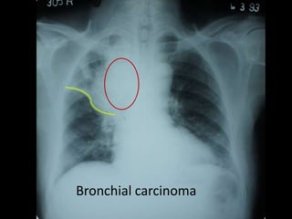
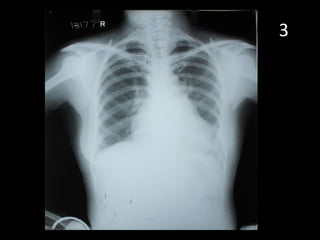
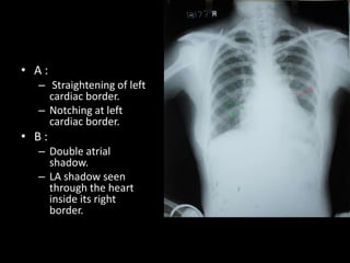
1. Chest x-ray showed free gas under the right hemidiaphragm.
2. This finding suggests a hiatal hernia, where part of the stomach protrudes through the diaphragm into the chest.
3. The 67-year old patient presented with chronic cough and mild heartburn, consistent with symptoms of a hiatal hernia.
The basics of Chest Radiology explained for the undergraduate students. The technical aspects including the various views, exposure, rotation and breath described.
The inside out approach of interpretation explained. The ABCDEFGH description includes Airway, Bones & soft tissue, Cardiac shadow, Diaphragm, Effusion (pleura), Fields (lungs), Gastric bubble and Hila & mediastinum.
The basic cardiac and lung pathologies discussed.
genitourinary tb - contains radiological findings of genitourinary tuberculosis including ivp,, hsg, usg and ct findings in kidney, ureter, urinary bladder, uterus and prostate
Couples presenting to the infertility clinic- Do they really have infertility...Sujoy Dasgupta
Dr Sujoy Dasgupta presented the study on "Couples presenting to the infertility clinic- Do they really have infertility? – The unexplored stories of non-consummation" in the 13th Congress of the Asia Pacific Initiative on Reproduction (ASPIRE 2024) at Manila on 24 May, 2024.
Factory Supply Best Quality Pmk Oil CAS 28578–16–7 PMK Powder in Stockrebeccabio
Factory Supply Best Quality Pmk Oil CAS 28578–16–7 PMK Powder in Stock
Telegram: bmksupplier
signal: +85264872720
threema: TUD4A6YC
You can contact me on Telegram or Threema
Communicate promptly and reply
Free of customs clearance, Double Clearance 100% pass delivery to USA, Canada, Spain, Germany, Netherland, Poland, Italy, Sweden, UK, Czech Republic, Australia, Mexico, Russia, Ukraine, Kazakhstan.Door to door service
Hot Selling Organic intermediates
These lecture slides, by Dr Sidra Arshad, offer a quick overview of physiological basis of a normal electrocardiogram.
Learning objectives:
1. Define an electrocardiogram (ECG) and electrocardiography
2. Describe how dipoles generated by the heart produce the waveforms of the ECG
3. Describe the components of a normal electrocardiogram of a typical bipolar leads (limb II)
4. Differentiate between intervals and segments
5. Enlist some common indications for obtaining an ECG
Study Resources:
1. Chapter 11, Guyton and Hall Textbook of Medical Physiology, 14th edition
2. Chapter 9, Human Physiology - From Cells to Systems, Lauralee Sherwood, 9th edition
3. Chapter 29, Ganong’s Review of Medical Physiology, 26th edition
4. Electrocardiogram, StatPearls - https://www.ncbi.nlm.nih.gov/books/NBK549803/
5. ECG in Medical Practice by ABM Abdullah, 4th edition
6. ECG Basics, http://www.nataliescasebook.com/tag/e-c-g-basics
Title: Sense of Taste
Presenter: Dr. Faiza, Assistant Professor of Physiology
Qualifications:
MBBS (Best Graduate, AIMC Lahore)
FCPS Physiology
ICMT, CHPE, DHPE (STMU)
MPH (GC University, Faisalabad)
MBA (Virtual University of Pakistan)
Learning Objectives:
Describe the structure and function of taste buds.
Describe the relationship between the taste threshold and taste index of common substances.
Explain the chemical basis and signal transduction of taste perception for each type of primary taste sensation.
Recognize different abnormalities of taste perception and their causes.
Key Topics:
Significance of Taste Sensation:
Differentiation between pleasant and harmful food
Influence on behavior
Selection of food based on metabolic needs
Receptors of Taste:
Taste buds on the tongue
Influence of sense of smell, texture of food, and pain stimulation (e.g., by pepper)
Primary and Secondary Taste Sensations:
Primary taste sensations: Sweet, Sour, Salty, Bitter, Umami
Chemical basis and signal transduction mechanisms for each taste
Taste Threshold and Index:
Taste threshold values for Sweet (sucrose), Salty (NaCl), Sour (HCl), and Bitter (Quinine)
Taste index relationship: Inversely proportional to taste threshold
Taste Blindness:
Inability to taste certain substances, particularly thiourea compounds
Example: Phenylthiocarbamide
Structure and Function of Taste Buds:
Composition: Epithelial cells, Sustentacular/Supporting cells, Taste cells, Basal cells
Features: Taste pores, Taste hairs/microvilli, and Taste nerve fibers
Location of Taste Buds:
Found in papillae of the tongue (Fungiform, Circumvallate, Foliate)
Also present on the palate, tonsillar pillars, epiglottis, and proximal esophagus
Mechanism of Taste Stimulation:
Interaction of taste substances with receptors on microvilli
Signal transduction pathways for Umami, Sweet, Bitter, Sour, and Salty tastes
Taste Sensitivity and Adaptation:
Decrease in sensitivity with age
Rapid adaptation of taste sensation
Role of Saliva in Taste:
Dissolution of tastants to reach receptors
Washing away the stimulus
Taste Preferences and Aversions:
Mechanisms behind taste preference and aversion
Influence of receptors and neural pathways
Impact of Sensory Nerve Damage:
Degeneration of taste buds if the sensory nerve fiber is cut
Abnormalities of Taste Detection:
Conditions: Ageusia, Hypogeusia, Dysgeusia (parageusia)
Causes: Nerve damage, neurological disorders, infections, poor oral hygiene, adverse drug effects, deficiencies, aging, tobacco use, altered neurotransmitter levels
Neurotransmitters and Taste Threshold:
Effects of serotonin (5-HT) and norepinephrine (NE) on taste sensitivity
Supertasters:
25% of the population with heightened sensitivity to taste, especially bitterness
Increased number of fungiform papillae
Title: Sense of Smell
Presenter: Dr. Faiza, Assistant Professor of Physiology
Qualifications:
MBBS (Best Graduate, AIMC Lahore)
FCPS Physiology
ICMT, CHPE, DHPE (STMU)
MPH (GC University, Faisalabad)
MBA (Virtual University of Pakistan)
Learning Objectives:
Describe the primary categories of smells and the concept of odor blindness.
Explain the structure and location of the olfactory membrane and mucosa, including the types and roles of cells involved in olfaction.
Describe the pathway and mechanisms of olfactory signal transmission from the olfactory receptors to the brain.
Illustrate the biochemical cascade triggered by odorant binding to olfactory receptors, including the role of G-proteins and second messengers in generating an action potential.
Identify different types of olfactory disorders such as anosmia, hyposmia, hyperosmia, and dysosmia, including their potential causes.
Key Topics:
Olfactory Genes:
3% of the human genome accounts for olfactory genes.
400 genes for odorant receptors.
Olfactory Membrane:
Located in the superior part of the nasal cavity.
Medially: Folds downward along the superior septum.
Laterally: Folds over the superior turbinate and upper surface of the middle turbinate.
Total surface area: 5-10 square centimeters.
Olfactory Mucosa:
Olfactory Cells: Bipolar nerve cells derived from the CNS (100 million), with 4-25 olfactory cilia per cell.
Sustentacular Cells: Produce mucus and maintain ionic and molecular environment.
Basal Cells: Replace worn-out olfactory cells with an average lifespan of 1-2 months.
Bowman’s Gland: Secretes mucus.
Stimulation of Olfactory Cells:
Odorant dissolves in mucus and attaches to receptors on olfactory cilia.
Involves a cascade effect through G-proteins and second messengers, leading to depolarization and action potential generation in the olfactory nerve.
Quality of a Good Odorant:
Small (3-20 Carbon atoms), volatile, water-soluble, and lipid-soluble.
Facilitated by odorant-binding proteins in mucus.
Membrane Potential and Action Potential:
Resting membrane potential: -55mV.
Action potential frequency in the olfactory nerve increases with odorant strength.
Adaptation Towards the Sense of Smell:
Rapid adaptation within the first second, with further slow adaptation.
Psychological adaptation greater than receptor adaptation, involving feedback inhibition from the central nervous system.
Primary Sensations of Smell:
Camphoraceous, Musky, Floral, Pepperminty, Ethereal, Pungent, Putrid.
Odor Detection Threshold:
Examples: Hydrogen sulfide (0.0005 ppm), Methyl-mercaptan (0.002 ppm).
Some toxic substances are odorless at lethal concentrations.
Characteristics of Smell:
Odor blindness for single substances due to lack of appropriate receptor protein.
Behavioral and emotional influences of smell.
Transmission of Olfactory Signals:
From olfactory cells to glomeruli in the olfactory bulb, involving lateral inhibition.
Primitive, less old, and new olfactory systems with different path
MANAGEMENT OF ATRIOVENTRICULAR CONDUCTION BLOCK.pdfJim Jacob Roy
Cardiac conduction defects can occur due to various causes.
Atrioventricular conduction blocks ( AV blocks ) are classified into 3 types.
This document describes the acute management of AV block.
Tom Selleck Health: A Comprehensive Look at the Iconic Actor’s Wellness Journeygreendigital
Tom Selleck, an enduring figure in Hollywood. has captivated audiences for decades with his rugged charm, iconic moustache. and memorable roles in television and film. From his breakout role as Thomas Magnum in Magnum P.I. to his current portrayal of Frank Reagan in Blue Bloods. Selleck's career has spanned over 50 years. But beyond his professional achievements. fans have often been curious about Tom Selleck Health. especially as he has aged in the public eye.
Follow us on: Pinterest
Introduction
Many have been interested in Tom Selleck health. not only because of his enduring presence on screen but also because of the challenges. and lifestyle choices he has faced and made over the years. This article delves into the various aspects of Tom Selleck health. exploring his fitness regimen, diet, mental health. and the challenges he has encountered as he ages. We'll look at how he maintains his well-being. the health issues he has faced, and his approach to ageing .
Early Life and Career
Childhood and Athletic Beginnings
Tom Selleck was born on January 29, 1945, in Detroit, Michigan, and grew up in Sherman Oaks, California. From an early age, he was involved in sports, particularly basketball. which played a significant role in his physical development. His athletic pursuits continued into college. where he attended the University of Southern California (USC) on a basketball scholarship. This early involvement in sports laid a strong foundation for his physical health and disciplined lifestyle.
Transition to Acting
Selleck's transition from an athlete to an actor came with its physical demands. His first significant role in "Magnum P.I." required him to perform various stunts and maintain a fit appearance. This role, which he played from 1980 to 1988. necessitated a rigorous fitness routine to meet the show's demands. setting the stage for his long-term commitment to health and wellness.
Fitness Regimen
Workout Routine
Tom Selleck health and fitness regimen has evolved. adapting to his changing roles and age. During his "Magnum, P.I." days. Selleck's workouts were intense and focused on building and maintaining muscle mass. His routine included weightlifting, cardiovascular exercises. and specific training for the stunts he performed on the show.
Selleck adjusted his fitness routine as he aged to suit his body's needs. Today, his workouts focus on maintaining flexibility, strength, and cardiovascular health. He incorporates low-impact exercises such as swimming, walking, and light weightlifting. This balanced approach helps him stay fit without putting undue strain on his joints and muscles.
Importance of Flexibility and Mobility
In recent years, Selleck has emphasized the importance of flexibility and mobility in his fitness regimen. Understanding the natural decline in muscle mass and joint flexibility with age. he includes stretching and yoga in his routine. These practices help prevent injuries, improve posture, and maintain mobilit
Prix Galien International 2024 Forum ProgramLevi Shapiro
June 20, 2024, Prix Galien International and Jerusalem Ethics Forum in ROME. Detailed agenda including panels:
- ADVANCES IN CARDIOLOGY: A NEW PARADIGM IS COMING
- WOMEN’S HEALTH: FERTILITY PRESERVATION
- WHAT’S NEW IN THE TREATMENT OF INFECTIOUS,
ONCOLOGICAL AND INFLAMMATORY SKIN DISEASES?
- ARTIFICIAL INTELLIGENCE AND ETHICS
- GENE THERAPY
- BEYOND BORDERS: GLOBAL INITIATIVES FOR DEMOCRATIZING LIFE SCIENCE TECHNOLOGIES AND PROMOTING ACCESS TO HEALTHCARE
- ETHICAL CHALLENGES IN LIFE SCIENCES
- Prix Galien International Awards Ceremony
ARTIFICIAL INTELLIGENCE IN HEALTHCARE.pdfAnujkumaranit
Artificial intelligence (AI) refers to the simulation of human intelligence processes by machines, especially computer systems. It encompasses tasks such as learning, reasoning, problem-solving, perception, and language understanding. AI technologies are revolutionizing various fields, from healthcare to finance, by enabling machines to perform tasks that typically require human intelligence.
- Video recording of this lecture in English language: https://youtu.be/lK81BzxMqdo
- Video recording of this lecture in Arabic language: https://youtu.be/Ve4P0COk9OI
- Link to download the book free: https://nephrotube.blogspot.com/p/nephrotube-nephrology-books.html
- Link to NephroTube website: www.NephroTube.com
- Link to NephroTube social media accounts: https://nephrotube.blogspot.com/p/join-nephrotube-on-social-media.html
4. • First look at the mediastinal contours—run your eye down the left side of the patient and
then up the right.
• The trachea should be central.
• The aortic arch is the first structure on the left, followed by the left pulmonary artery; notice
how you can trace the pulmonary artery branches fanning out through the lung
• Two thirds of the heart lies on the left side of the chest, with one third on the right. The heart
should take up no more than half of the thoracic cavity.
• The left border of the heart is made up by the left atrium and left ventricle.
• The right border is made up by the right atrium alone. Above the right heart border lies the
edge of the superior vena cava.
• The pulmonary arteries and main bronchi arise at the left and right hila. Enlarged lymph
nodes can also occur here, as can primary tumours. These make the hilum seem bulky.
• Now look at the lungs.
• Apart from the pulmonary vessels (arteries and veins),they should be black (because they are
full of air).
• Scan both lungs, starting at the apices and working down, comparing left with right at the
same level. The lungs extend behind the heart, so look here too. Force your eye to look at
the periphery of the lungs—you should not see many lung markings here; if you do then
there may be disease of the air spaces or interstitium.
• Don’t forget to look for a pneumothorax.
• Make sure you can see the surface of the hemidiaphragms curving downwards, and that the
costophrenic and cardiophrenic angles are not blunted—suggesting an effusion.
• Check there is no free air under the hemidiaphragm
5. The Normal Cardiac Borders in the PA View:
A) The left cardiac border is formed from above down by:
1. The aortic arch (= knob, knuckle).
2. The main pulmonary artery.
3. The left atrial appendage.
4. The left ventricle.
B) The right border of the heart is formed by:
1. In the lower two thirds by the right atrium.
2. In the upper third by the superior vena cava.
6.
7.
8. How to read
• This is a chest radiograph of a young male patient
PA view.
• The patient has taken a good inspiration and is
not rotated; the film is well penetrated.
• The trachea is central, the mediastinum is not
displaced.
• The mediastinal contours and hila seem normal.
• The lungs seem clear, with no pneumothorax.
• There is no free air under the diaphragm.
• The bones and soft tissues seem normal
15. Features to look for Present features
1. Opacification Dense opacification of the right upper
zone, V shaped
2. Horizontal fissure Has been displaced upwards from its
original position
3. Hilum enlarged or elevated Right hilum is elevated than left hilum
4.Smaller lung, smaller hemithorax Not present
5. Compensatory hyperinflation and Present
hyperlucency in other lobes
6. Tracheal deviation Absent
7. Elevated ipsilateral hemidiaphragm Present
25. Pneumothorax
Increased volume of the right hemithorax
with tracheal shift to the OPPOSITE side-
”PUSH” effect
Hyperlucency on the right side
Absence of lung markings
Presence of margin of collapsed lung at the
hilum
D/D:Unilateral emphysema
27. Hydropneumothorax
Horizontal fluid level with hyperlucent area
above it
Hyperlucent area with no lung markings
Presence of air+fluid = Hydropneumothorax
Trachea shifted to the SAME side-”PULL “
effect
D/D:lung abscess
29. Lung abscess
Loculated cavity with thin wall(maybe post
infective cyst, wall made up of a fibrous
tissue)
Presence of air fluid level
Mild trachea shift to the left(probably due
to rotation)
Fundic air bubbles separately seen
30. Lung abscess VS
Hydropneumothorax
Feature Lung Abscess Hydropneumothorax
Definition Intrapulmonary air- Intrapleural air-
fluid collection fluid collection
Shape of fluid Round, take up the Take up shape of the
shape of cavity wall thoracic cavity &
margin of collapsed
lung
Cavity Wall Clearly seen Not seen
Margin of collapsed Not seen May be seen
lung
Length of air-fluid Equal regardless of Length varies with
level radiographic radiographic
projection projection
32. Pleural effusion
Homogenous opacity of right middle &
lower zone = almost totally WHITE OUT
LUNG
Minimal tracheal shift to the left
Cardiac shift to the opposite side with
reduced left hemithorax size.
Obliteration of right costophrenic &
cardiophrenic angles
D/D:consolidation, pleural
thickening(heterogenous opacity), collapse.
39. • Double aortic
knuckle.
• 3 sign at left margin
of the aorta at the
level of coarctation.
Coarctation of Aorta.
40. Coarctation of Aorta.
• Rib notching:
– Due to pressure from tortuous intercostal arteries
acting as collaterals.
– Becomes evident after the age of 8 years.
• Cardiomegaly.
43. • A:
– Straightening of left
cardiac border.
– Notching at left
cardiac border.
• B:
– Double atrial
shadow.
– LA shadow seen
through the heart
inside its right
border.
46. Pulmonary Edema
Bat
wings/butterfly
distribution
Pleural effusion
47. Pulmonary Edema
• Produces air space opacities with variable
distribution.
• Sparing of the apices and extreme lung bases.
• “Butterfly” or “Bat wings” distribution – central
lungs affected more.
• With progression – opacities coalesce to form a
“white-out” on chest radiograph.
• Blurring of blood vessels occurs.
• Air bronchogram – indicating intra alveolar
edema.
48. Sequence of events on CXR in acute
pulmonary edema.
Early changes. Late changes.
1. Prominent pulmonary artery. 7. Fluffy shadows.
2. Prominent pulmonary lobe 8. Air bronchogram.
veins.
3. Interlobar fissure. 9. Bat’s wing appearance.
4. Perivascular cuffing. 10. Cardiomegaly .
5. Peribronchial cuffing. 11. Pleural effusion.
6. Kerley B lines.
49.
50. Differences..
Criteria. Cardiogenic (LVH) Non cardiogenic(ARDS)
1. Cardiomegaly . Comman +++ Uncomman+
2. Alveolar edema. +++ +++
3. Appearance of Bat’s wing /butterfly More patchy
shadow . appearance
4. Perivascular and More often seen Less likely
peribronchial cuffing.
5. Tendency for Yes No
gravitational
distribution.
6. Pleural effusion. May be present Unlikely
51. Silhouette Sign
• On the radiograph, loss of normal border of
the heart, aorta, or diaphragm by
intrathoracic lesions known as silhouette
sign.
Silhouete signs seen as Intrathoracic lesions
Upper right heart
Anterior segment of RUL
border/ascending aorta
Right heart border RML (medial)
Upper left heart border Anterior segment of LUL
Left heart border Lingula (anterior)
Apical portion of LUL
Aortic knob
(posterior)
Anterior hemidiaphragms Lower lobes (anterior)
54. Hiatal Hernia.
67 years old
patient
complained of
chronic cough and
mild heartburn
with no other
symptoms.
Editor's Notes
most common symptoms being dyspnoea and / or a non-productive cough. In patients who are profoundly immunocompromised, onset may be more dramatic and resemble other pulmonary infectionsbilateral, diffuse, often perihilar, fine, reticular interstitial opacification, which may appear somewhat granularBat's wing or butterfly pulmonary opacities
Nodules are either sharply or poorly defined 1–4-mm in size Diffuse, random distributionImaging Findings Discrete Distinctive Pin-point opacities Nodule size 1 – 2 mm in diameter - SPHERICAL LESION IN Miliary ( millet seed ) Pattern INTERSTITIUM B/L even distribution - WELL CIRCUMSCRIBED Basal Predominance HOMOGENOUS PATTERN Rare or non-existent calcifications Upto 30 % no radiological signs Thickening of intralobar fissure / interlobular septa Nodular irregularity of vessels HRCT – more sensitiveAlveolar microlithiasisPNEUMOCONIOSIS - Coal Workers Pneumoconiosis - Silicosis - Siderosis - StannosisSARCOIDOSIS METASTATIC LUNG DISEASES NON TB INFECTIONS – Histoplasmosis - Blastomycosis - Cryptococcosis - Nocardiosis – Coccidiodomycosis BRONCHIOLITIS OBLITERANS
Many thoracic aneurysms are visible on chest x ray and are characterised by widening of the mediastinal silhouette, enlargement of the aortic knob or displacement of the trachea of the midline.
Tracheal shift to the left-PUSH effect, increased volume of the right hemithoraxHyperlucency of right hemithorax + absence of lung markings + margin of collapsed lung(compression collapsed) = pneumothoraxTotal collapse=collapse like a cricket ball towards the hilum
Loculated cavity with thin wall(myb post infective cyst, wall made upof a fibrous tissue)Presence of air fluid levelMild trachea shift to the left(probably due to rotation)Fundic air bubbles separately seend/d:lung abscess(unresolved pneumonia), primary tumor (?blood clot)
Trachea remains central-why?Cardiac shift to the opposite side ,lefthemithorax size reducedHomogenous opacity starts from right middle zone downwards +obliteration of right costophrenic & cardiophrenic angles = almost totally WHITE OUT LUNGNot WHITE OUT LUNG because,not involve entire lungd/d:consolidation, pleural effusion/thickening(heterogenous opacity), collapse, upward enlargement of the liver(r/o)Notes:left 4th rib…cavities???
Not WHITE OUT LUNG because,not involve entire lungWhy notConsolidation-no air bronchogramPleural thickening-heterogenous opacityCollapse-presence of lung markings
Changes of 1-6 indicates interstitial edema.7-9 indicates alveolar edema.10-11 likely present in the cardiogenic causes of pulmonary edema (LVH)
left border of heart obliterated:lesion in lingulaRight hemidiaphragmobliterated:lower lobe lesionParatracheal stripes obliteration :paratrachealdzIt can be either consolidation or superimposed mass lesion.p/s :tram track appearance near arrow 2?? : thickened bronchial wall..(d/d:bronchiectasis ,cystic fibrosis,allergicaspergillosis,recurrent asthma/bronchitis in child)Air under left diaphragm : colonic gas shadow overlapping fundic gas bubble…..you can see the haustration!! Volvulus??
Air under diaphragm:In normal x-ray :there will be a fundic bubble gas on right side.Presence of air under diaphragm here may be due to perforation of hollow viscus (stomach @ intestine) following procedures.examples following laparatomy.Laparoscopy??
As u can see a curved white line lying behind the heart.