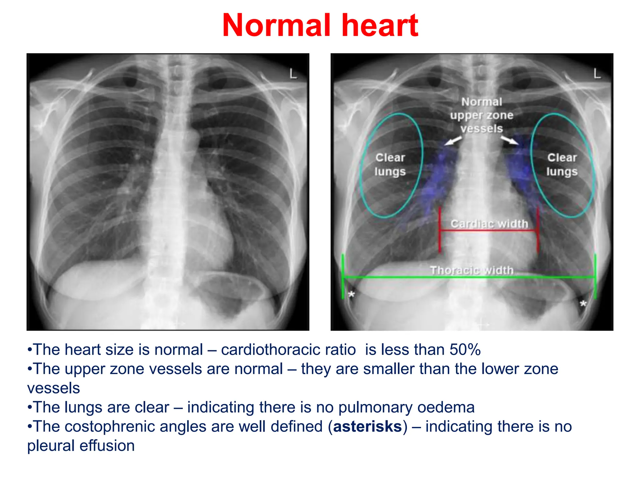
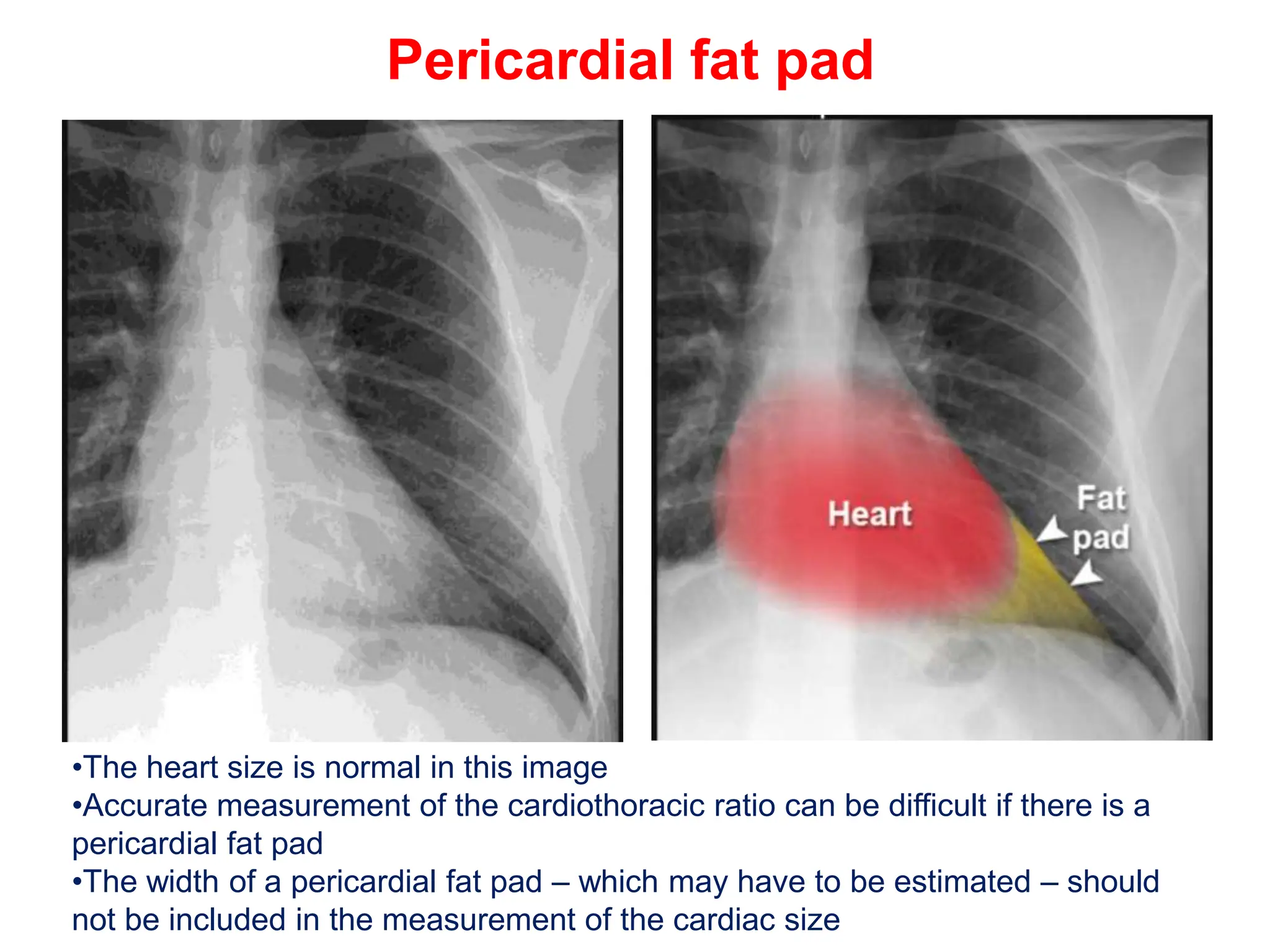
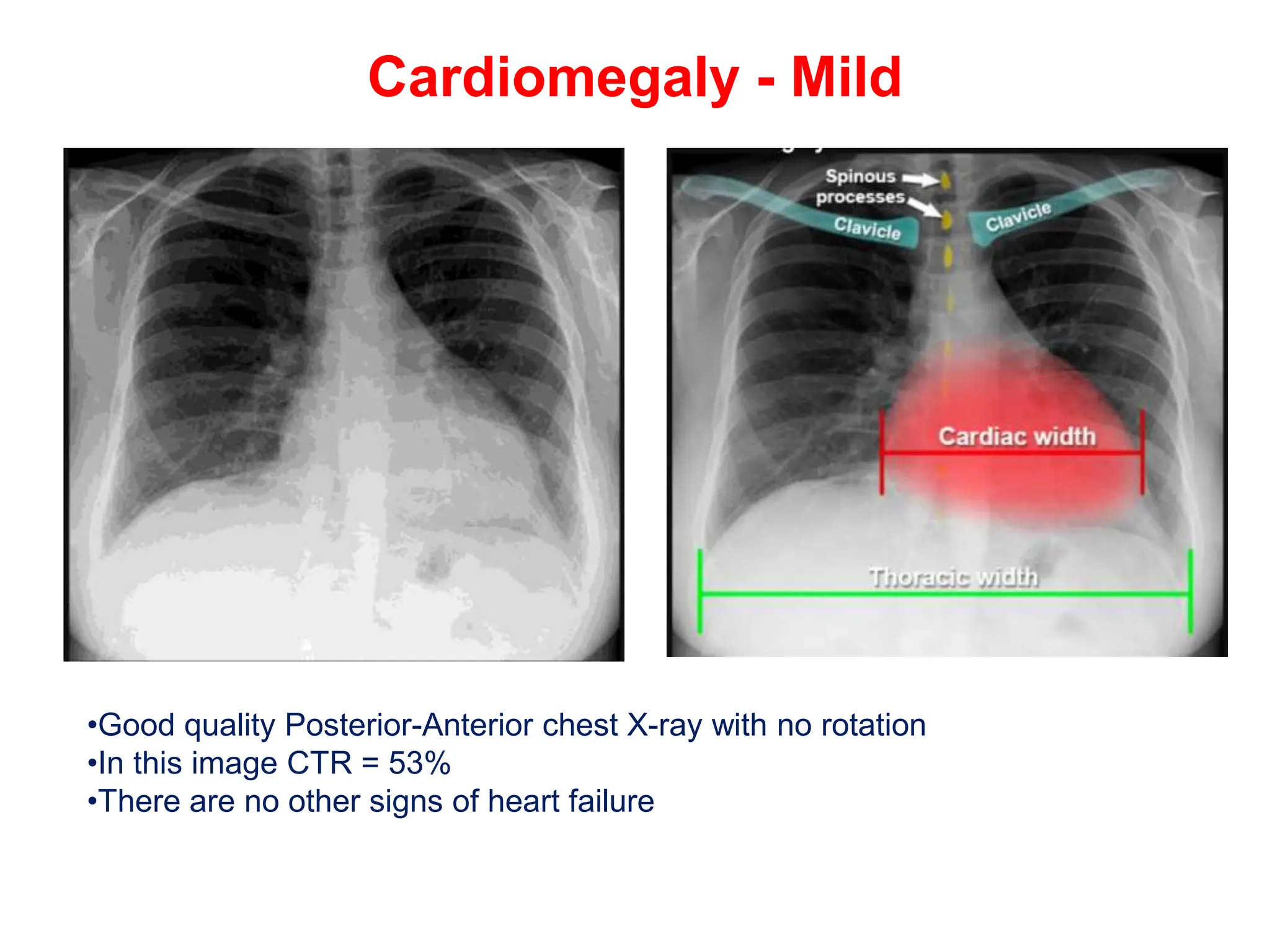
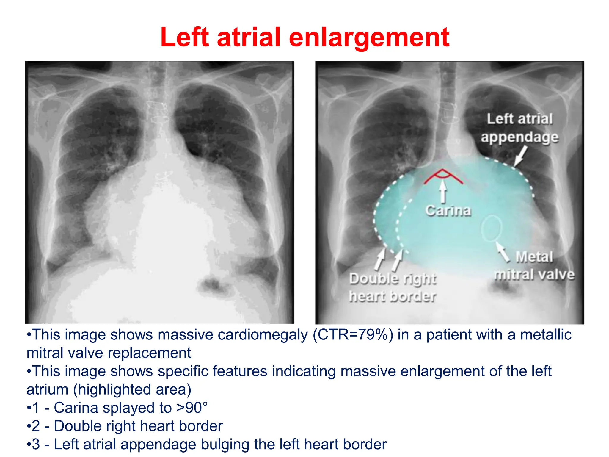
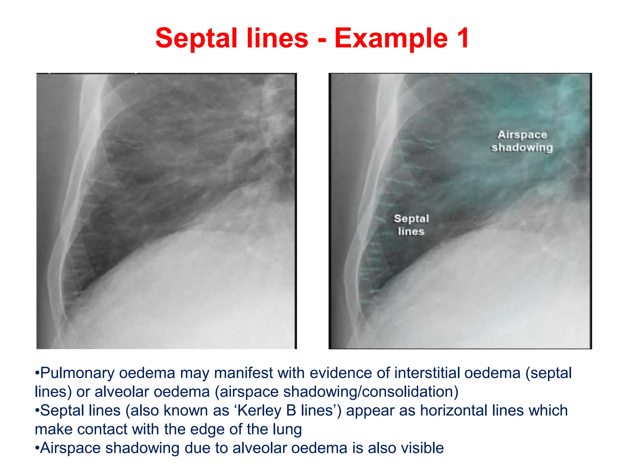
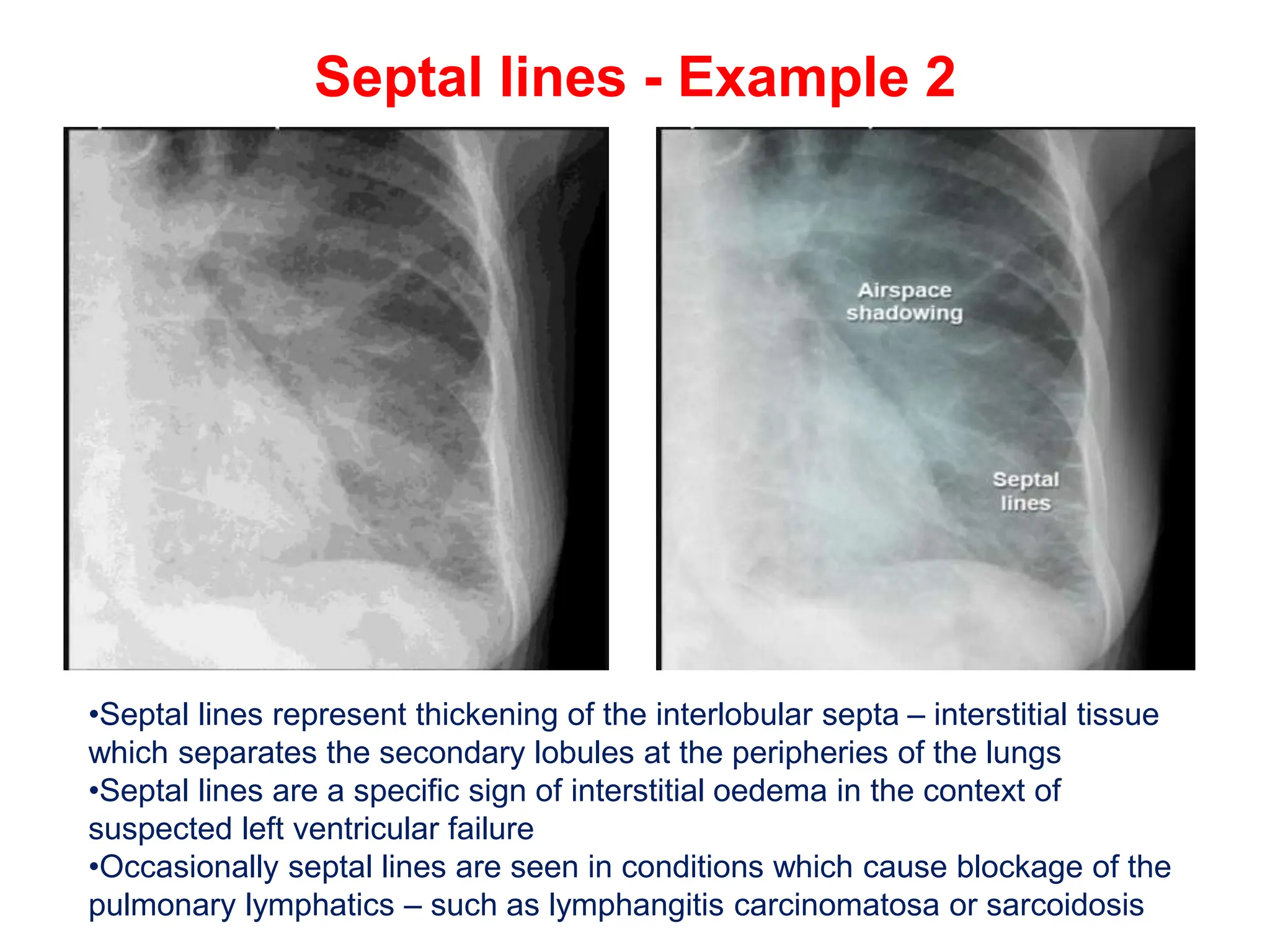
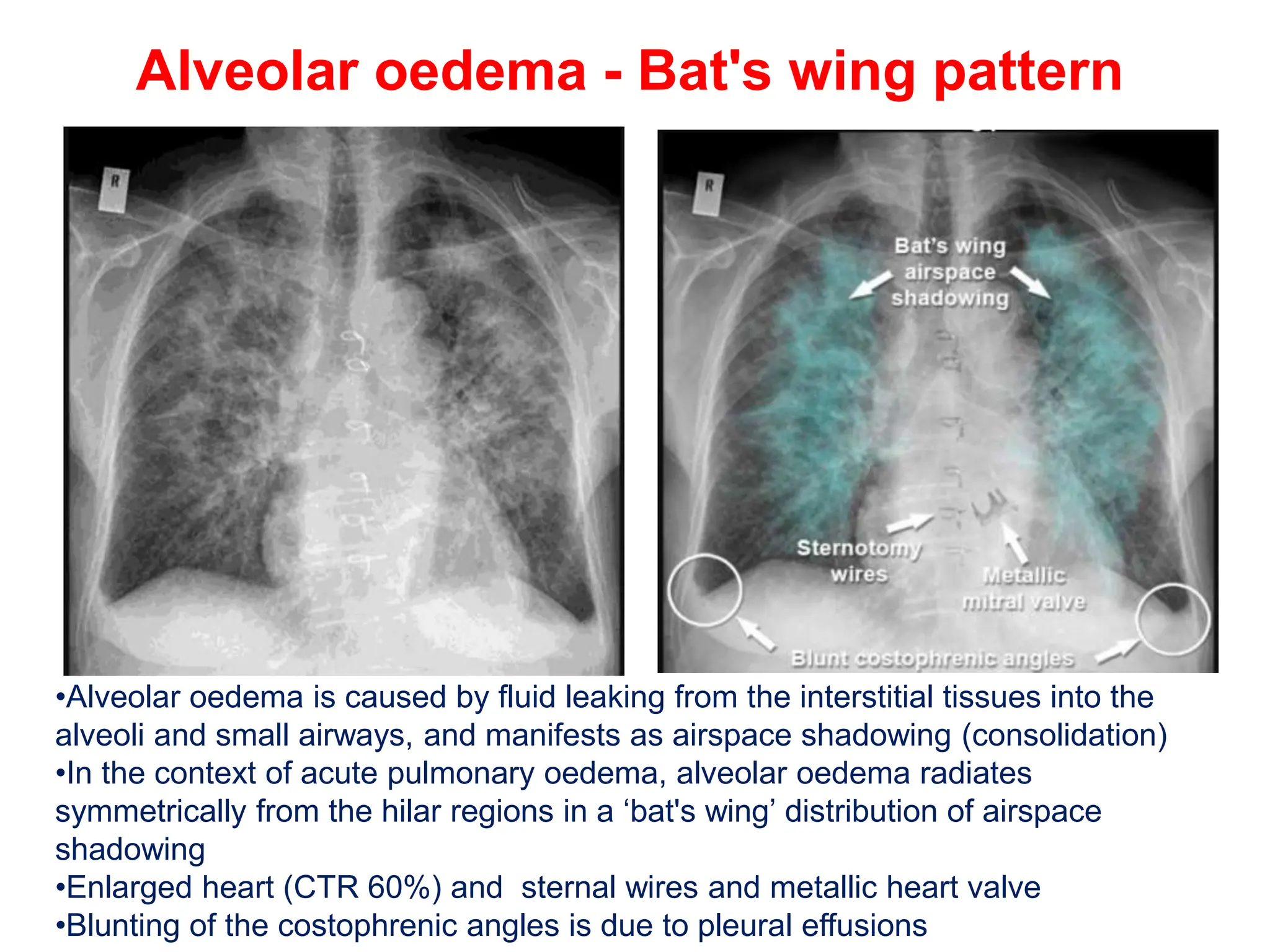
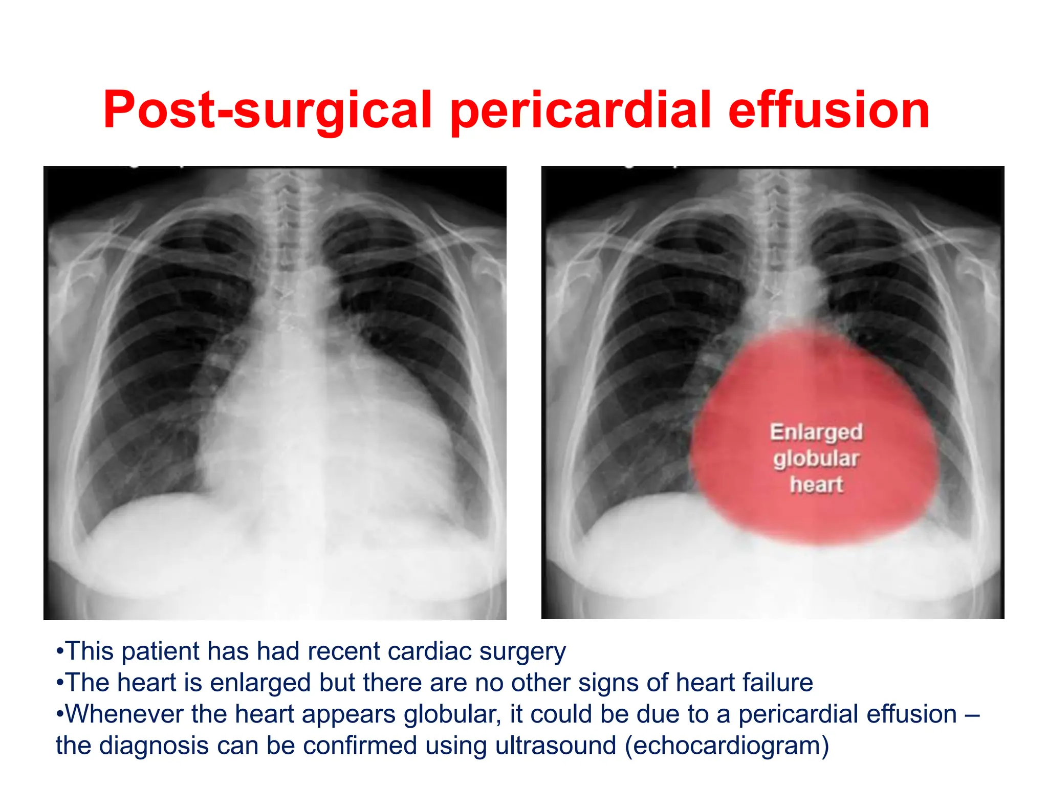
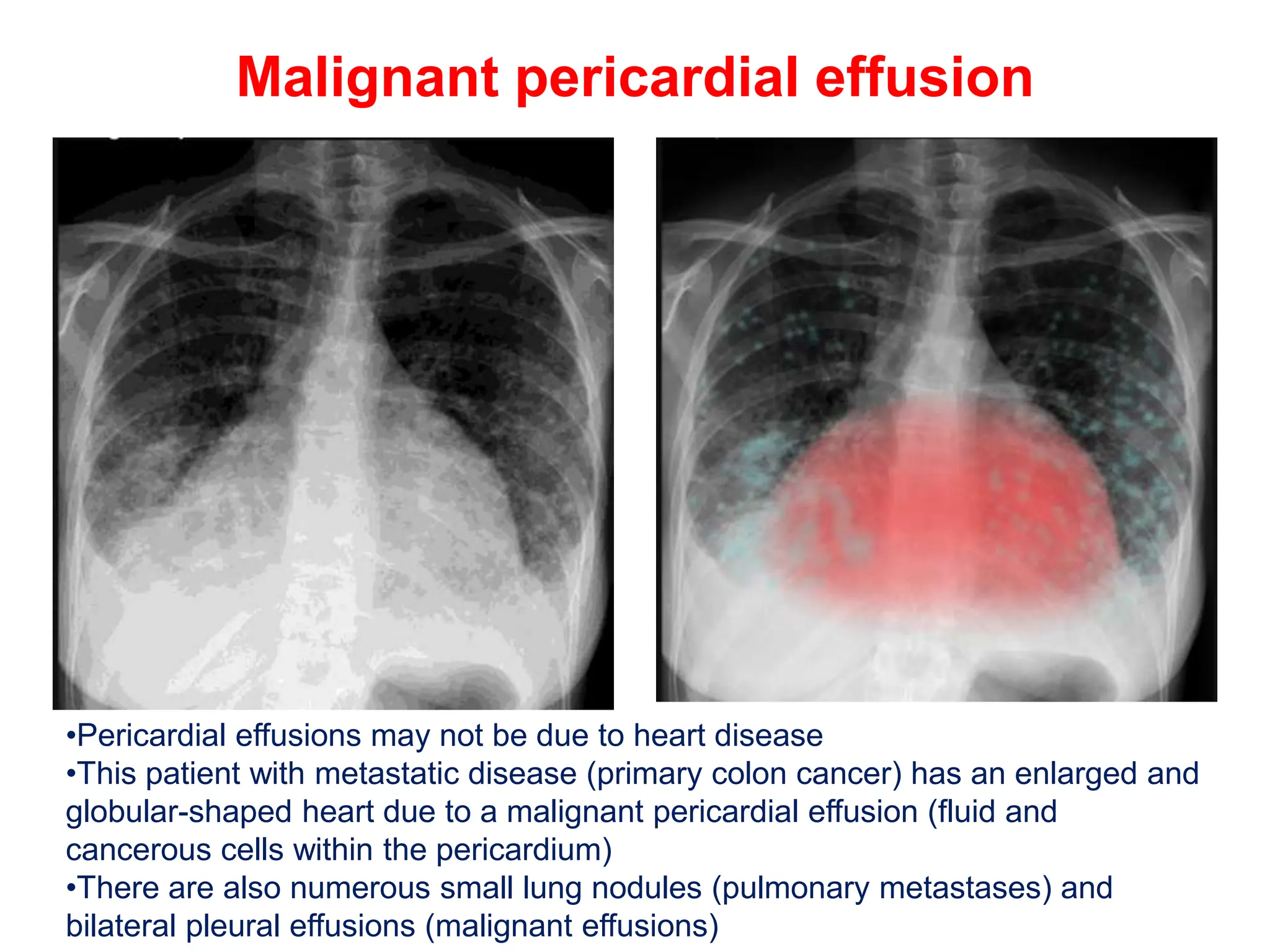
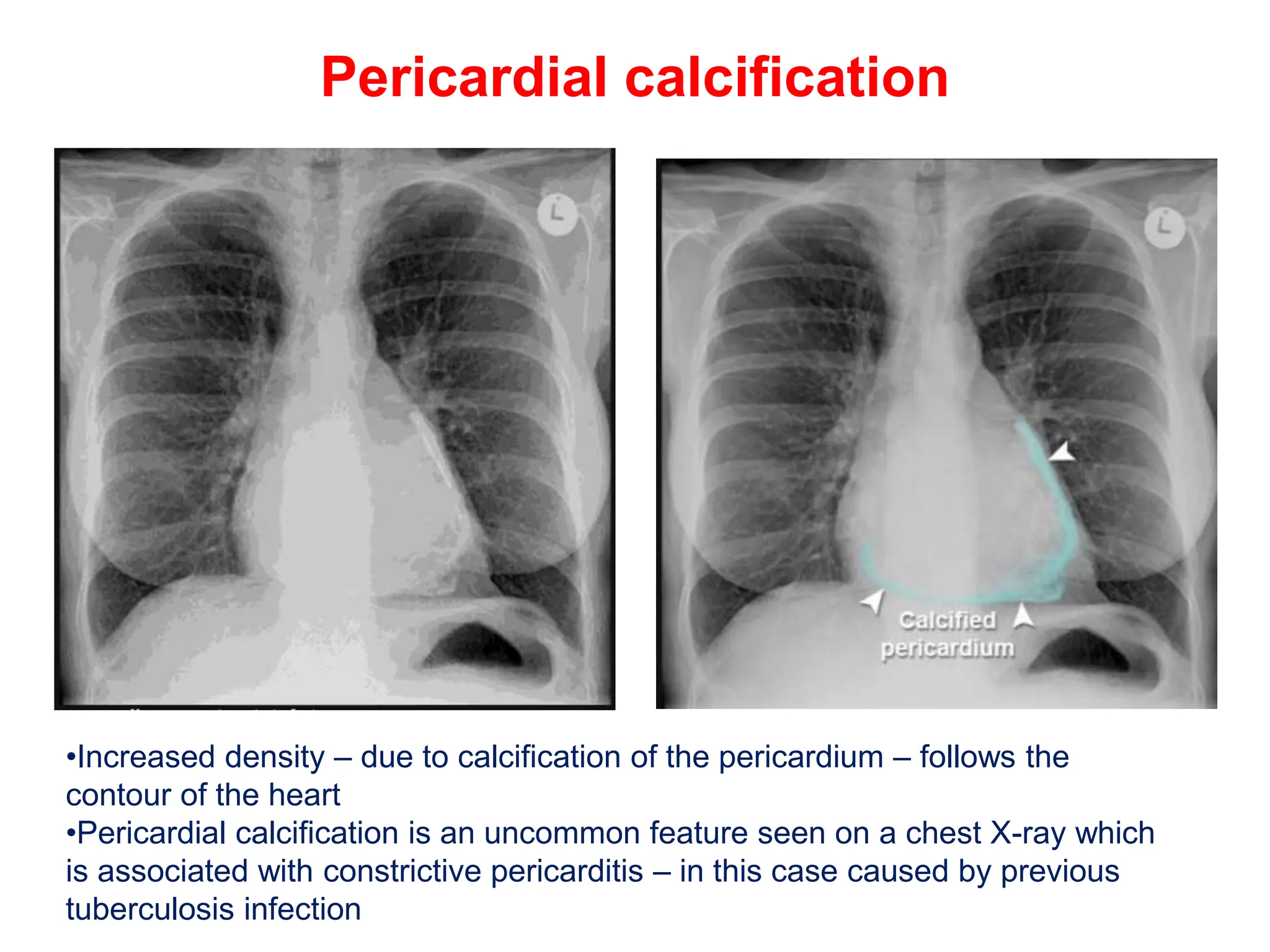
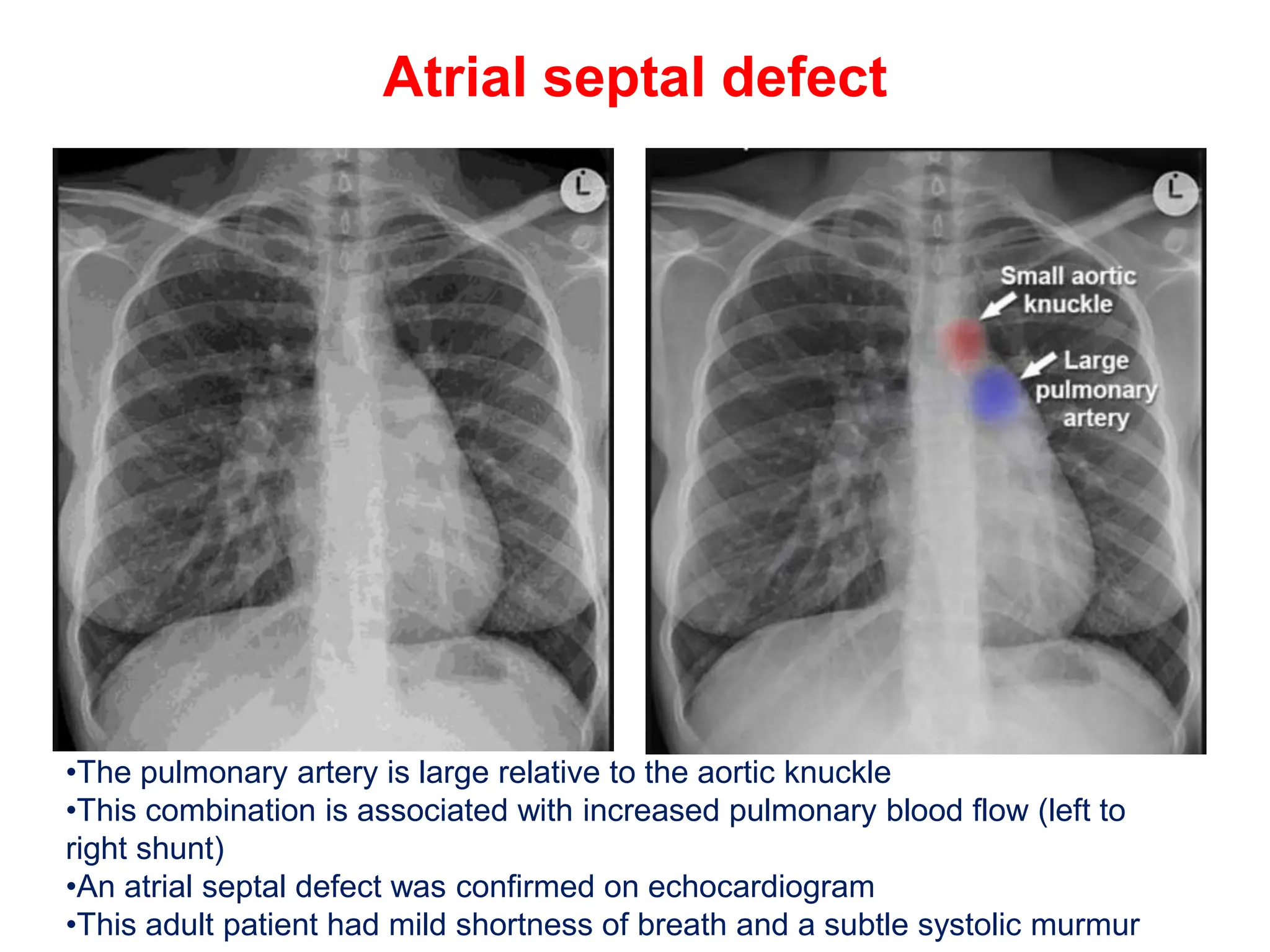
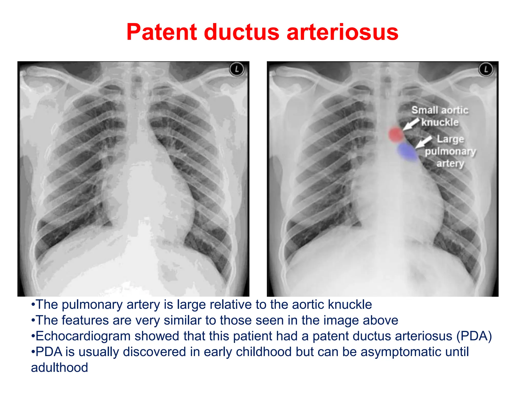
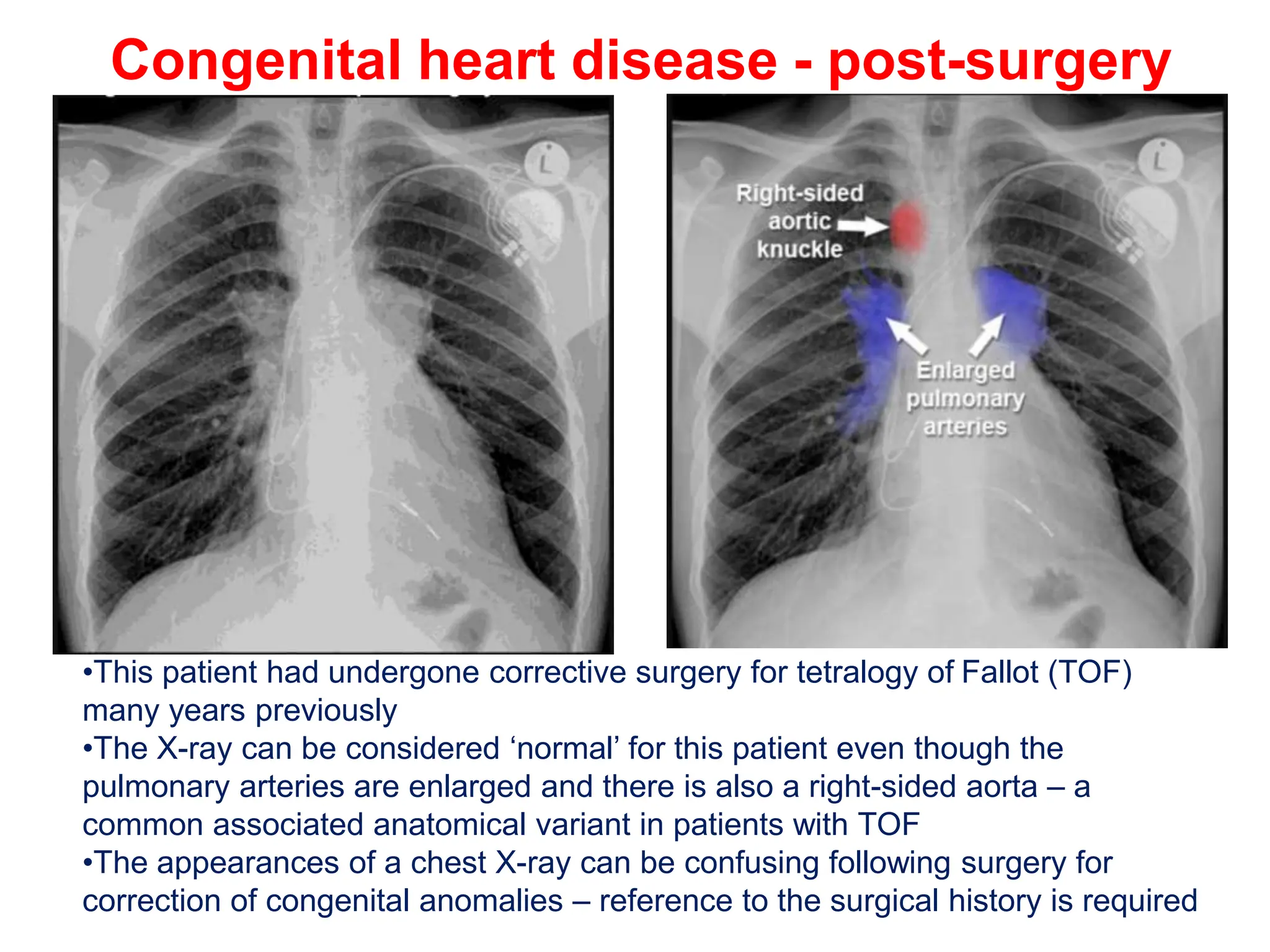
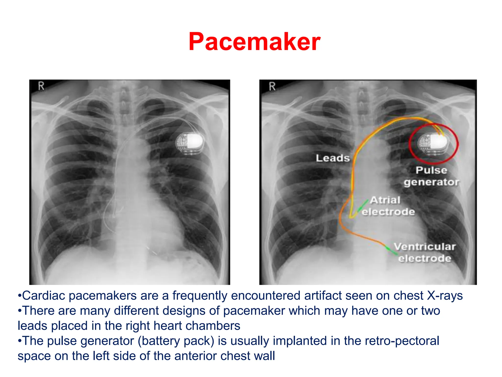
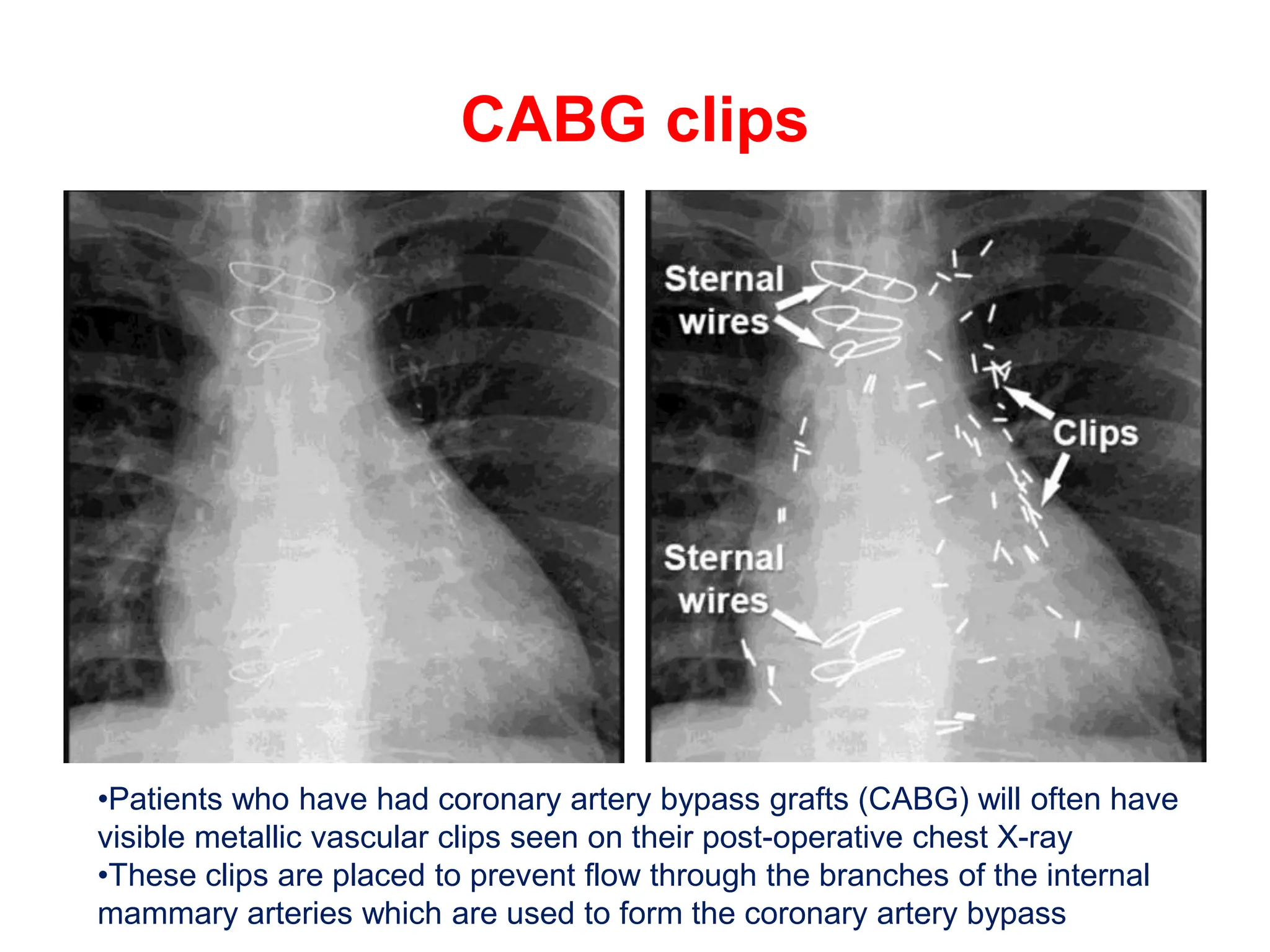
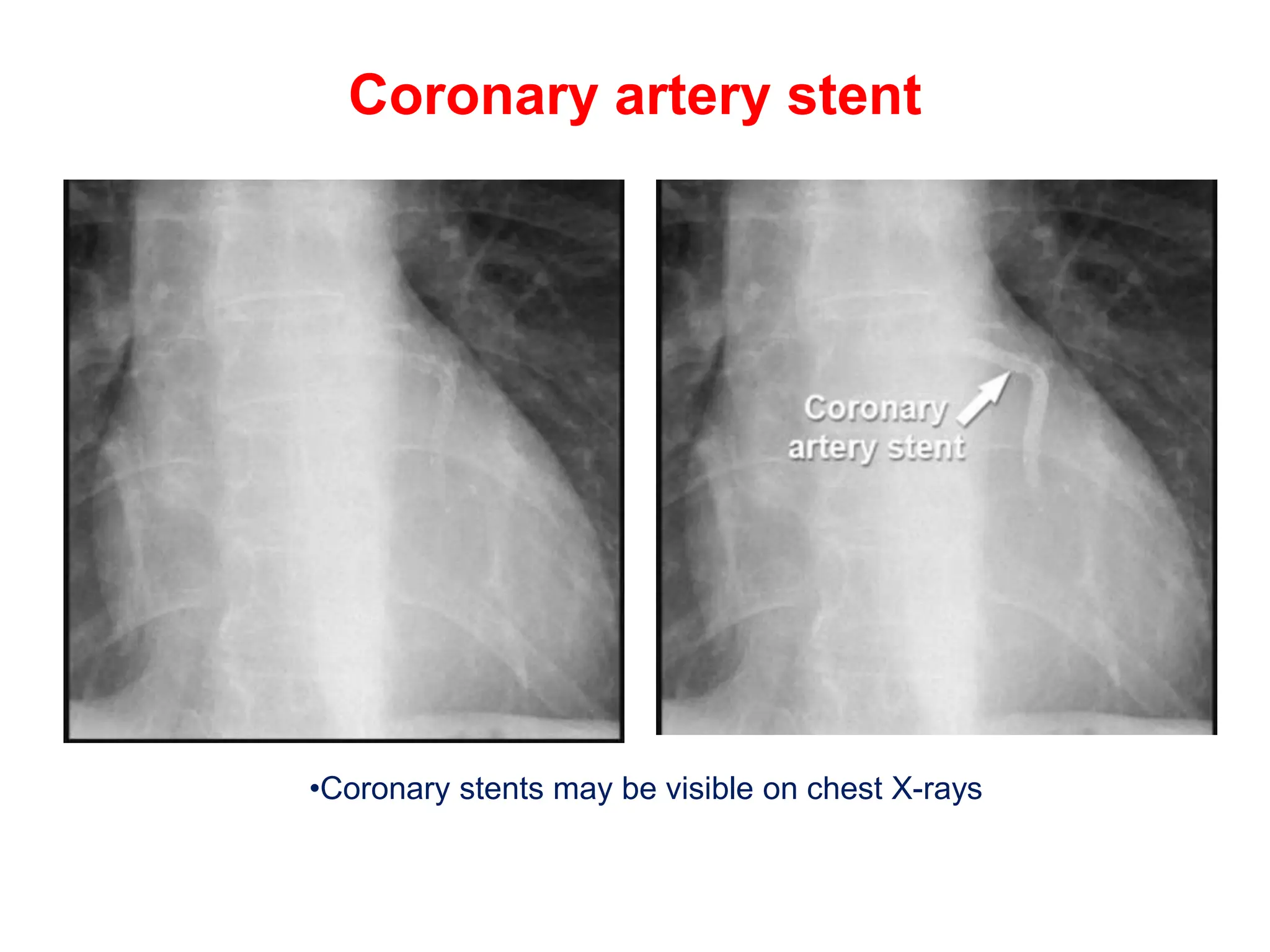
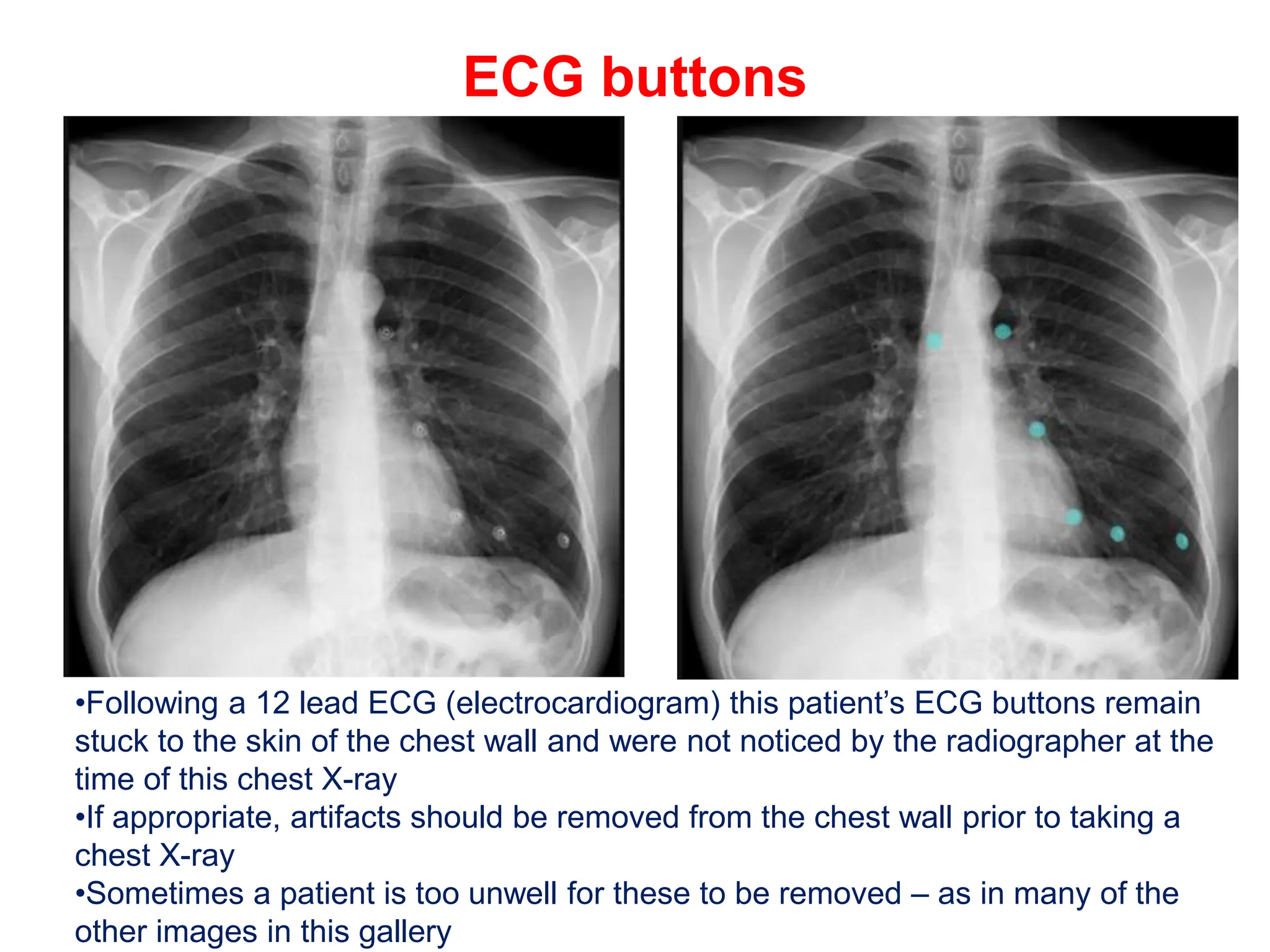
SlideShare a Scribd company logo
1 of 34
CHEST X-RAY
CARDIAC DISEASE
Normal heart
•The heart size is normal – cardiothoracic ratio is less than 50%
•The upper zone vessels are normal – they are smaller than the lower zone
vessels
•The lungs are clear – indicating there is no pulmonary oedema
•The costophrenic angles are well defined (asterisks) – indicating there is no
pleural effusion
Pericardial fat pad
•The heart size is normal in this image
•Accurate measurement of the cardiothoracic ratio can be difficult if there is a
pericardial fat pad
•The width of a pericardial fat pad – which may have to be estimated – should
not be included in the measurement of the cardiac size
Cardiomegaly - Mild
•Good quality Posterior-Anterior chest X-ray with no rotation
•In this image CTR = 53%
•There are no other signs of heart failure
Cardiomegaly
•History of hypertension and angina
•This patient has an enlarged heart
•CTR = 68%
Left atrial enlargement
•This image shows massive cardiomegaly (CTR=79%) in a patient with a metallic
mitral valve replacement
•This image shows specific features indicating massive enlargement of the left
atrium (highlighted area)
•1 - Carina splayed to >90°
•2 - Double right heart border
•3 - Left atrial appendage bulging the left heart border
Upper zone vascular prominence
•Enlarged heart (CTR = 55%)
•The upper zone vessels are prominent – an indicator of increased pulmonary
venous pressure
•Signs of pulmonary oedema have also developed – septal lines (Kerley B lines)
due to interstitial oedema, and airspace shadowing due to alveolar oedema
•The costophrenic angles are blunt due to bilateral pleural effusions
Septal lines - Example 1
•Pulmonary oedema may manifest with evidence of interstitial oedema (septal
lines) or alveolar oedema (airspace shadowing/consolidation)
•Septal lines (also known as ‘Kerley B lines’) appear as horizontal lines which
make contact with the edge of the lung
•Airspace shadowing due to alveolar oedema is also visible )
Septal lines - Example 2
•Septal lines represent thickening of the interlobular septa – interstitial tissue
which separates the secondary lobules at the peripheries of the lungs
•Septal lines are a specific sign of interstitial oedema in the context of
suspected left ventricular failure
•Occasionally septal lines are seen in conditions which cause blockage of the
pulmonary lymphatics – such as lymphangitis carcinomatosa or sarcoidosis
Septal lines - Example 3
•Septal lines may be very subtle but if present are a clear indicator of
interstitial oedema
•Look carefully at the lung bases near the costophrenic angles whenever heart
failure is suspected clinically
•Septal lines may be present with or without alveolar oedema
Alveolar oedema - Bat's wing pattern
•Alveolar oedema is caused by fluid leaking from the interstitial tissues into the
alveoli and small airways, and manifests as airspace shadowing (consolidation)
•In the context of acute pulmonary oedema, alveolar oedema radiates
symmetrically from the hilar regions in a ‘bat's wing’ distribution of airspace
shadowing
•Enlarged heart (CTR 60%) and sternal wires and metallic heart valve
•Blunting of the costophrenic angles is due to pleural effusions
Asymmetric bat's wing shadowing
•Bat's wing pulmonary oedema may not be symmetrical
•Note the septal lines on the right (interstitial oedema) and blunting of the
costophrenic angles bilaterally (pleural effusions)
•The oxygen tubing and ECG buttons have not been removed – indicating the
patient is acutely unwell
Pulmonary oedema
•Images which show pulmonary oedema are frequently of poor quality
because the patient is too unwell to stand or hold their breath
•This is a common appearance of acute pulmonary oedema
•Remember that bilateral air space shadowing may also be caused by other
disease processes such as infection – it is usually the clinical features that
indicate the diagnosis
Non-cardiogenic pulmonary oedema
•This patient had pulmonary oedema secondary to nephrotic syndrome –
albumin was very low
•Note that the heart size is normal (CTR <50%)
•If the heart size is normal, then heart disease may still be the cause of
pulmonary oedema, but non-cardiogenic causes should also be considered
•The converse is also true – if the heart is enlarged, then the cause of
pulmonary oedema is not always cardiac
Pleural effusions
•This patient with left ventricular failure has developed pleural effusions
•Note that the heart is enlarged and the upper zone vessels appear prominent –
if these features are absent then other causes of pleural effusions become more
likely
Asymmetric pleural effusions
•Pleural effusions caused by heart failure may not be symmetrical
•This patient with heart failure had been nursed lying on their right side before this
X-ray was taken
•Fluid has accumulated in the right pleural space – the right costophrenic angle is
not visible
•No effusion is present in the left pleural space – the left costophrenic angle
remains visible
•The left heart border is not distinct because there is pulmonary oedema of the
adjacent lung
Pericardial effusion
•This image shows some of the features of heart failure
•1 - Upper zone vascular prominence
•2 - Airspace shadowing (alveolar oedema)
•3 - Septal lines (interstitial oedema)
•4 - Pleural effusion
•The heart is also enlarged and has a globular (rounded) appearance due to a
pericardial effusion (fluid accumulation within the pericardial sac)
Post-surgical pericardial effusion
•This patient has had recent cardiac surgery
•The heart is enlarged but there are no other signs of heart failure
•Whenever the heart appears globular, it could be due to a pericardial effusion –
the diagnosis can be confirmed using ultrasound (echocardiogram)
Malignant pericardial effusion
•Pericardial effusions may not be due to heart disease
•This patient with metastatic disease (primary colon cancer) has an enlarged and
globular-shaped heart due to a malignant pericardial effusion (fluid and
cancerous cells within the pericardium)
•There are also numerous small lung nodules (pulmonary metastases) and
bilateral pleural effusions (malignant effusions)
Left ventricular aneurysm
•Aneurysms of the left ventricle are an uncommon complication of previous
myocardial infarction
•They may calcify and appear as a smooth eggshell-like line near the left
heart border
Pericardial calcification
•Increased density – due to calcification of the pericardium – follows the
contour of the heart
•Pericardial calcification is an uncommon feature seen on a chest X-ray which
is associated with constrictive pericarditis – in this case caused by previous
tuberculosis infection
Mitral valve calcification
•Calcification of the mitral valve is common in elderly patients – occasionally this
is heavy enough to be seen on a chest X-ray
•Mitral valve calcification is often asymptomatic but may be associated with
arrhythmias or mitral valve incompetence
Atrial septal defect
•The pulmonary artery is large relative to the aortic knuckle
•This combination is associated with increased pulmonary blood flow (left to
right shunt)
•An atrial septal defect was confirmed on echocardiogram
•This adult patient had mild shortness of breath and a subtle systolic murmur
Patent ductus arteriosus
•The pulmonary artery is large relative to the aortic knuckle
•The features are very similar to those seen in the image above
•Echocardiogram showed that this patient had a patent ductus arteriosus (PDA)
•PDA is usually discovered in early childhood but can be asymptomatic until
adulthood
Congenital heart disease - post-surgery
•This patient had undergone corrective surgery for tetralogy of Fallot (TOF)
many years previously
•The X-ray can be considered ‘normal’ for this patient even though the
pulmonary arteries are enlarged and there is also a right-sided aorta – a
common associated anatomical variant in patients with TOF
•The appearances of a chest X-ray can be confusing following surgery for
correction of congenital anomalies – reference to the surgical history is required
Pacemaker
•Cardiac pacemakers are a frequently encountered artifact seen on chest X-rays
•There are many different designs of pacemaker which may have one or two
leads placed in the right heart chambers
•The pulse generator (battery pack) is usually implanted in the retro-pectoral
space on the left side of the anterior chest wall
Pneumothorax following pacemaker
insertion
•After pacemaker insertion a chest X-ray is used to check for a pneumothorax
Implantable cardioverter defibrillator
•Implantable cardioverter defibrillators (ICDs) have a similar appearance to
pacemakers
•The ventricular lead has two thicker segments – these are the shocking
electrodes which automatically defibrillate the heart if an arrhythmia is detected
•The proximal shocking electrode is located in the superior vena cava and left
brachiocephalic vein
•The distal shocking electrode is located in the right ventricle
Cardiac surgery artifact
•Surgical artifacts such as sternotomy wires and metallic heart valves are
common artifacts seen on chest X-rays
•This patient also has a single chamber pacemaker
•Note the signs of heart failure – large heart, prominent upper zone vessels and
pulmonary oedema
CABG clips
•Patients who have had coronary artery bypass grafts (CABG) will often have
visible metallic vascular clips seen on their post-operative chest X-ray
•These clips are placed to prevent flow through the branches of the internal
mammary arteries which are used to form the coronary artery bypass
Prosthetic heart valves
•This patient had previously undergone mitral and aortic valve replacement
surgery – see the metallic heart valve artifact
•New signs of heart failure are evident – large heart, prominent upper zone
vessels, septal lines (Kerley B lines), and pleural effusions
Coronary artery stent
•Coronary stents may be visible on chest X-rays
ECG buttons
•Following a 12 lead ECG (electrocardiogram) this patient’s ECG buttons remain
stuck to the skin of the chest wall and were not noticed by the radiographer at the
time of this chest X-ray
•If appropriate, artifacts should be removed from the chest wall prior to taking a
chest X-ray
•Sometimes a patient is too unwell for these to be removed – as in many of the
other images in this gallery
Other medical artifacts
•This X-ray was acquired to verify the position of the temporary pacing wire –
the only internal artifact visible in this image
•Very sick patients frequently have a large number of lines, tubes and other
artifacts projected over the chest X-ray
•It is often not appropriate for these to be removed prior to acquisition of a chest
X-ray
•If you cannot identify an artifact on a chest X-ray then its identity can be
checked by examining the patient or checking the notes

More Related Content

Similar to CHEST X-RAY CARDIAC DISEASE.........pptx

PULMONARY VALVE DISEASES (2).pptx
PULMONARY VALVE DISEASES (2).pptxPULMONARY VALVE DISEASES (2).pptx
PULMONARY VALVE DISEASES (2).pptxSYED ALI AFRIN
 
cardiac bypass.pptx iulhihuliuFHQIULHFLIhiluhI
cardiac bypass.pptx iulhihuliuFHQIULHFLIhiluhIcardiac bypass.pptx iulhihuliuFHQIULHFLIhiluhI
cardiac bypass.pptx iulhihuliuFHQIULHFLIhiluhIAAZIZ13
 
Radiographic approach to cardiac enlargement
Radiographic approach to cardiac enlargementRadiographic approach to cardiac enlargement
Radiographic approach to cardiac enlargementMilan Silwal
 
PULMONARY VALVE DISEASES.pptx
PULMONARY VALVE DISEASES.pptxPULMONARY VALVE DISEASES.pptx
PULMONARY VALVE DISEASES.pptxSYED ALI AFRIN
 
Tetralogy of Fallot.pdf
Tetralogy of Fallot.pdfTetralogy of Fallot.pdf
Tetralogy of Fallot.pdfKararSurgery
 
Congenital cardiac ...... lecture 61 18 4-2016
Congenital cardiac ...... lecture 61 18 4-2016Congenital cardiac ...... lecture 61 18 4-2016
Congenital cardiac ...... lecture 61 18 4-2016pathologydept
 
Anatomy & physiology for the EP professional part I 8.4.14
Anatomy & physiology for the EP professional part I 8.4.14Anatomy & physiology for the EP professional part I 8.4.14
Anatomy & physiology for the EP professional part I 8.4.14lpesbens
 
Clinical Anatomy of the Heart, Pericardium and.pptx
Clinical Anatomy of the Heart, Pericardium and.pptxClinical Anatomy of the Heart, Pericardium and.pptx
Clinical Anatomy of the Heart, Pericardium and.pptxHafizMohd21
 
Chapter 6- Chest & Cardiovascular radiology.pdf
Chapter 6- Chest & Cardiovascular radiology.pdfChapter 6- Chest & Cardiovascular radiology.pdf
Chapter 6- Chest & Cardiovascular radiology.pdfBereketMathewosGeleb
 
HYPERCALCEMIA-V-HYPOCALCEMIA.pptx
HYPERCALCEMIA-V-HYPOCALCEMIA.pptxHYPERCALCEMIA-V-HYPOCALCEMIA.pptx
HYPERCALCEMIA-V-HYPOCALCEMIA.pptxNTGaMinG8
 
congenital heart diseases.pptx
congenital heart diseases.pptxcongenital heart diseases.pptx
congenital heart diseases.pptxCHANDAN PADHAN
 
radiology.CVS 2 .(dr.abeer)
radiology.CVS 2 .(dr.abeer)radiology.CVS 2 .(dr.abeer)
radiology.CVS 2 .(dr.abeer)student
 
Ischemic and valvular heart disease
Ischemic and valvular heart diseaseIschemic and valvular heart disease
Ischemic and valvular heart diseaseMilan Silwal
 
Xray in Congenital Heart Disease - .pptx
Xray in Congenital Heart Disease - .pptxXray in Congenital Heart Disease - .pptx
Xray in Congenital Heart Disease - .pptxruhailbhat
 
Congenital heart diseases
Congenital heart diseasesCongenital heart diseases
Congenital heart diseasesZaid Ansari
 

Similar to CHEST X-RAY CARDIAC DISEASE.........pptx (20)

PULMONARY VALVE DISEASES (2).pptx
PULMONARY VALVE DISEASES (2).pptxPULMONARY VALVE DISEASES (2).pptx
PULMONARY VALVE DISEASES (2).pptx
 
cardiac bypass.pptx iulhihuliuFHQIULHFLIhiluhI
cardiac bypass.pptx iulhihuliuFHQIULHFLIhiluhIcardiac bypass.pptx iulhihuliuFHQIULHFLIhiluhI
cardiac bypass.pptx iulhihuliuFHQIULHFLIhiluhI
 
Radiographic approach to cardiac enlargement
Radiographic approach to cardiac enlargementRadiographic approach to cardiac enlargement
Radiographic approach to cardiac enlargement
 
PULMONARY VALVE DISEASES.pptx
PULMONARY VALVE DISEASES.pptxPULMONARY VALVE DISEASES.pptx
PULMONARY VALVE DISEASES.pptx
 
Tetralogy of Fallot.pdf
Tetralogy of Fallot.pdfTetralogy of Fallot.pdf
Tetralogy of Fallot.pdf
 
Congenital cardiac ...... lecture 61 18 4-2016
Congenital cardiac ...... lecture 61 18 4-2016Congenital cardiac ...... lecture 61 18 4-2016
Congenital cardiac ...... lecture 61 18 4-2016
 
Valvular diseases
Valvular diseasesValvular diseases
Valvular diseases
 
Pare
ParePare
Pare
 
Anatomy & physiology for the EP professional part I 8.4.14
Anatomy & physiology for the EP professional part I 8.4.14Anatomy & physiology for the EP professional part I 8.4.14
Anatomy & physiology for the EP professional part I 8.4.14
 
Clinical Anatomy of the Heart, Pericardium and.pptx
Clinical Anatomy of the Heart, Pericardium and.pptxClinical Anatomy of the Heart, Pericardium and.pptx
Clinical Anatomy of the Heart, Pericardium and.pptx
 
Chapter 6- Chest & Cardiovascular radiology.pdf
Chapter 6- Chest & Cardiovascular radiology.pdfChapter 6- Chest & Cardiovascular radiology.pdf
Chapter 6- Chest & Cardiovascular radiology.pdf
 
HYPERCALCEMIA-V-HYPOCALCEMIA.pptx
HYPERCALCEMIA-V-HYPOCALCEMIA.pptxHYPERCALCEMIA-V-HYPOCALCEMIA.pptx
HYPERCALCEMIA-V-HYPOCALCEMIA.pptx
 
Radiology 5th year, 13th lecture (Dr. Abeer)
Radiology 5th year, 13th lecture (Dr. Abeer)Radiology 5th year, 13th lecture (Dr. Abeer)
Radiology 5th year, 13th lecture (Dr. Abeer)
 
congenital heart diseases.pptx
congenital heart diseases.pptxcongenital heart diseases.pptx
congenital heart diseases.pptx
 
radiology.CVS 2 .(dr.abeer)
radiology.CVS 2 .(dr.abeer)radiology.CVS 2 .(dr.abeer)
radiology.CVS 2 .(dr.abeer)
 
Pericardial diseases
Pericardial diseases Pericardial diseases
Pericardial diseases
 
Ischemic and valvular heart disease
Ischemic and valvular heart diseaseIschemic and valvular heart disease
Ischemic and valvular heart disease
 
Xray in Congenital Heart Disease - .pptx
Xray in Congenital Heart Disease - .pptxXray in Congenital Heart Disease - .pptx
Xray in Congenital Heart Disease - .pptx
 
Congenital heart diseases
Congenital heart diseasesCongenital heart diseases
Congenital heart diseases
 
Cardiac Tamponade.pdf
Cardiac Tamponade.pdfCardiac Tamponade.pdf
Cardiac Tamponade.pdf
 

More from DR Venkata Ramana

1.MITRAL STENOSIS AND ITS MANAGEMENT....
1.MITRAL STENOSIS AND ITS MANAGEMENT....1.MITRAL STENOSIS AND ITS MANAGEMENT....
1.MITRAL STENOSIS AND ITS MANAGEMENT....DR Venkata Ramana
 
CHEST X-RAY PULMONARY DISEASE pptx.pptx
CHEST X-RAY PULMONARY DISEASE  pptx.pptxCHEST X-RAY PULMONARY DISEASE  pptx.pptx
CHEST X-RAY PULMONARY DISEASE pptx.pptxDR Venkata Ramana
 
CHEST X-RAY MEDIASTINUM AND HILUMpptx.pptx
CHEST X-RAY MEDIASTINUM AND HILUMpptx.pptxCHEST X-RAY MEDIASTINUM AND HILUMpptx.pptx
CHEST X-RAY MEDIASTINUM AND HILUMpptx.pptxDR Venkata Ramana
 
CHEST X-RAY DEVICES AND ARTIFACTS pptx.pptx
CHEST X-RAY DEVICES AND ARTIFACTS pptx.pptxCHEST X-RAY DEVICES AND ARTIFACTS pptx.pptx
CHEST X-RAY DEVICES AND ARTIFACTS pptx.pptxDR Venkata Ramana
 
CHESTX-RAY ANATOMICAL VARIANTS......pptx
CHESTX-RAY ANATOMICAL VARIANTS......pptxCHESTX-RAY ANATOMICAL VARIANTS......pptx
CHESTX-RAY ANATOMICAL VARIANTS......pptxDR Venkata Ramana
 
CHEST X-RAYS OF LUNGCANCER.........pptx
CHEST X-RAYS OF  LUNGCANCER.........pptxCHEST X-RAYS OF  LUNGCANCER.........pptx
CHEST X-RAYS OF LUNGCANCER.........pptxDR Venkata Ramana
 
PNEUMOTHORAX IN CHEST XRAY INTERPRETATIONpptx
PNEUMOTHORAX IN CHEST XRAY INTERPRETATIONpptxPNEUMOTHORAX IN CHEST XRAY INTERPRETATIONpptx
PNEUMOTHORAX IN CHEST XRAY INTERPRETATIONpptxDR Venkata Ramana
 
CHEST XRAYS SYSTEMATIC APPROACH,,,,,,,,,
CHEST XRAYS SYSTEMATIC APPROACH,,,,,,,,,CHEST XRAYS SYSTEMATIC APPROACH,,,,,,,,,
CHEST XRAYS SYSTEMATIC APPROACH,,,,,,,,,DR Venkata Ramana
 
CHEST XRAY TUBES.pptx,,,,,,,,,,,,,,,,,,,
CHEST XRAY TUBES.pptx,,,,,,,,,,,,,,,,,,,CHEST XRAY TUBES.pptx,,,,,,,,,,,,,,,,,,,
CHEST XRAY TUBES.pptx,,,,,,,,,,,,,,,,,,,DR Venkata Ramana
 
CHEST X-RAYS QUALITY,,,,,,,,,,,,,,,, pptx
CHEST X-RAYS QUALITY,,,,,,,,,,,,,,,, pptxCHEST X-RAYS QUALITY,,,,,,,,,,,,,,,, pptx
CHEST X-RAYS QUALITY,,,,,,,,,,,,,,,, pptxDR Venkata Ramana
 
ST SEGMENT IN ECG,ST ELEVATION AND ST DEPRESSION
ST SEGMENT IN ECG,ST ELEVATION AND ST DEPRESSIONST SEGMENT IN ECG,ST ELEVATION AND ST DEPRESSION
ST SEGMENT IN ECG,ST ELEVATION AND ST DEPRESSIONDR Venkata Ramana
 
J POINT IN ECG AND ITS INERPRETATION IN ECG
J POINT IN ECG AND ITS INERPRETATION IN ECGJ POINT IN ECG AND ITS INERPRETATION IN ECG
J POINT IN ECG AND ITS INERPRETATION IN ECGDR Venkata Ramana
 
OSBORN WAVE (J Wave) IN ECG AND ITS INTERPRETATION
OSBORN WAVE (J Wave) IN ECG AND ITS INTERPRETATIONOSBORN WAVE (J Wave) IN ECG AND ITS INTERPRETATION
OSBORN WAVE (J Wave) IN ECG AND ITS INTERPRETATIONDR Venkata Ramana
 
EPSILON WAVE IN ECG AND ITS INTERPRETATION
EPSILON WAVE IN ECG AND ITS INTERPRETATIONEPSILON WAVE IN ECG AND ITS INTERPRETATION
EPSILON WAVE IN ECG AND ITS INTERPRETATIONDR Venkata Ramana
 
DDELTA WAVE IN ECG AND ITS INTERPRETATION IN ECG
DDELTA WAVE IN ECG AND ITS INTERPRETATION IN ECGDDELTA WAVE IN ECG AND ITS INTERPRETATION IN ECG
DDELTA WAVE IN ECG AND ITS INTERPRETATION IN ECGDR Venkata Ramana
 
HEART BLOCKS IN ECG AND HOW TO INTEPRET IN ECG?
HEART BLOCKS IN ECG AND HOW TO INTEPRET IN ECG?HEART BLOCKS IN ECG AND HOW TO INTEPRET IN ECG?
HEART BLOCKS IN ECG AND HOW TO INTEPRET IN ECG?DR Venkata Ramana
 
U WAVE IN ECG AND ITS ABNORMALITIES IN ECG
U WAVE IN ECG AND ITS ABNORMALITIES IN ECGU WAVE IN ECG AND ITS ABNORMALITIES IN ECG
U WAVE IN ECG AND ITS ABNORMALITIES IN ECGDR Venkata Ramana
 
QT INTERVAL IN ECG,CAUSES OF SHORT AND LONG QT INTERVAL
QT INTERVAL IN ECG,CAUSES OF SHORT AND LONG QT INTERVALQT INTERVAL IN ECG,CAUSES OF SHORT AND LONG QT INTERVAL
QT INTERVAL IN ECG,CAUSES OF SHORT AND LONG QT INTERVALDR Venkata Ramana
 
T WAVE IN ECG AND ITS ABNORMALITIES IN ECG
T WAVE IN ECG AND ITS ABNORMALITIES IN ECGT WAVE IN ECG AND ITS ABNORMALITIES IN ECG
T WAVE IN ECG AND ITS ABNORMALITIES IN ECGDR Venkata Ramana
 
ST SEGMENT IN ECG,ST ELEVATION AND ST DEPRESSION
ST SEGMENT IN ECG,ST ELEVATION AND ST DEPRESSIONST SEGMENT IN ECG,ST ELEVATION AND ST DEPRESSION
ST SEGMENT IN ECG,ST ELEVATION AND ST DEPRESSIONDR Venkata Ramana
 

More from DR Venkata Ramana (20)

1.MITRAL STENOSIS AND ITS MANAGEMENT....
1.MITRAL STENOSIS AND ITS MANAGEMENT....1.MITRAL STENOSIS AND ITS MANAGEMENT....
1.MITRAL STENOSIS AND ITS MANAGEMENT....
 
CHEST X-RAY PULMONARY DISEASE pptx.pptx
CHEST X-RAY PULMONARY DISEASE  pptx.pptxCHEST X-RAY PULMONARY DISEASE  pptx.pptx
CHEST X-RAY PULMONARY DISEASE pptx.pptx
 
CHEST X-RAY MEDIASTINUM AND HILUMpptx.pptx
CHEST X-RAY MEDIASTINUM AND HILUMpptx.pptxCHEST X-RAY MEDIASTINUM AND HILUMpptx.pptx
CHEST X-RAY MEDIASTINUM AND HILUMpptx.pptx
 
CHEST X-RAY DEVICES AND ARTIFACTS pptx.pptx
CHEST X-RAY DEVICES AND ARTIFACTS pptx.pptxCHEST X-RAY DEVICES AND ARTIFACTS pptx.pptx
CHEST X-RAY DEVICES AND ARTIFACTS pptx.pptx
 
CHESTX-RAY ANATOMICAL VARIANTS......pptx
CHESTX-RAY ANATOMICAL VARIANTS......pptxCHESTX-RAY ANATOMICAL VARIANTS......pptx
CHESTX-RAY ANATOMICAL VARIANTS......pptx
 
CHEST X-RAYS OF LUNGCANCER.........pptx
CHEST X-RAYS OF  LUNGCANCER.........pptxCHEST X-RAYS OF  LUNGCANCER.........pptx
CHEST X-RAYS OF LUNGCANCER.........pptx
 
PNEUMOTHORAX IN CHEST XRAY INTERPRETATIONpptx
PNEUMOTHORAX IN CHEST XRAY INTERPRETATIONpptxPNEUMOTHORAX IN CHEST XRAY INTERPRETATIONpptx
PNEUMOTHORAX IN CHEST XRAY INTERPRETATIONpptx
 
CHEST XRAYS SYSTEMATIC APPROACH,,,,,,,,,
CHEST XRAYS SYSTEMATIC APPROACH,,,,,,,,,CHEST XRAYS SYSTEMATIC APPROACH,,,,,,,,,
CHEST XRAYS SYSTEMATIC APPROACH,,,,,,,,,
 
CHEST XRAY TUBES.pptx,,,,,,,,,,,,,,,,,,,
CHEST XRAY TUBES.pptx,,,,,,,,,,,,,,,,,,,CHEST XRAY TUBES.pptx,,,,,,,,,,,,,,,,,,,
CHEST XRAY TUBES.pptx,,,,,,,,,,,,,,,,,,,
 
CHEST X-RAYS QUALITY,,,,,,,,,,,,,,,, pptx
CHEST X-RAYS QUALITY,,,,,,,,,,,,,,,, pptxCHEST X-RAYS QUALITY,,,,,,,,,,,,,,,, pptx
CHEST X-RAYS QUALITY,,,,,,,,,,,,,,,, pptx
 
ST SEGMENT IN ECG,ST ELEVATION AND ST DEPRESSION
ST SEGMENT IN ECG,ST ELEVATION AND ST DEPRESSIONST SEGMENT IN ECG,ST ELEVATION AND ST DEPRESSION
ST SEGMENT IN ECG,ST ELEVATION AND ST DEPRESSION
 
J POINT IN ECG AND ITS INERPRETATION IN ECG
J POINT IN ECG AND ITS INERPRETATION IN ECGJ POINT IN ECG AND ITS INERPRETATION IN ECG
J POINT IN ECG AND ITS INERPRETATION IN ECG
 
OSBORN WAVE (J Wave) IN ECG AND ITS INTERPRETATION
OSBORN WAVE (J Wave) IN ECG AND ITS INTERPRETATIONOSBORN WAVE (J Wave) IN ECG AND ITS INTERPRETATION
OSBORN WAVE (J Wave) IN ECG AND ITS INTERPRETATION
 
EPSILON WAVE IN ECG AND ITS INTERPRETATION
EPSILON WAVE IN ECG AND ITS INTERPRETATIONEPSILON WAVE IN ECG AND ITS INTERPRETATION
EPSILON WAVE IN ECG AND ITS INTERPRETATION
 
DDELTA WAVE IN ECG AND ITS INTERPRETATION IN ECG
DDELTA WAVE IN ECG AND ITS INTERPRETATION IN ECGDDELTA WAVE IN ECG AND ITS INTERPRETATION IN ECG
DDELTA WAVE IN ECG AND ITS INTERPRETATION IN ECG
 
HEART BLOCKS IN ECG AND HOW TO INTEPRET IN ECG?
HEART BLOCKS IN ECG AND HOW TO INTEPRET IN ECG?HEART BLOCKS IN ECG AND HOW TO INTEPRET IN ECG?
HEART BLOCKS IN ECG AND HOW TO INTEPRET IN ECG?
 
U WAVE IN ECG AND ITS ABNORMALITIES IN ECG
U WAVE IN ECG AND ITS ABNORMALITIES IN ECGU WAVE IN ECG AND ITS ABNORMALITIES IN ECG
U WAVE IN ECG AND ITS ABNORMALITIES IN ECG
 
QT INTERVAL IN ECG,CAUSES OF SHORT AND LONG QT INTERVAL
QT INTERVAL IN ECG,CAUSES OF SHORT AND LONG QT INTERVALQT INTERVAL IN ECG,CAUSES OF SHORT AND LONG QT INTERVAL
QT INTERVAL IN ECG,CAUSES OF SHORT AND LONG QT INTERVAL
 
T WAVE IN ECG AND ITS ABNORMALITIES IN ECG
T WAVE IN ECG AND ITS ABNORMALITIES IN ECGT WAVE IN ECG AND ITS ABNORMALITIES IN ECG
T WAVE IN ECG AND ITS ABNORMALITIES IN ECG
 
ST SEGMENT IN ECG,ST ELEVATION AND ST DEPRESSION
ST SEGMENT IN ECG,ST ELEVATION AND ST DEPRESSIONST SEGMENT IN ECG,ST ELEVATION AND ST DEPRESSION
ST SEGMENT IN ECG,ST ELEVATION AND ST DEPRESSION
 

Recently uploaded

TEST BANK For Porth's Essentials of Pathophysiology, 5th Edition by Tommie L ...
TEST BANK For Porth's Essentials of Pathophysiology, 5th Edition by Tommie L ...TEST BANK For Porth's Essentials of Pathophysiology, 5th Edition by Tommie L ...
TEST BANK For Porth's Essentials of Pathophysiology, 5th Edition by Tommie L ...rightmanforbloodline
 
Dr. A Sumathi - LINEARITY CONCEPT OF SIGNIFICANCE.pdf
Dr. A Sumathi - LINEARITY CONCEPT OF SIGNIFICANCE.pdfDr. A Sumathi - LINEARITY CONCEPT OF SIGNIFICANCE.pdf
Dr. A Sumathi - LINEARITY CONCEPT OF SIGNIFICANCE.pdfSumathi Arumugam
 
Face and Muscles of facial expression.pptx
Face and Muscles of facial expression.pptxFace and Muscles of facial expression.pptx
Face and Muscles of facial expression.pptxDr. Rabia Inam Gandapore
 
VIP ℂall Girls Arekere Bangalore 6378878445 WhatsApp: Me All Time Serviℂe Ava...
VIP ℂall Girls Arekere Bangalore 6378878445 WhatsApp: Me All Time Serviℂe Ava...VIP ℂall Girls Arekere Bangalore 6378878445 WhatsApp: Me All Time Serviℂe Ava...
VIP ℂall Girls Arekere Bangalore 6378878445 WhatsApp: Me All Time Serviℂe Ava...deepakkumar115120
 
Jual Obat Aborsi Di Dubai UAE Wa 0838-4800-7379 Obat Penggugur Kandungan Cytotec
Jual Obat Aborsi Di Dubai UAE Wa 0838-4800-7379 Obat Penggugur Kandungan CytotecJual Obat Aborsi Di Dubai UAE Wa 0838-4800-7379 Obat Penggugur Kandungan Cytotec
Jual Obat Aborsi Di Dubai UAE Wa 0838-4800-7379 Obat Penggugur Kandungan Cytotecjualobat34
 
ABO Blood grouping in-compatibility in pregnancy
ABO Blood grouping in-compatibility in pregnancyABO Blood grouping in-compatibility in pregnancy
ABO Blood grouping in-compatibility in pregnancyMs. Sapna Pal
 
7 steps How to prevent Thalassemia : Dr Sharda Jain & Vandana Gupta
7 steps How to prevent Thalassemia : Dr Sharda Jain & Vandana Gupta7 steps How to prevent Thalassemia : Dr Sharda Jain & Vandana Gupta
7 steps How to prevent Thalassemia : Dr Sharda Jain & Vandana GuptaLifecare Centre
 
VIP ℂall Girls Kothanur {{ Bangalore }} 6378878445 WhatsApp: Me 24/7 Hours Se...
VIP ℂall Girls Kothanur {{ Bangalore }} 6378878445 WhatsApp: Me 24/7 Hours Se...VIP ℂall Girls Kothanur {{ Bangalore }} 6378878445 WhatsApp: Me 24/7 Hours Se...
VIP ℂall Girls Kothanur {{ Bangalore }} 6378878445 WhatsApp: Me 24/7 Hours Se...deepakkumar115120
 
Physicochemical properties (descriptors) in QSAR.pdf
Physicochemical properties (descriptors) in QSAR.pdfPhysicochemical properties (descriptors) in QSAR.pdf
Physicochemical properties (descriptors) in QSAR.pdfRAJ K. MAURYA
 
ANATOMY AND PHYSIOLOGY OF RESPIRATORY SYSTEM.pptx
ANATOMY AND PHYSIOLOGY OF RESPIRATORY SYSTEM.pptxANATOMY AND PHYSIOLOGY OF RESPIRATORY SYSTEM.pptx
ANATOMY AND PHYSIOLOGY OF RESPIRATORY SYSTEM.pptxSwetaba Besh
 
Creeping Stroke - Venous thrombosis presenting with pc-stroke.pptx
Creeping Stroke - Venous thrombosis presenting with pc-stroke.pptxCreeping Stroke - Venous thrombosis presenting with pc-stroke.pptx
Creeping Stroke - Venous thrombosis presenting with pc-stroke.pptxYasser Alzainy
 
Physiologic Anatomy of Heart_AntiCopy.pdf
Physiologic Anatomy of Heart_AntiCopy.pdfPhysiologic Anatomy of Heart_AntiCopy.pdf
Physiologic Anatomy of Heart_AntiCopy.pdfMedicoseAcademics
 
Obat Aborsi Ampuh Usia 1,2,3,4,5,6,7 Bulan 081901222272 Obat Penggugur Kandu...
Obat Aborsi Ampuh Usia 1,2,3,4,5,6,7 Bulan  081901222272 Obat Penggugur Kandu...Obat Aborsi Ampuh Usia 1,2,3,4,5,6,7 Bulan  081901222272 Obat Penggugur Kandu...
Obat Aborsi Ampuh Usia 1,2,3,4,5,6,7 Bulan 081901222272 Obat Penggugur Kandu...Halo Docter
 
Cardiac Output, Venous Return, and Their Regulation
Cardiac Output, Venous Return, and Their RegulationCardiac Output, Venous Return, and Their Regulation
Cardiac Output, Venous Return, and Their RegulationMedicoseAcademics
 
Intro to disinformation and public health
Intro to disinformation and public healthIntro to disinformation and public health
Intro to disinformation and public healthTina Purnat
 
Test bank for critical care nursing a holistic approach 11th edition morton f...
Test bank for critical care nursing a holistic approach 11th edition morton f...Test bank for critical care nursing a holistic approach 11th edition morton f...
Test bank for critical care nursing a holistic approach 11th edition morton f...robinsonayot
 
See it and Catch it! Recognizing the Thought Traps that Negatively Impact How...
See it and Catch it! Recognizing the Thought Traps that Negatively Impact How...See it and Catch it! Recognizing the Thought Traps that Negatively Impact How...
See it and Catch it! Recognizing the Thought Traps that Negatively Impact How...bkling
 
Circulatory Shock, types and stages, compensatory mechanisms
Circulatory Shock, types and stages, compensatory mechanismsCirculatory Shock, types and stages, compensatory mechanisms
Circulatory Shock, types and stages, compensatory mechanismsMedicoseAcademics
 
Shazia Iqbal 2024 - Bioorganic Chemistry.pdf
Shazia Iqbal 2024 - Bioorganic Chemistry.pdfShazia Iqbal 2024 - Bioorganic Chemistry.pdf
Shazia Iqbal 2024 - Bioorganic Chemistry.pdfTrustlife
 
Cara Menggugurkan Kandungan Dengan Cepat Selesai Dalam 24 Jam Secara Alami Bu...
Cara Menggugurkan Kandungan Dengan Cepat Selesai Dalam 24 Jam Secara Alami Bu...Cara Menggugurkan Kandungan Dengan Cepat Selesai Dalam 24 Jam Secara Alami Bu...
Cara Menggugurkan Kandungan Dengan Cepat Selesai Dalam 24 Jam Secara Alami Bu...Cara Menggugurkan Kandungan 087776558899
 

Recently uploaded (20)

TEST BANK For Porth's Essentials of Pathophysiology, 5th Edition by Tommie L ...
TEST BANK For Porth's Essentials of Pathophysiology, 5th Edition by Tommie L ...TEST BANK For Porth's Essentials of Pathophysiology, 5th Edition by Tommie L ...
TEST BANK For Porth's Essentials of Pathophysiology, 5th Edition by Tommie L ...
 
Dr. A Sumathi - LINEARITY CONCEPT OF SIGNIFICANCE.pdf
Dr. A Sumathi - LINEARITY CONCEPT OF SIGNIFICANCE.pdfDr. A Sumathi - LINEARITY CONCEPT OF SIGNIFICANCE.pdf
Dr. A Sumathi - LINEARITY CONCEPT OF SIGNIFICANCE.pdf
 
Face and Muscles of facial expression.pptx
Face and Muscles of facial expression.pptxFace and Muscles of facial expression.pptx
Face and Muscles of facial expression.pptx
 
VIP ℂall Girls Arekere Bangalore 6378878445 WhatsApp: Me All Time Serviℂe Ava...
VIP ℂall Girls Arekere Bangalore 6378878445 WhatsApp: Me All Time Serviℂe Ava...VIP ℂall Girls Arekere Bangalore 6378878445 WhatsApp: Me All Time Serviℂe Ava...
VIP ℂall Girls Arekere Bangalore 6378878445 WhatsApp: Me All Time Serviℂe Ava...
 
Jual Obat Aborsi Di Dubai UAE Wa 0838-4800-7379 Obat Penggugur Kandungan Cytotec
Jual Obat Aborsi Di Dubai UAE Wa 0838-4800-7379 Obat Penggugur Kandungan CytotecJual Obat Aborsi Di Dubai UAE Wa 0838-4800-7379 Obat Penggugur Kandungan Cytotec
Jual Obat Aborsi Di Dubai UAE Wa 0838-4800-7379 Obat Penggugur Kandungan Cytotec
 
ABO Blood grouping in-compatibility in pregnancy
ABO Blood grouping in-compatibility in pregnancyABO Blood grouping in-compatibility in pregnancy
ABO Blood grouping in-compatibility in pregnancy
 
7 steps How to prevent Thalassemia : Dr Sharda Jain & Vandana Gupta
7 steps How to prevent Thalassemia : Dr Sharda Jain & Vandana Gupta7 steps How to prevent Thalassemia : Dr Sharda Jain & Vandana Gupta
7 steps How to prevent Thalassemia : Dr Sharda Jain & Vandana Gupta
 
VIP ℂall Girls Kothanur {{ Bangalore }} 6378878445 WhatsApp: Me 24/7 Hours Se...
VIP ℂall Girls Kothanur {{ Bangalore }} 6378878445 WhatsApp: Me 24/7 Hours Se...VIP ℂall Girls Kothanur {{ Bangalore }} 6378878445 WhatsApp: Me 24/7 Hours Se...
VIP ℂall Girls Kothanur {{ Bangalore }} 6378878445 WhatsApp: Me 24/7 Hours Se...
 
Physicochemical properties (descriptors) in QSAR.pdf
Physicochemical properties (descriptors) in QSAR.pdfPhysicochemical properties (descriptors) in QSAR.pdf
Physicochemical properties (descriptors) in QSAR.pdf
 
ANATOMY AND PHYSIOLOGY OF RESPIRATORY SYSTEM.pptx
ANATOMY AND PHYSIOLOGY OF RESPIRATORY SYSTEM.pptxANATOMY AND PHYSIOLOGY OF RESPIRATORY SYSTEM.pptx
ANATOMY AND PHYSIOLOGY OF RESPIRATORY SYSTEM.pptx
 
Creeping Stroke - Venous thrombosis presenting with pc-stroke.pptx
Creeping Stroke - Venous thrombosis presenting with pc-stroke.pptxCreeping Stroke - Venous thrombosis presenting with pc-stroke.pptx
Creeping Stroke - Venous thrombosis presenting with pc-stroke.pptx
 
Physiologic Anatomy of Heart_AntiCopy.pdf
Physiologic Anatomy of Heart_AntiCopy.pdfPhysiologic Anatomy of Heart_AntiCopy.pdf
Physiologic Anatomy of Heart_AntiCopy.pdf
 
Obat Aborsi Ampuh Usia 1,2,3,4,5,6,7 Bulan 081901222272 Obat Penggugur Kandu...
Obat Aborsi Ampuh Usia 1,2,3,4,5,6,7 Bulan  081901222272 Obat Penggugur Kandu...Obat Aborsi Ampuh Usia 1,2,3,4,5,6,7 Bulan  081901222272 Obat Penggugur Kandu...
Obat Aborsi Ampuh Usia 1,2,3,4,5,6,7 Bulan 081901222272 Obat Penggugur Kandu...
 
Cardiac Output, Venous Return, and Their Regulation
Cardiac Output, Venous Return, and Their RegulationCardiac Output, Venous Return, and Their Regulation
Cardiac Output, Venous Return, and Their Regulation
 
Intro to disinformation and public health
Intro to disinformation and public healthIntro to disinformation and public health
Intro to disinformation and public health
 
Test bank for critical care nursing a holistic approach 11th edition morton f...
Test bank for critical care nursing a holistic approach 11th edition morton f...Test bank for critical care nursing a holistic approach 11th edition morton f...
Test bank for critical care nursing a holistic approach 11th edition morton f...
 
See it and Catch it! Recognizing the Thought Traps that Negatively Impact How...
See it and Catch it! Recognizing the Thought Traps that Negatively Impact How...See it and Catch it! Recognizing the Thought Traps that Negatively Impact How...
See it and Catch it! Recognizing the Thought Traps that Negatively Impact How...
 
Circulatory Shock, types and stages, compensatory mechanisms
Circulatory Shock, types and stages, compensatory mechanismsCirculatory Shock, types and stages, compensatory mechanisms
Circulatory Shock, types and stages, compensatory mechanisms
 
Shazia Iqbal 2024 - Bioorganic Chemistry.pdf
Shazia Iqbal 2024 - Bioorganic Chemistry.pdfShazia Iqbal 2024 - Bioorganic Chemistry.pdf
Shazia Iqbal 2024 - Bioorganic Chemistry.pdf
 
Cara Menggugurkan Kandungan Dengan Cepat Selesai Dalam 24 Jam Secara Alami Bu...
Cara Menggugurkan Kandungan Dengan Cepat Selesai Dalam 24 Jam Secara Alami Bu...Cara Menggugurkan Kandungan Dengan Cepat Selesai Dalam 24 Jam Secara Alami Bu...
Cara Menggugurkan Kandungan Dengan Cepat Selesai Dalam 24 Jam Secara Alami Bu...
 

CHEST X-RAY CARDIAC DISEASE.........pptx

  • 2. Normal heart •The heart size is normal – cardiothoracic ratio is less than 50% •The upper zone vessels are normal – they are smaller than the lower zone vessels •The lungs are clear – indicating there is no pulmonary oedema •The costophrenic angles are well defined (asterisks) – indicating there is no pleural effusion
  • 3. Pericardial fat pad •The heart size is normal in this image •Accurate measurement of the cardiothoracic ratio can be difficult if there is a pericardial fat pad •The width of a pericardial fat pad – which may have to be estimated – should not be included in the measurement of the cardiac size
  • 4. Cardiomegaly - Mild •Good quality Posterior-Anterior chest X-ray with no rotation •In this image CTR = 53% •There are no other signs of heart failure
  • 5. Cardiomegaly •History of hypertension and angina •This patient has an enlarged heart •CTR = 68%
  • 6. Left atrial enlargement •This image shows massive cardiomegaly (CTR=79%) in a patient with a metallic mitral valve replacement •This image shows specific features indicating massive enlargement of the left atrium (highlighted area) •1 - Carina splayed to >90° •2 - Double right heart border •3 - Left atrial appendage bulging the left heart border
  • 7. Upper zone vascular prominence •Enlarged heart (CTR = 55%) •The upper zone vessels are prominent – an indicator of increased pulmonary venous pressure •Signs of pulmonary oedema have also developed – septal lines (Kerley B lines) due to interstitial oedema, and airspace shadowing due to alveolar oedema •The costophrenic angles are blunt due to bilateral pleural effusions
  • 8. Septal lines - Example 1 •Pulmonary oedema may manifest with evidence of interstitial oedema (septal lines) or alveolar oedema (airspace shadowing/consolidation) •Septal lines (also known as ‘Kerley B lines’) appear as horizontal lines which make contact with the edge of the lung •Airspace shadowing due to alveolar oedema is also visible )
  • 9. Septal lines - Example 2 •Septal lines represent thickening of the interlobular septa – interstitial tissue which separates the secondary lobules at the peripheries of the lungs •Septal lines are a specific sign of interstitial oedema in the context of suspected left ventricular failure •Occasionally septal lines are seen in conditions which cause blockage of the pulmonary lymphatics – such as lymphangitis carcinomatosa or sarcoidosis
  • 10. Septal lines - Example 3 •Septal lines may be very subtle but if present are a clear indicator of interstitial oedema •Look carefully at the lung bases near the costophrenic angles whenever heart failure is suspected clinically •Septal lines may be present with or without alveolar oedema
  • 11. Alveolar oedema - Bat's wing pattern •Alveolar oedema is caused by fluid leaking from the interstitial tissues into the alveoli and small airways, and manifests as airspace shadowing (consolidation) •In the context of acute pulmonary oedema, alveolar oedema radiates symmetrically from the hilar regions in a ‘bat's wing’ distribution of airspace shadowing •Enlarged heart (CTR 60%) and sternal wires and metallic heart valve •Blunting of the costophrenic angles is due to pleural effusions
  • 12. Asymmetric bat's wing shadowing •Bat's wing pulmonary oedema may not be symmetrical •Note the septal lines on the right (interstitial oedema) and blunting of the costophrenic angles bilaterally (pleural effusions) •The oxygen tubing and ECG buttons have not been removed – indicating the patient is acutely unwell
  • 13. Pulmonary oedema •Images which show pulmonary oedema are frequently of poor quality because the patient is too unwell to stand or hold their breath •This is a common appearance of acute pulmonary oedema •Remember that bilateral air space shadowing may also be caused by other disease processes such as infection – it is usually the clinical features that indicate the diagnosis
  • 14. Non-cardiogenic pulmonary oedema •This patient had pulmonary oedema secondary to nephrotic syndrome – albumin was very low •Note that the heart size is normal (CTR <50%) •If the heart size is normal, then heart disease may still be the cause of pulmonary oedema, but non-cardiogenic causes should also be considered •The converse is also true – if the heart is enlarged, then the cause of pulmonary oedema is not always cardiac
  • 15. Pleural effusions •This patient with left ventricular failure has developed pleural effusions •Note that the heart is enlarged and the upper zone vessels appear prominent – if these features are absent then other causes of pleural effusions become more likely
  • 16. Asymmetric pleural effusions •Pleural effusions caused by heart failure may not be symmetrical •This patient with heart failure had been nursed lying on their right side before this X-ray was taken •Fluid has accumulated in the right pleural space – the right costophrenic angle is not visible •No effusion is present in the left pleural space – the left costophrenic angle remains visible •The left heart border is not distinct because there is pulmonary oedema of the adjacent lung
  • 17. Pericardial effusion •This image shows some of the features of heart failure •1 - Upper zone vascular prominence •2 - Airspace shadowing (alveolar oedema) •3 - Septal lines (interstitial oedema) •4 - Pleural effusion •The heart is also enlarged and has a globular (rounded) appearance due to a pericardial effusion (fluid accumulation within the pericardial sac)
  • 18. Post-surgical pericardial effusion •This patient has had recent cardiac surgery •The heart is enlarged but there are no other signs of heart failure •Whenever the heart appears globular, it could be due to a pericardial effusion – the diagnosis can be confirmed using ultrasound (echocardiogram)
  • 19. Malignant pericardial effusion •Pericardial effusions may not be due to heart disease •This patient with metastatic disease (primary colon cancer) has an enlarged and globular-shaped heart due to a malignant pericardial effusion (fluid and cancerous cells within the pericardium) •There are also numerous small lung nodules (pulmonary metastases) and bilateral pleural effusions (malignant effusions)
  • 20. Left ventricular aneurysm •Aneurysms of the left ventricle are an uncommon complication of previous myocardial infarction •They may calcify and appear as a smooth eggshell-like line near the left heart border
  • 21. Pericardial calcification •Increased density – due to calcification of the pericardium – follows the contour of the heart •Pericardial calcification is an uncommon feature seen on a chest X-ray which is associated with constrictive pericarditis – in this case caused by previous tuberculosis infection
  • 22. Mitral valve calcification •Calcification of the mitral valve is common in elderly patients – occasionally this is heavy enough to be seen on a chest X-ray •Mitral valve calcification is often asymptomatic but may be associated with arrhythmias or mitral valve incompetence
  • 23. Atrial septal defect •The pulmonary artery is large relative to the aortic knuckle •This combination is associated with increased pulmonary blood flow (left to right shunt) •An atrial septal defect was confirmed on echocardiogram •This adult patient had mild shortness of breath and a subtle systolic murmur
  • 24. Patent ductus arteriosus •The pulmonary artery is large relative to the aortic knuckle •The features are very similar to those seen in the image above •Echocardiogram showed that this patient had a patent ductus arteriosus (PDA) •PDA is usually discovered in early childhood but can be asymptomatic until adulthood
  • 25. Congenital heart disease - post-surgery •This patient had undergone corrective surgery for tetralogy of Fallot (TOF) many years previously •The X-ray can be considered ‘normal’ for this patient even though the pulmonary arteries are enlarged and there is also a right-sided aorta – a common associated anatomical variant in patients with TOF •The appearances of a chest X-ray can be confusing following surgery for correction of congenital anomalies – reference to the surgical history is required
  • 26. Pacemaker •Cardiac pacemakers are a frequently encountered artifact seen on chest X-rays •There are many different designs of pacemaker which may have one or two leads placed in the right heart chambers •The pulse generator (battery pack) is usually implanted in the retro-pectoral space on the left side of the anterior chest wall
  • 27. Pneumothorax following pacemaker insertion •After pacemaker insertion a chest X-ray is used to check for a pneumothorax
  • 28. Implantable cardioverter defibrillator •Implantable cardioverter defibrillators (ICDs) have a similar appearance to pacemakers •The ventricular lead has two thicker segments – these are the shocking electrodes which automatically defibrillate the heart if an arrhythmia is detected •The proximal shocking electrode is located in the superior vena cava and left brachiocephalic vein •The distal shocking electrode is located in the right ventricle
  • 29. Cardiac surgery artifact •Surgical artifacts such as sternotomy wires and metallic heart valves are common artifacts seen on chest X-rays •This patient also has a single chamber pacemaker •Note the signs of heart failure – large heart, prominent upper zone vessels and pulmonary oedema
  • 30. CABG clips •Patients who have had coronary artery bypass grafts (CABG) will often have visible metallic vascular clips seen on their post-operative chest X-ray •These clips are placed to prevent flow through the branches of the internal mammary arteries which are used to form the coronary artery bypass
  • 31. Prosthetic heart valves •This patient had previously undergone mitral and aortic valve replacement surgery – see the metallic heart valve artifact •New signs of heart failure are evident – large heart, prominent upper zone vessels, septal lines (Kerley B lines), and pleural effusions
  • 32. Coronary artery stent •Coronary stents may be visible on chest X-rays
  • 33. ECG buttons •Following a 12 lead ECG (electrocardiogram) this patient’s ECG buttons remain stuck to the skin of the chest wall and were not noticed by the radiographer at the time of this chest X-ray •If appropriate, artifacts should be removed from the chest wall prior to taking a chest X-ray •Sometimes a patient is too unwell for these to be removed – as in many of the other images in this gallery
  • 34. Other medical artifacts •This X-ray was acquired to verify the position of the temporary pacing wire – the only internal artifact visible in this image •Very sick patients frequently have a large number of lines, tubes and other artifacts projected over the chest X-ray •It is often not appropriate for these to be removed prior to acquisition of a chest X-ray •If you cannot identify an artifact on a chest X-ray then its identity can be checked by examining the patient or checking the notes