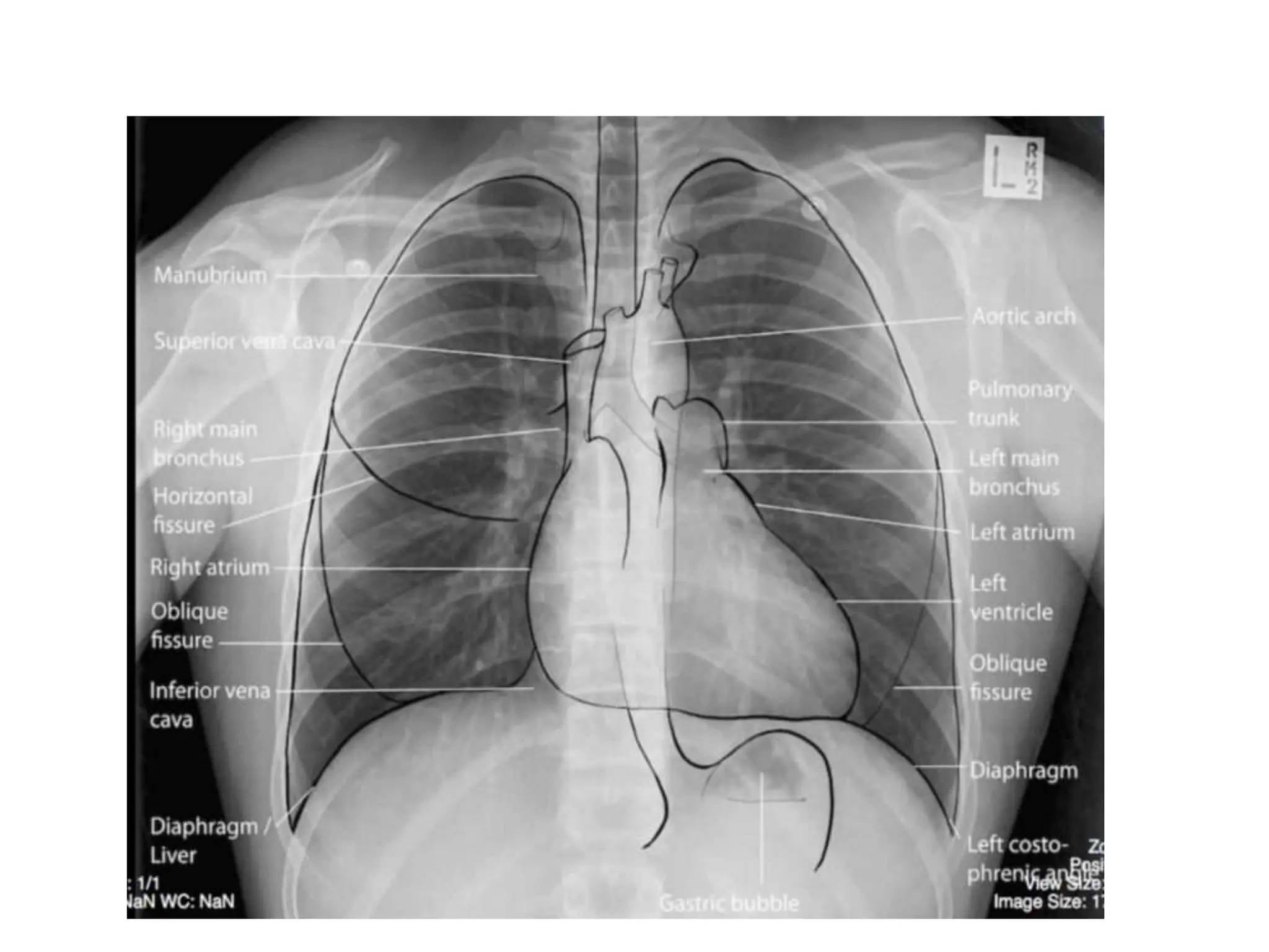
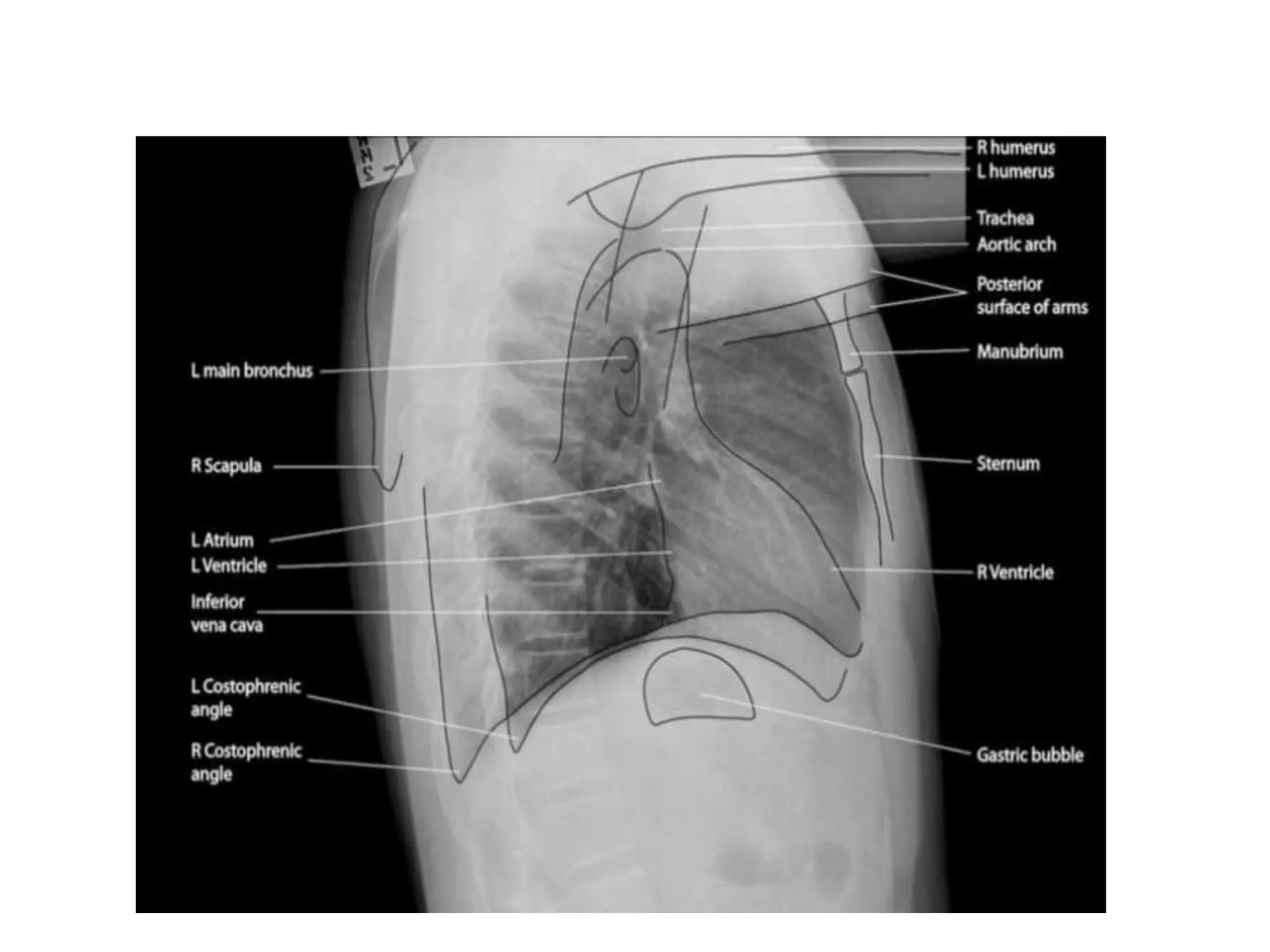
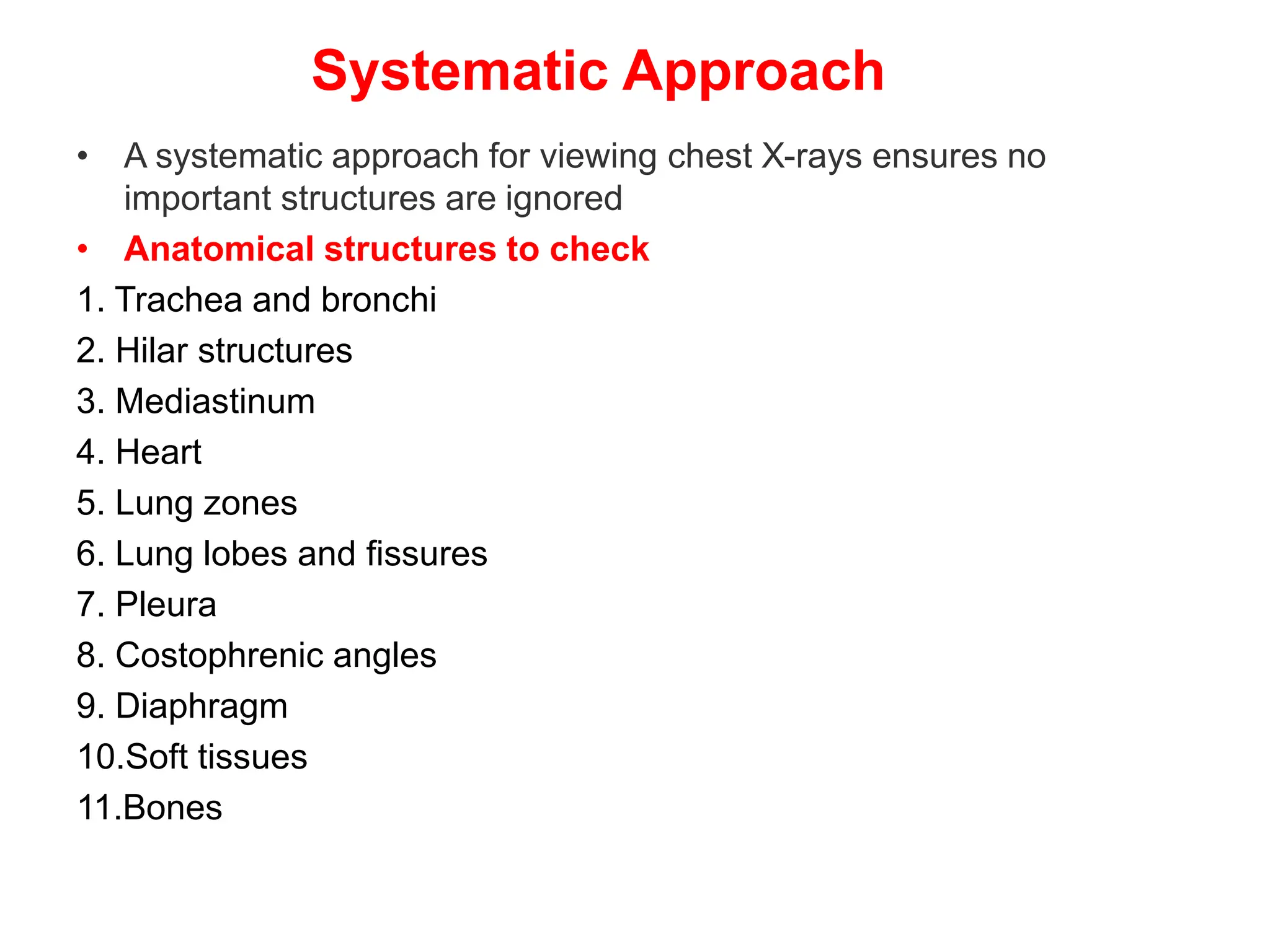
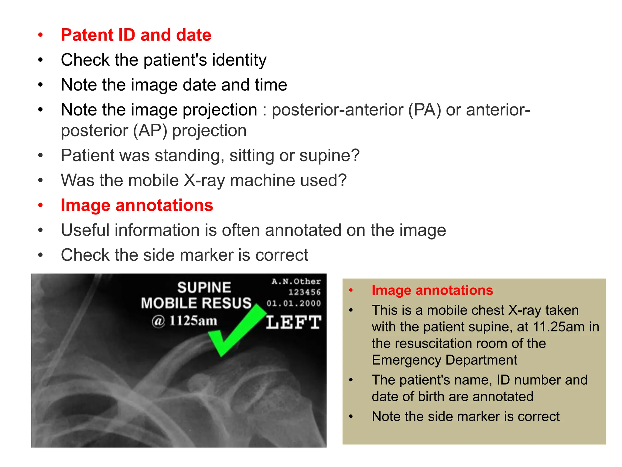
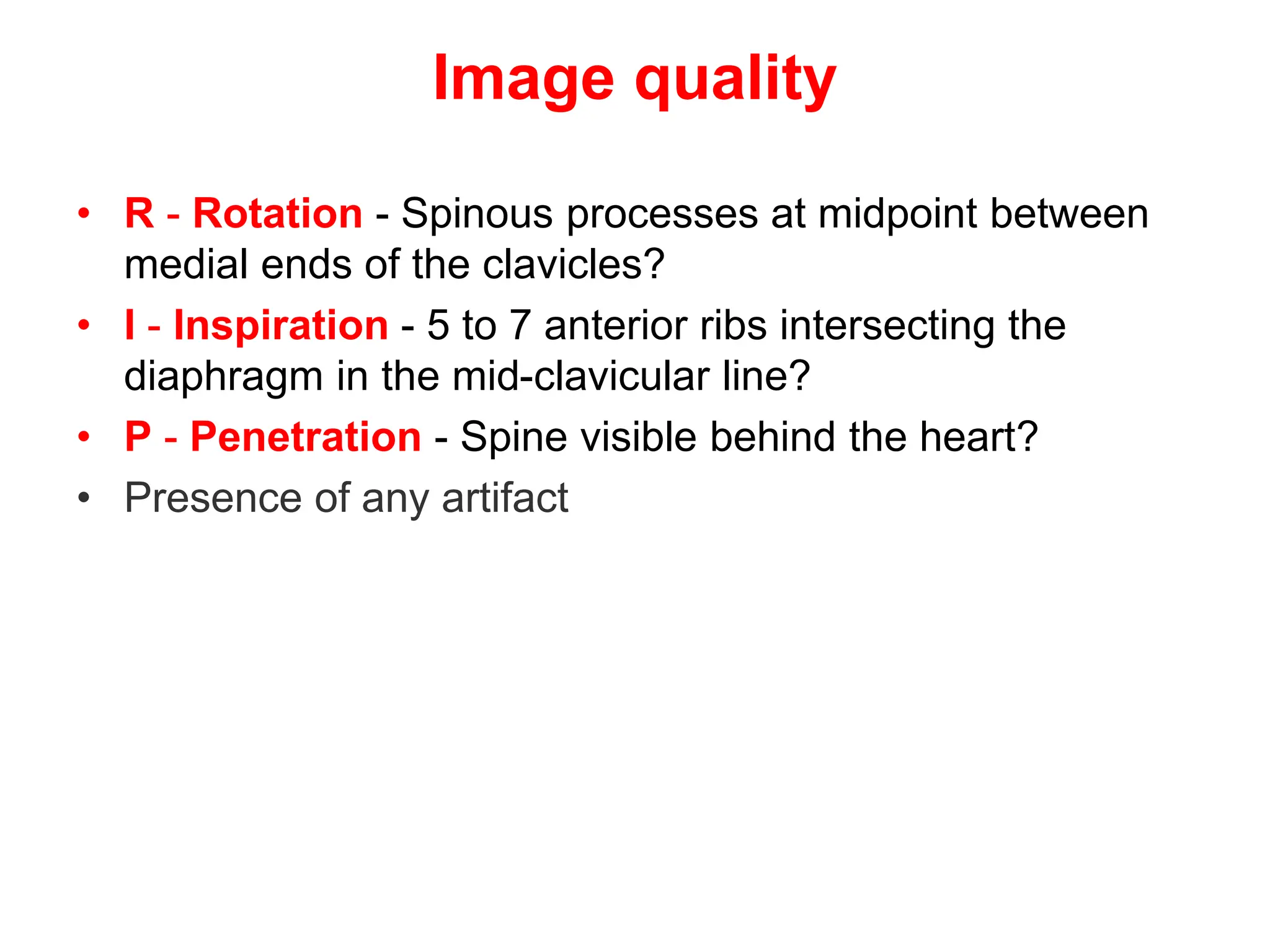
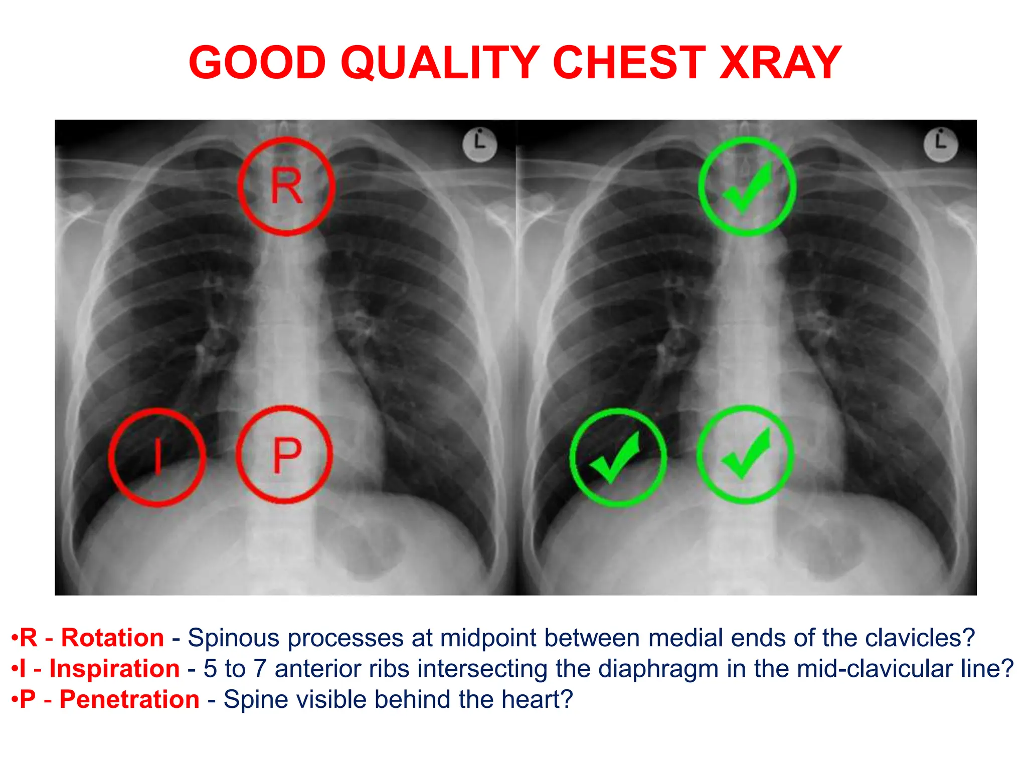
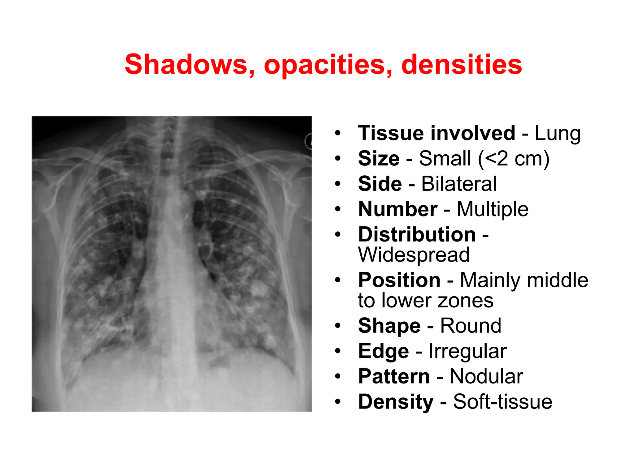
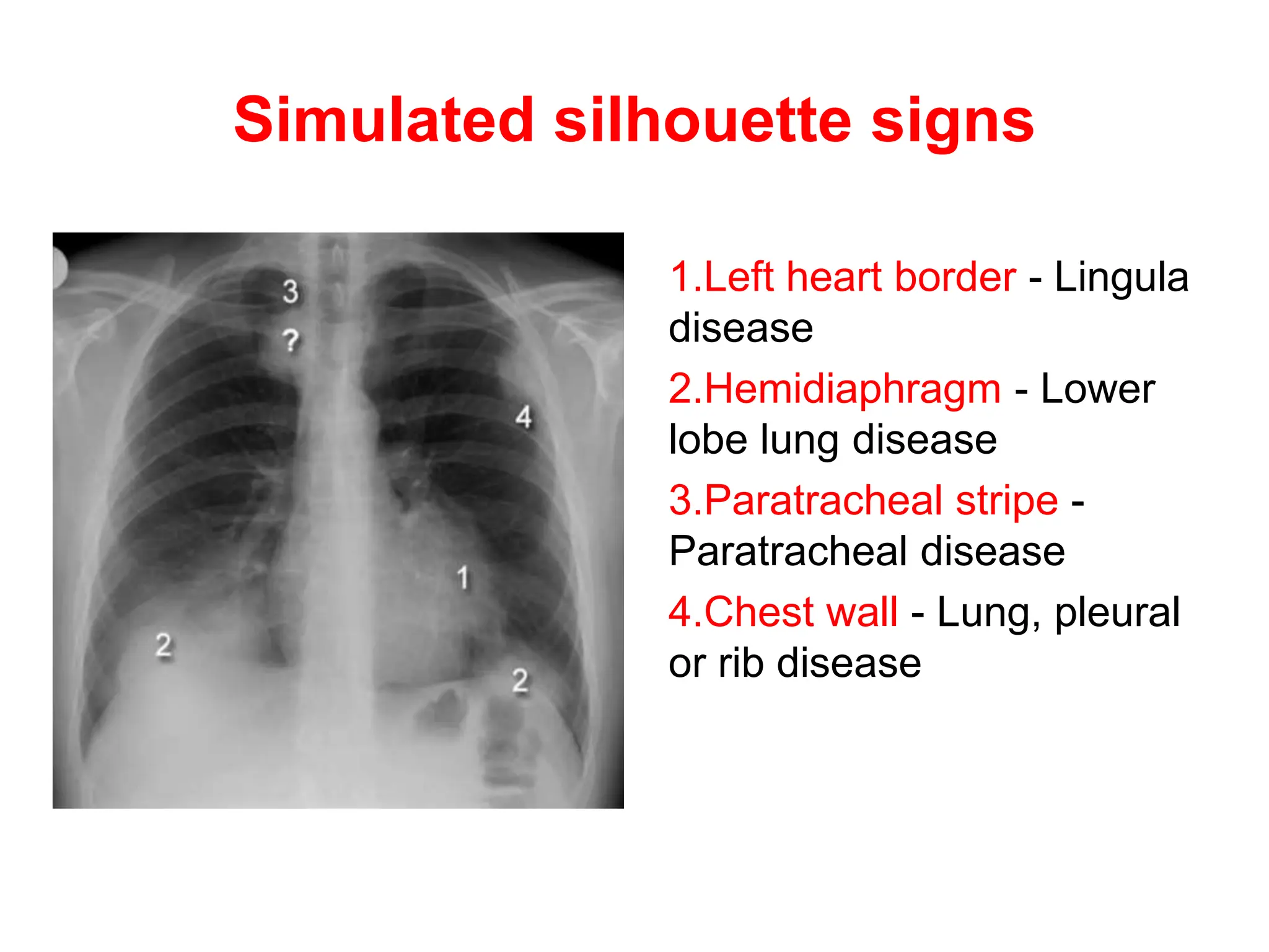
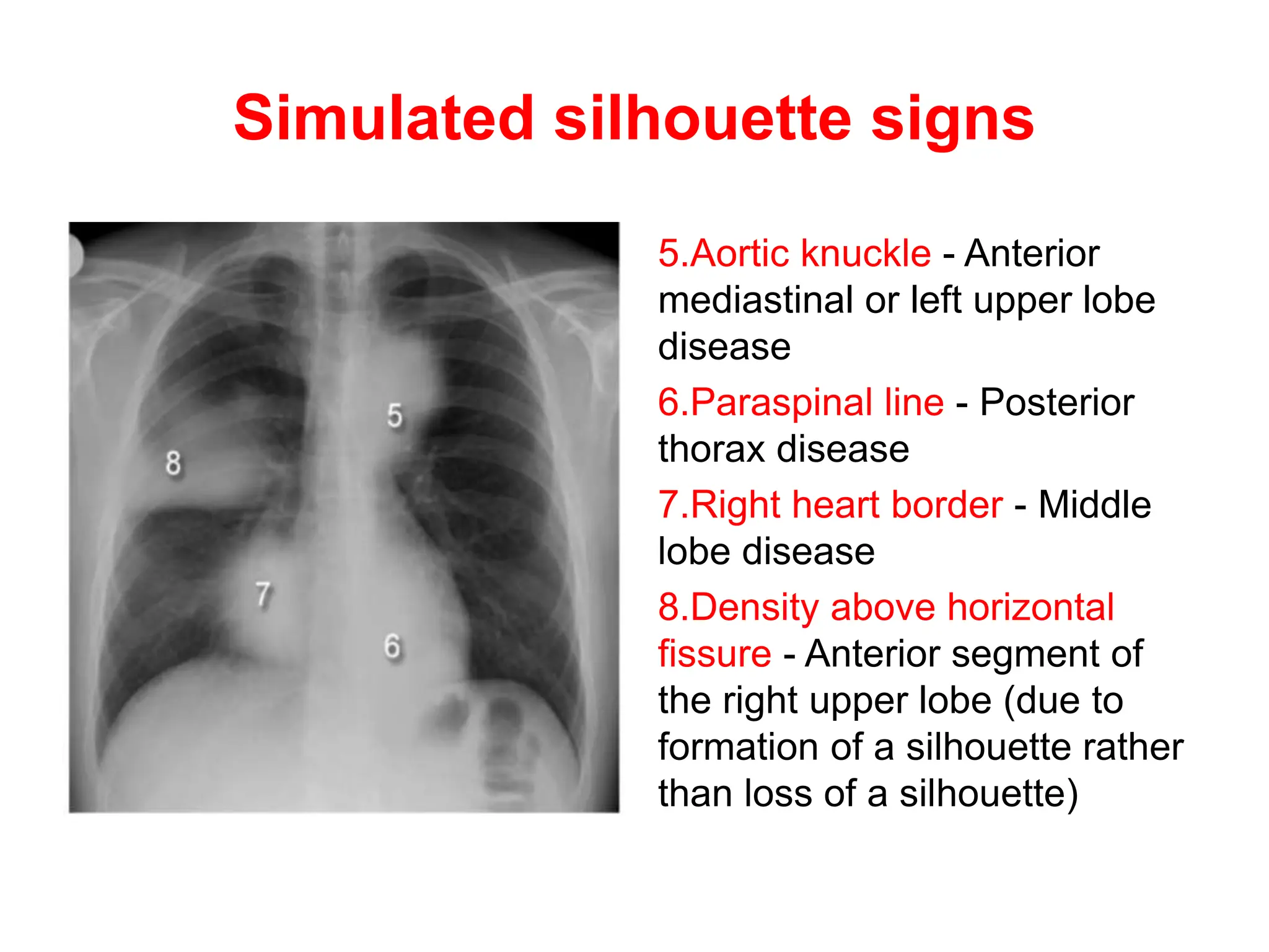
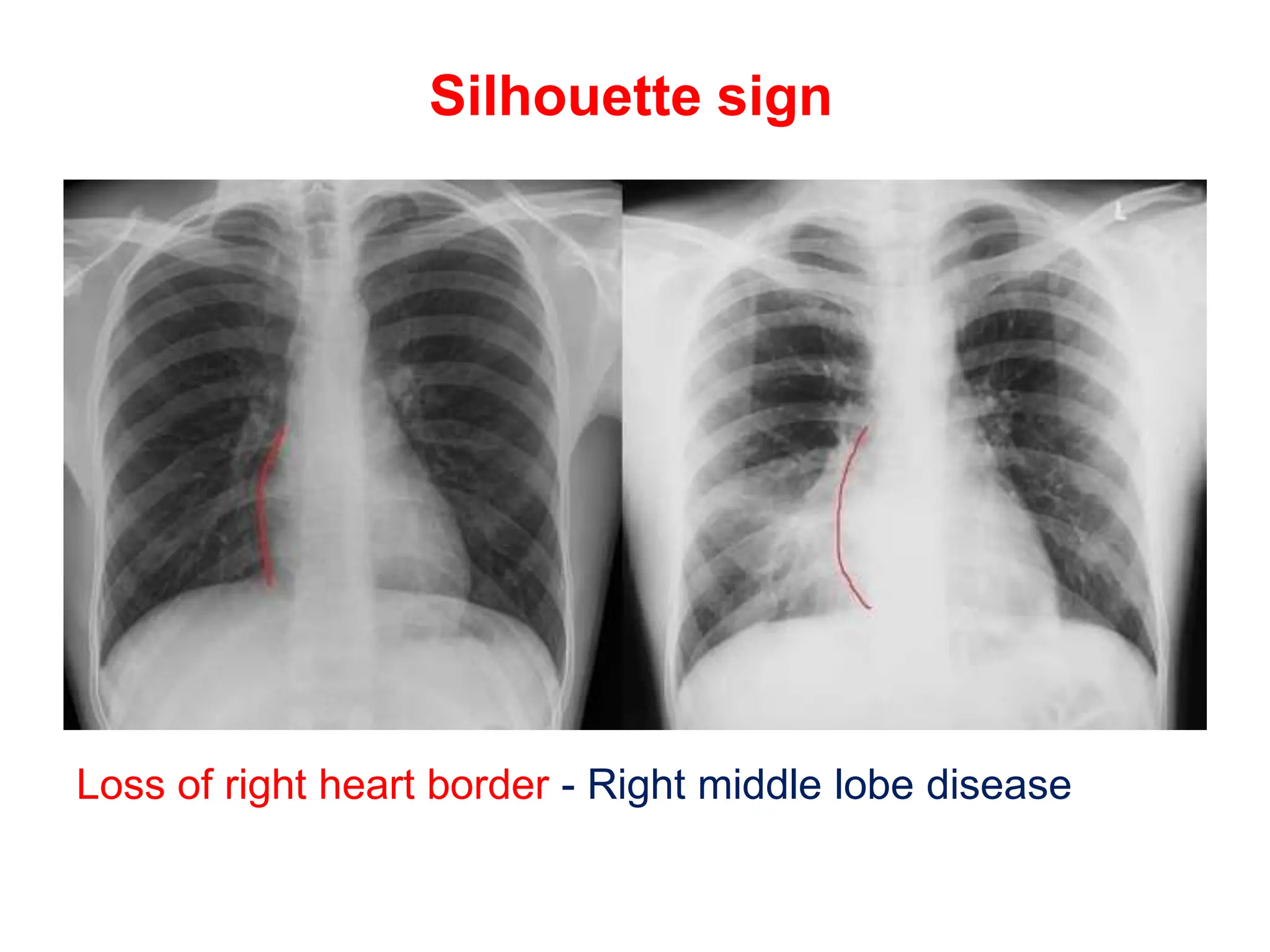
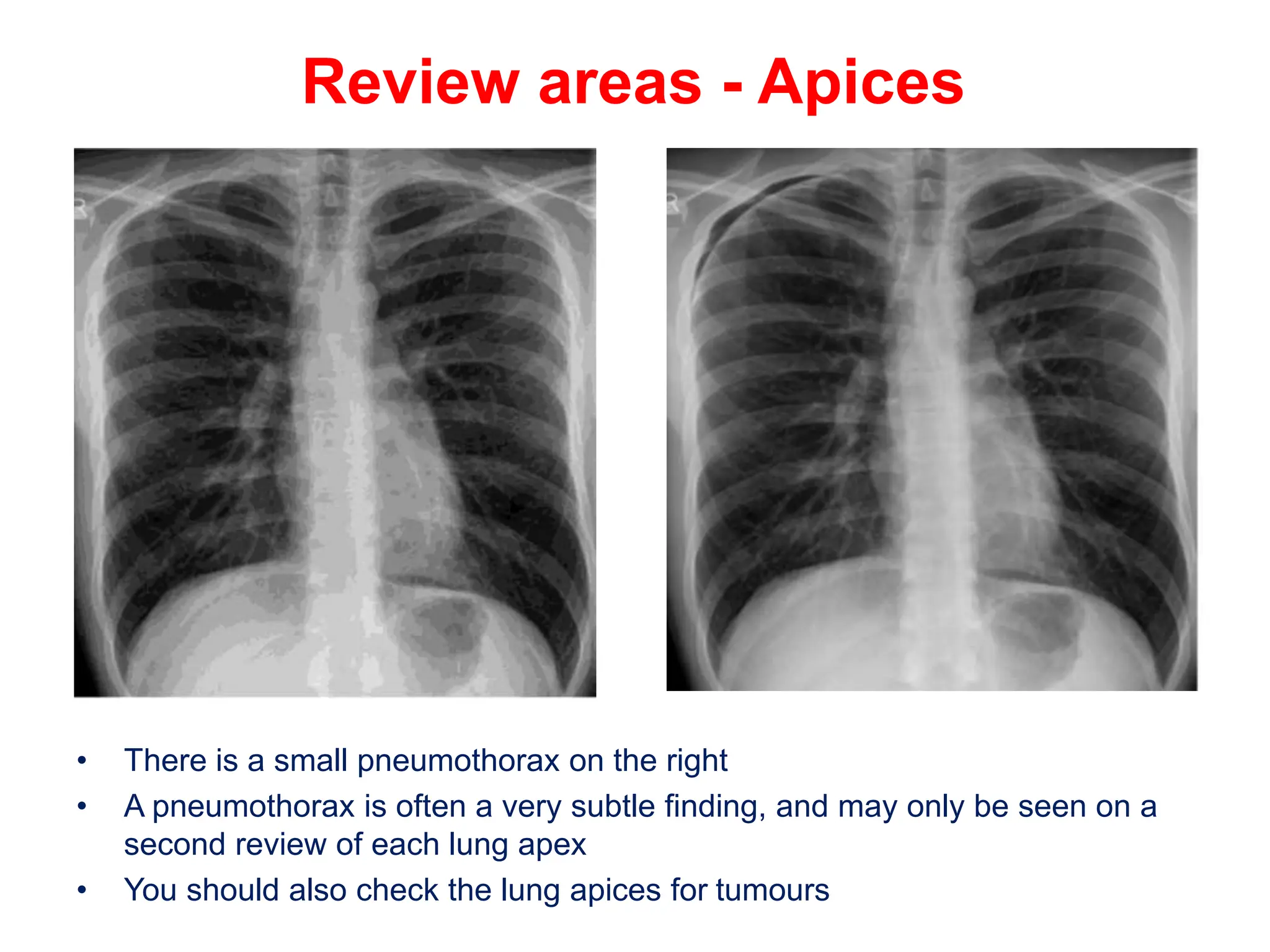
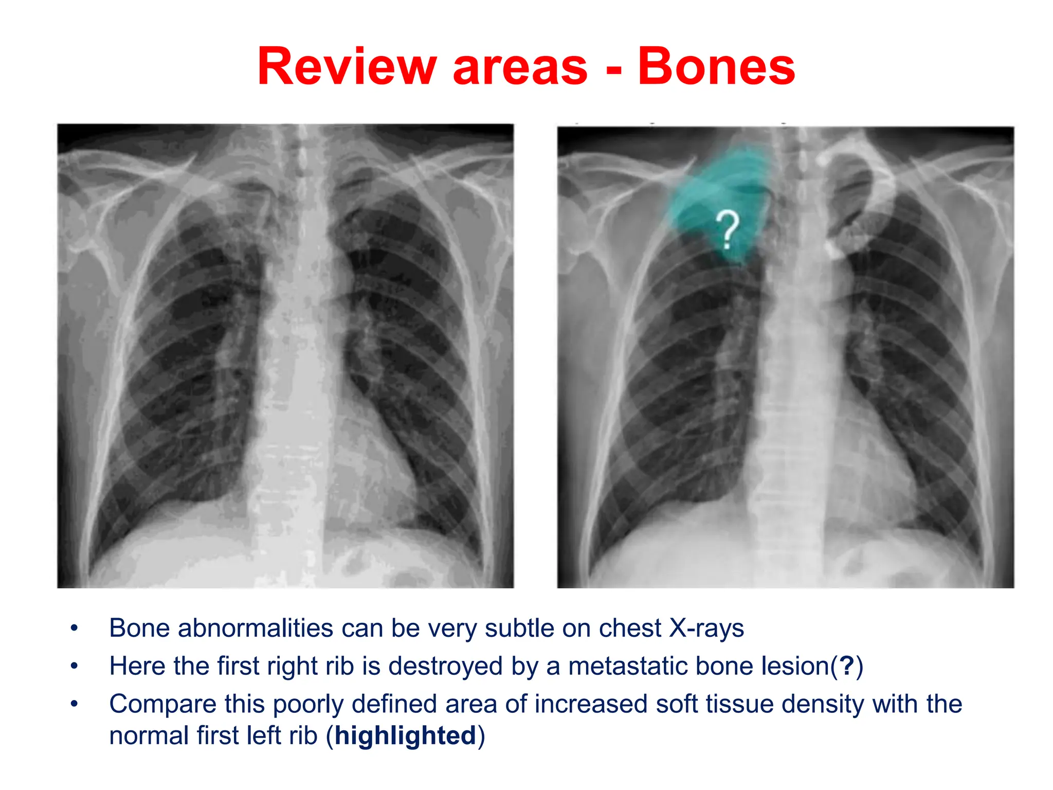
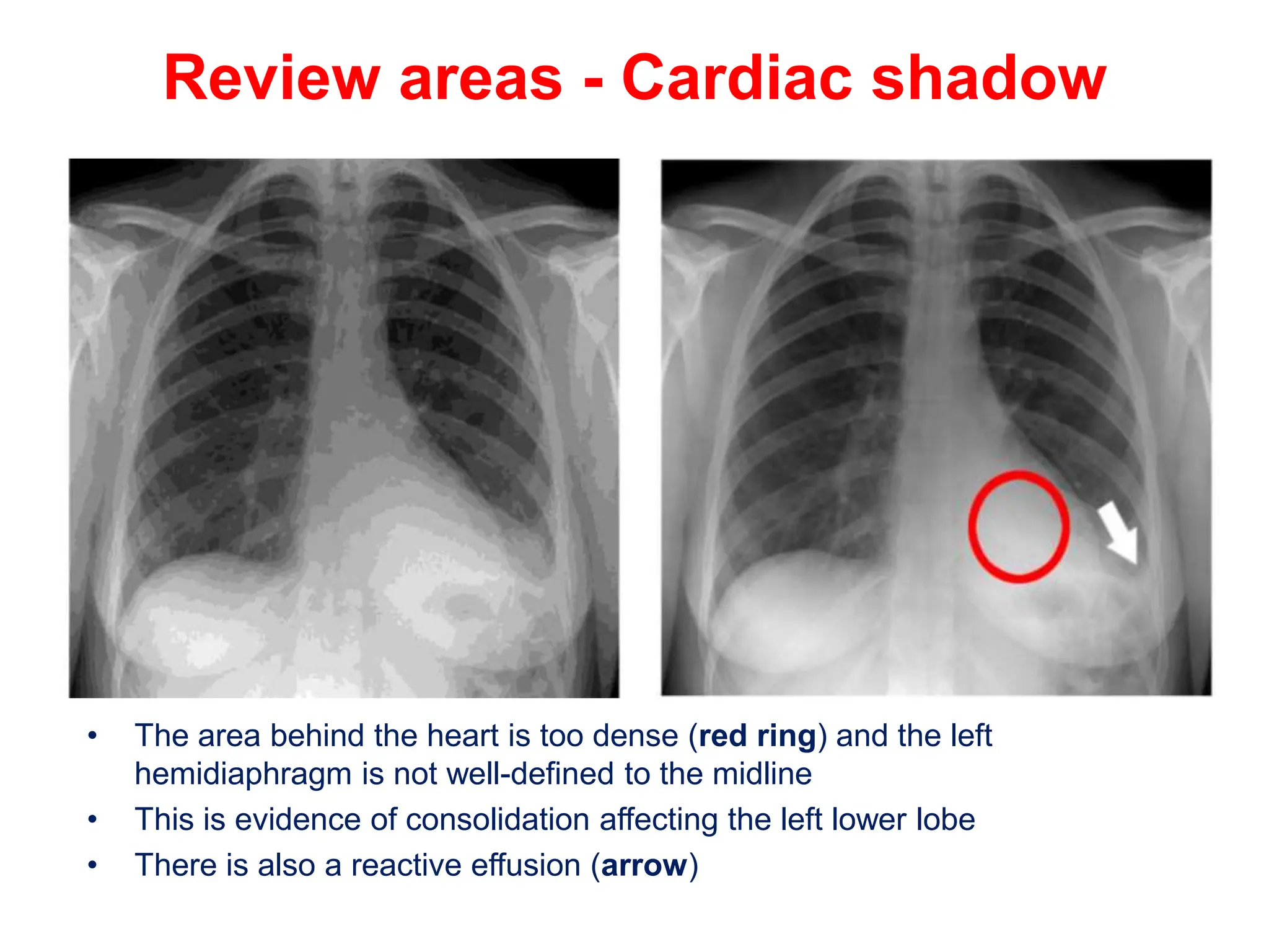
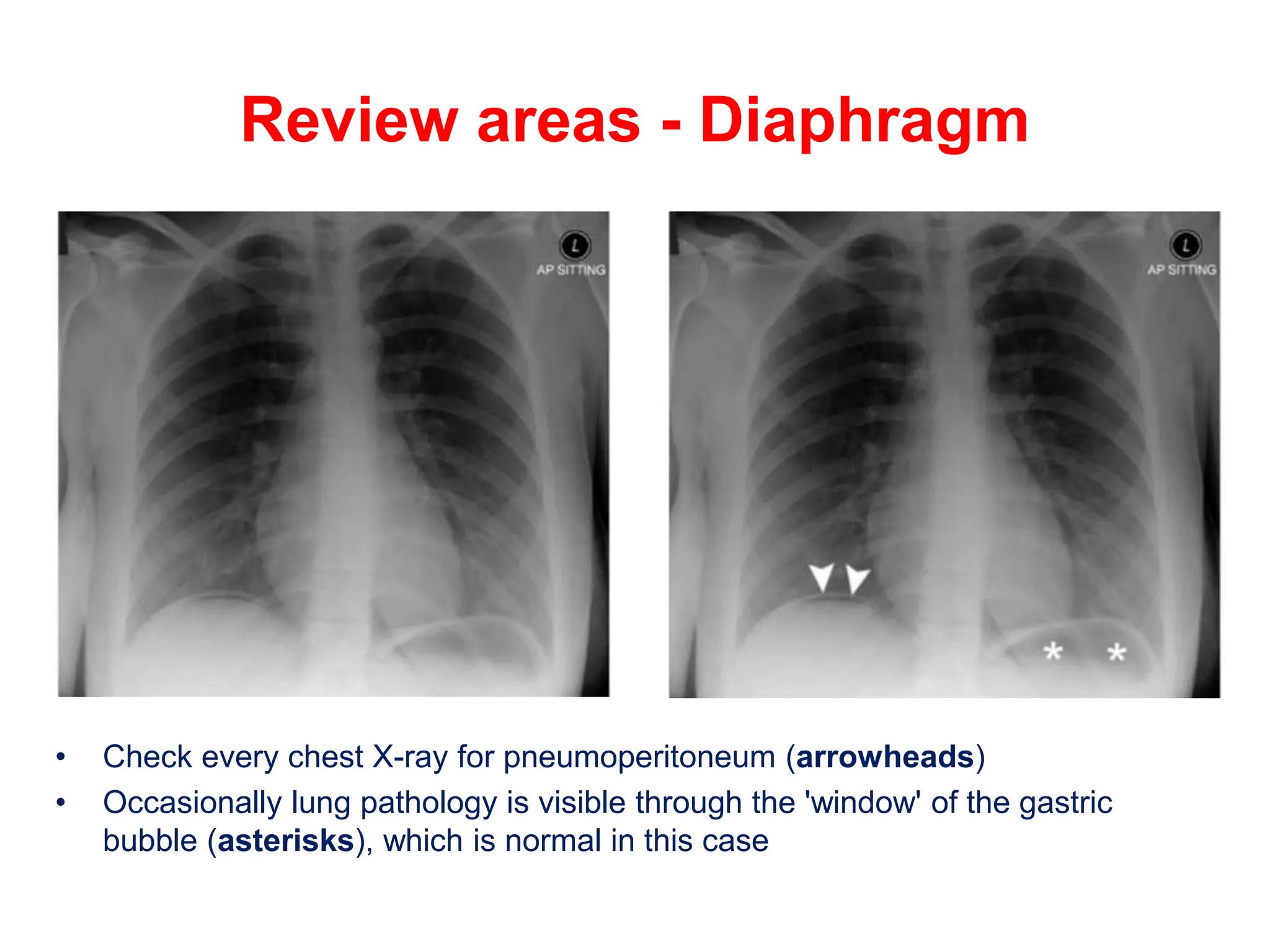
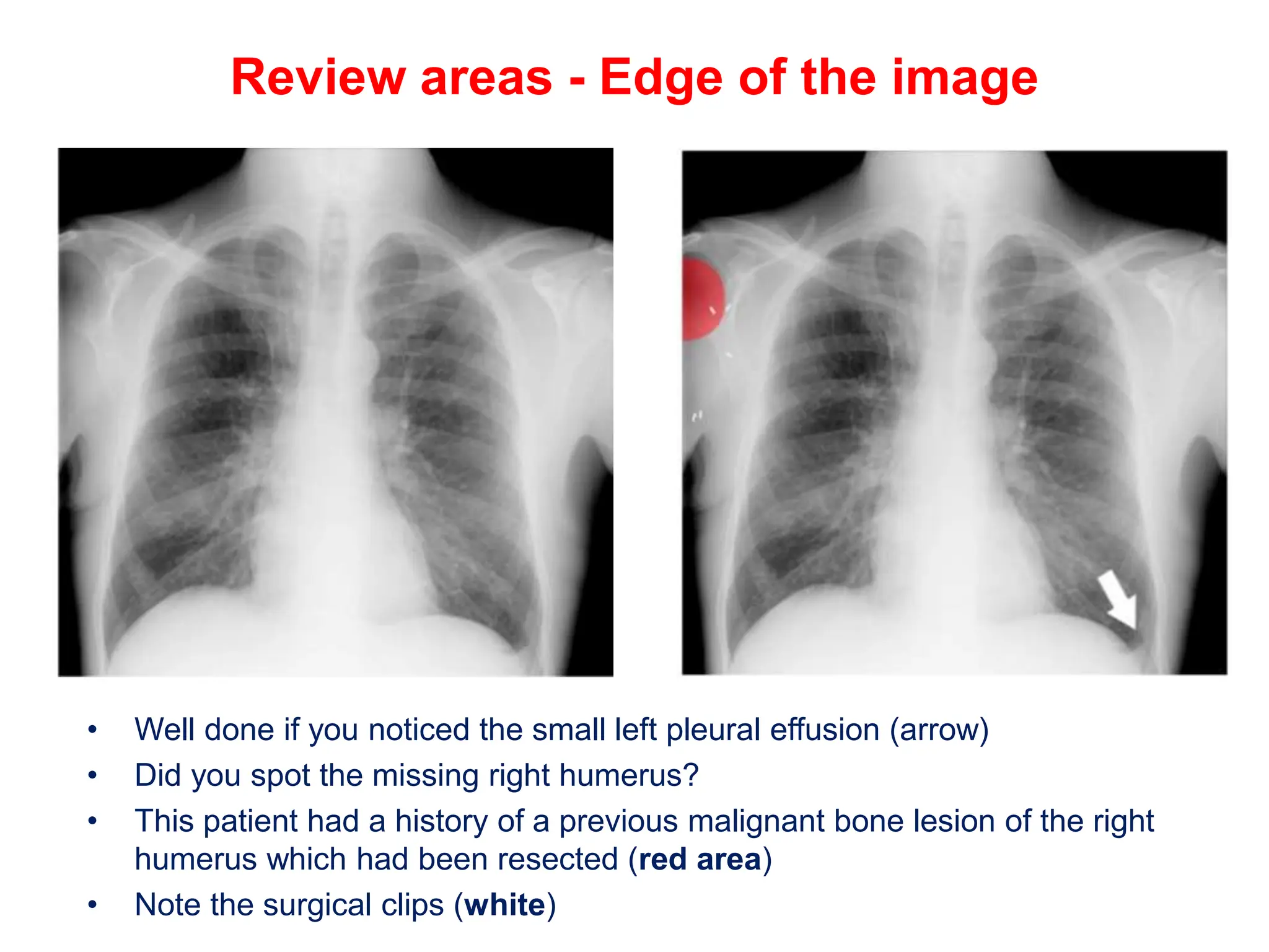
4. Systematic Approach
• A systematic approach for viewing chest X-rays ensures no
important structures are ignored
• Anatomical structures to check
1. Trachea and bronchi
2. Hilar structures
3. Mediastinum
4. Heart
5. Lung zones
6. Lung lobes and fissures
7. Pleura
8. Costophrenic angles
9. Diaphragm
10.Soft tissues
11.Bones
5. • Patent ID and date
• Check the patient's identity
• Note the image date and time
• Note the image projection : posterior-anterior (PA) or anterior-
posterior (AP) projection
• Patient was standing, sitting or supine?
• Was the mobile X-ray machine used?
• Image annotations
• Useful information is often annotated on the image
• Check the side marker is correct
• Image annotations
• This is a mobile chest X-ray taken
with the patient supine, at 11.25am in
the resuscitation room of the
Emergency Department
• The patient's name, ID number and
date of birth are annotated
• Note the side marker is correct
6. Image quality
• R - Rotation - Spinous processes at midpoint between
medial ends of the clavicles?
• I - Inspiration - 5 to 7 anterior ribs intersecting the
diaphragm in the mid-clavicular line?
• P - Penetration - Spine visible behind the heart?
• Presence of any artifact
7. GOOD QUALITY CHEST XRAY
•R - Rotation - Spinous processes at midpoint between medial ends of the clavicles?
•I - Inspiration - 5 to 7 anterior ribs intersecting the diaphragm in the mid-clavicular line?
•P - Penetration - Spine visible behind the heart?
8. Describing abnormalities
• 'Shadowing', 'opacification', 'increased density', 'increased
whiteness' are all acceptable terms
• 'Lesion descriptors' may lead you towards a diagnosis
• Be descriptive rather than jumping to a diagnosis
• Lesion descriptors
• Tissue involved - Lung, heart, aorta, bone etc
• Size - Large/Small/Varied
• Side - Right/Left - Unilateral/Bilateral
• Number - Single/Multiple
• Distribution - Focal/Widespread
• Position - Anterior/Posterior/Lung zone etc
• Shape - Round/Crescentic/etc
• Edge - Smooth/Irregular/Spiculated
• Pattern - Nodular/Reticular(net-like)
• Density - Air/Fat/Soft-tissue/Calcium/Metal
9. Shadows, opacities, densities
• Tissue involved - Lung
• Size - Small (<2 cm)
• Side - Bilateral
• Number - Multiple
• Distribution -
Widespread
• Position - Mainly middle
to lower zones
• Shape - Round
• Edge - Irregular
• Pattern - Nodular
• Density - Soft-tissue
10. Locating abnormalities
• The silhouette sign
• Normal adjacent anatomical structures of differing densities form a
crisp contour or 'silhouette‘
• Loss of a specific contour can help determine the position of a
disease process
• This phenomenon is known as the silhouette sign
• The silhouette sign is a misnomer - it should really be called the
'loss of silhouette' sign
• For example, the heart (a soft tissue density structure - near white)
lies adjacent to lung tissue (near air density - near black)
• A crisp contour or 'silhouette' is formed at the interface of these two
tissue densities
• Loss of clarity of the right heart contour (formed by the right atrium)
implies disease of the right middle lobe which lies next to the right
atrium
• Loss of distinction of the left heart contour indicates an abnormality
of the lingula (part of the left upper lobe which wraps over the left
ventricle)
11. • Silhouettes' and their adjacent tissues
• Left heart border (left ventricle) - Lingula
• Right heart border (right atrium) - Right middle lobe
• Left hemidiaphragm - Left lower lobe
• Right hemidiaphragm - Right lower lobe
• Aortic knuckle - Left upper lobe/middle mediastinum
• Descending aorta - Left lower lobe
• Right paratracheal stripe - Right upper lobe/anterior
mediastinum
• Paraspinal lines - Medial lung/Posterior mediastinum
13. Simulated silhouette signs
5.Aortic knuckle - Anterior
mediastinal or left upper lobe
disease
6.Paraspinal line - Posterior
thorax disease
7.Right heart border - Middle
lobe disease
8.Density above horizontal
fissure - Anterior segment of
the right upper lobe (due to
formation of a silhouette rather
than loss of a silhouette)
15. Review areas
• After a systematic look at the whole chest X-ray, it is
worth re-checking areas that may conceal important
pathology
• Apices - Pneumothorax?
• Bones/soft-tissues - Fractures/density?
• Cardiac shadow- Consolidation/mass?
• Diaphragm - Pneumoperitoneum?
• Edge of the image - Unexpected findings?
16. Review areas - Apices
• There is a small pneumothorax on the right
• A pneumothorax is often a very subtle finding, and may only be seen on a
second review of each lung apex
• You should also check the lung apices for tumours
17. Review areas - Bones
• Bone abnormalities can be very subtle on chest X-rays
• Here the first right rib is destroyed by a metastatic bone lesion(?)
• Compare this poorly defined area of increased soft tissue density with the
normal first left rib (highlighted)
18. Review areas - Cardiac shadow
• The area behind the heart is too dense (red ring) and the left
hemidiaphragm is not well-defined to the midline
• This is evidence of consolidation affecting the left lower lobe
• There is also a reactive effusion (arrow)
19. Review areas - Diaphragm
• Check every chest X-ray for pneumoperitoneum (arrowheads)
• Occasionally lung pathology is visible through the 'window' of the gastric
bubble (asterisks), which is normal in this case
20. Review areas - Edge of the image
• Well done if you noticed the small left pleural effusion (arrow)
• Did you spot the missing right humerus?
• This patient had a history of a previous malignant bone lesion of the right
humerus which had been resected (red area)
• Note the surgical clips (white)
21. The clinical question
• Interpret chest X-rays only in view of the clinical setting
• Treat the patient - not the X-ray!
• When requesting a chest X-ray always provide specific
clinical information
22. • No clinical
information provided
• Without clinical information
this patient may be
considered to have a
pneumonia in the left
upper zone, and started on
antibiotics
23. • Clinical information provided
• Recent increase in shortness of
breath
• No fever or productive cough
• Left shoulder and arm pain
• Heavy smoker
• Weight loss
• Findings
• Left apical shadowing
• Raised left hemidiaphragm
• Increased extra-thoracic soft
tissue density (asterisks) with
displacement of the scapula on
the left (arrowheads) - compare
with right
24. • Interpretation in view of clinical details
• Cancer - Smoker with weight loss and left apical
consolidation/mass and no clinical features of
infection
• Phrenic nerve palsy - Increased shortness of
breath and raised left hemidiaphragm
• Brachial plexopathy - Arm pain and axillary soft
tissue swelling