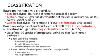
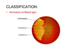
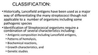
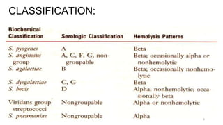
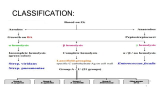
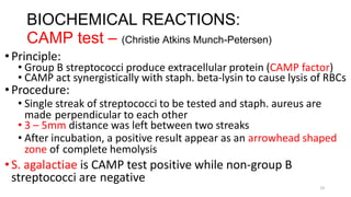
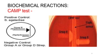
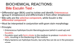
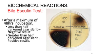
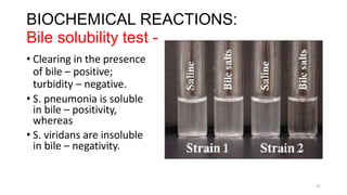
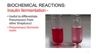
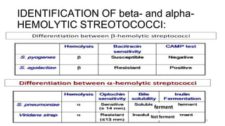
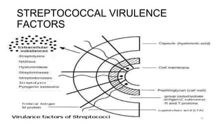
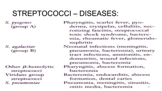
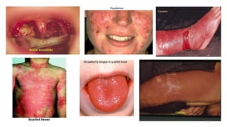
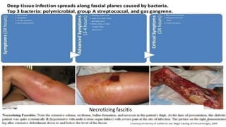
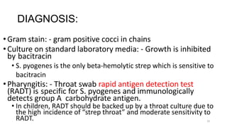
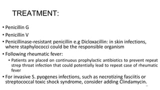
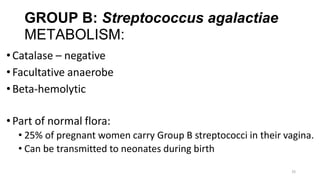
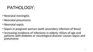
SlideShare a Scribd company logo
STREPTOCOCCAL
INFECTIONS
1
2
INTRODUCTION:
•STREPTOCOCCI are widely distributed in nature and
some are members of the normal flora while others are
pathogenic
•Pathogenicity can be attributed in part to
• Infection by the organism, and
• Sensitization to them
•They elaborate a variety of extracellular substances and
enzymes
•They are a large and heterogeneous group of bacteria
impossible to classify into one system
•Classification by various properties is key to understanding
their medical importance.
3
IDENTIFICATION:
•Gram positive spheres (cocci) like staphylococci
•But unlike staphylococci that appear in clusters, streptococci
appear in strips (chains) on gram stain – determined by their
planes of division
•1µm in diameter and usually capsulated
•Facultative anaerobes (some species – microaerophilic)
• Some – Capnophilic
•For most – growth and hemolysis are aided by incubation in 10% CO2
•They are catalase negative
•Non-sporing bacteria and non-motile.
•Growth requires enriched media containing blood or serum.
IDENTIFICATION:
• Catalase test:
•Catalase converts H2O2(which is used by macrophages and neutrophils) to
Water and O2
4
Catalase +ve
Staphylococci
Catalase –ve
Streptococci
5
CLASSIFICATION:
• Based on the hemolytic properties:
▪ Beta-hemolytic: - clear zone of hemolysis around the colony
▪ Alpha-hemolytic: - greenish discolouration of the culture medium around the
colony (partial hemolysis)
▪ Gamma-hemolytic: - no hemolysis of RBCs (Non-hemolytic streptococci)
• Based on antigenic characteristics of cell wall CHO (C – carbohydrate)
–Lancefield Antigens (Serologic Classification from A to V):
▪ Out of over 30 species of streptococci, only 5 are significant human
pathogens
▪ 3 have Lancefield Antigens:
▪ Group A – (S. pyogenes),
▪ Group B – (S. agalactiae), and
▪ Group D – (Enteroccoci + Non-enterococci)
▪ 2 have no Lancefield antigens – Lancefield Non-groupable:
▪ S. pnuemoiae, and
▪ Viridans group Streptoccoci
CLASSIFICATION:
• Hemolysis on Blood agar:
6
7
CLASSIFICATION:
•Historically, Lancefield antigens have been used as a major
way of differentiating the many streptococci though not
applicable to a number of organisms including some
pathogenic species
•Identification of Streptococcal organisms require a
combination of several characteristics including:
• Antigenic composition including Lancefield antigens,
• Patterns of hemolysis,
• Biochemical reactions,
• Growth characteristics, and
• Genetic studies
CLASSIFICATION:
8
CLASSIFICATION:
9
10
BIOCHEMICAL REACTIONS:
Differentiation between beta-hemolytic
streptococci:
•Bacitracin susceptibility test
•CAMP test
•Bile Esculin Test
11
BIOCHEMICAL REACTIONS:
Bacitracin Test -
• Bacitracin susceptibility Test:
• Specific for S. pyogenes (Group A) – for its presumptive identification
• Principle:
• To distinguish between S. pyogenes (susceptible to B) & non group A such
as S. agalactiae (resistant to B)
• Bacitracin will inhibit the growth of Group A – S. pyogenes giving zone of
inhibition around the disk
• Procedure:
• Inoculate BAP with heavy suspension of tested organism
• Bacitracin disk (0.04 U) is applied to inoculated BAP
• After incubation, any zone of inhibition around the disk is considered as
susceptible
BIOCHEMICAL REACTIONS:
Bacitracin Test:
12
13
BIOCHEMICAL REACTIONS:
CAMP test – (Christie Atkins Munch-Petersen)
•Principle:
• Group B streptococci produce extracellular protein (CAMP factor)
• CAMP act synergistically with staph. beta-lysin to cause lysis of RBCs
•Procedure:
• Single streak of streptococci to be tested and staph. aureus are
made perpendicular to each other
• 3 – 5mm distance was left between two streaks
• After incubation, a positive result appear as an arrowhead shaped
zone of complete hemolysis
•S. agalactiae is CAMP test positive while non-group B
streptococci are negative
BIOCHEMICAL REACTIONS:
CAMP test -
14
15
BIOCHEMICAL REACTIONS:
Bile Esculin Test -
• Differential agar (BEA) used to isolate and identify Enterococcus
(group D streptococci) and differentiate it from other streptococci
• Bile salts are the selective component, while Esculin is the
differential component
• Must be interpreted in conjunction with gram stain morphology
• Principle:
• Enterococcus hydrolyze Esculin liberating glucose (which is used up) and
Esculetin.
• Esculetin react with ferric citrate in the medium to produce insoluble iron
salts, resulting in the blackening of the medium
• Many bacteria can hydrolyze Esculin, but only few can do so in the presence
BIOCHEMICAL REACTIONS:
Bile Esculin Test:
•After a maximum of
48hrs incubation,
•Less than half
darkened agar slant –
Negative result.
•Greater than half
darkened agar slant –
Positive result.
16
17
BIOCHEMICAL REACTIONS:
Differentiation between alpha-hemolytic
Streptococci
•Optochin test
•Bile solubility test
•Inulin fermentation
18
BIOCHEMICAL REACTIONS:
Optochin test -
•Principle:
• S. pneumonia is inhibited by Optochin reagent (<5 μg/mL) giving
an inhibition zone of ≥14mm in diameter.
•Procedure:
• BAP is inoculated with the organism to be tested and an Optochin
disc placed in the center of the plate
• After incubation at 37oC for 18hrs, carefully measure the
diameter of the inhibition zone with a ruler
• ≥14mm is positive; ≤ 13mm is negative
•S. pneumonia is positive (S); S. viridans is negative (R)
BIOCHEMICAL REACTIONS:
Optochin test -
19
20
BIOCHEMICAL REACTIONS:
Bile solubility test -
•Principle:
• S. pneumonia produce a self-lysing enzyme capable of inhibiting its
growth and this is accelerated in the presence of bile.
•Procedure:
• Add 10 parts of the broth culture of the organism to be tested to
one part of 2% Na-deoxycholate (bile) in a test-tube
• Negative control is made by adding saline instead of bile to the
culture
• Incubate at 37oC for 15mins
• Observe and record your findings.
BIOCHEMICAL REACTIONS:
Bile solubility test -
• Clearing in the presence
of bile – positive;
turbidity – negative.
• S. pneumonia is soluble
in bile – positivity,
whereas
• S. viridans are insoluble
in bile – negativity.
21
BIOCHEMICAL REACTIONS:
Insulin fermentation -
• Useful to differentiate
Pneumococci from
other Streptcocci:
• Pneumococci ferments
inulin
22
IDENTIFICATION OF beta- and alpha-
HEMOLYTIC STREOTOCOCCI:
ferment
23
Not ferment
STREPTOCOCCAL VIRULENCE
FACTORS
24
STREPTOCOCCI – DISEASES:
25
26
GROUP A: Streptococcus pyogenes
METABOLISM:
•Catalase – negative
•Microaerophilic
•Beta-hemolytic – due to enzymes that destroy blood cells
• Streptolysin-O:
• Oxygen labile
• Antigenic
• Streptolysin-S:
• Oxygen stable
• Non-antigenic
27
VIRULENCE:
• M-protein (70 types) – major virulence factor
• Adherence factor
• Antiphagocytic
• Antigenic: induces antibodies which can lead to phagocytosis
• Lipoteichoic acid:
• Adherence factor
• Streptokinase - FIBRINOLYSIN
• Hyaluronidase – destroys CT and aids spread of the organism
• DNAase (Streptodornase)
• Anti-C5a peptidase – prevents C5a mediated phagocytic activity
• Protein F -
• Streptolysin O – also antigenic; (but Streptolysin S – not antigenic)
• Hence Anti-Streptolysin O (ASO) antibody titer rises in recent infections
• Skin infection does not induce ASO
28
TOXINS:
•Erythrogenic or pyrogenic toxin (produced only by
lysogenized Group A Streptococci): responsible for
scarlet fever
•More than 4 serologically distinct toxins (Spe - A, B, C and F).
•Dick Test: once commonly used to confirm Scarlet Fever
diagnosis.
•Some strains produce pyrogenic exotoxins which act as
superantigens that superstimulate T cell leading to release
of cytokine which cause the Toxic shock syndrome.
•Toxic shock syndrome toxin (similar to, but different
from the staph exotoxin TSST-1)
29
PATHOLOGY:
DIRECT INVASION/TOXIN:
•Pharyngitis:
• Red, Swollen tonsils and pharynx
• Purulent exudate on tonsils
• Fever
• Swollen lymph nodes
•Skin infections:
• Folliculitis, Erysipelas, pyoderma
• Cellulitis
• Impetigo
• Necrotizing fasciitis
•Scarlet fever: - fever and scarlet red rash on body
•Toxic shock syndrome
30
Necrotizing fascitis
31
32
PATHOLOGY:
ANTIBODY MEDIATED (Delayed):
•Rheumatic fever (may follow streptococcal pharyngitis):
• Fever
• Myocarditis: heart inflammation >> Rhuematic valvular heart disease many
years later.
• Arthritis: migratory polyarthritis
• Chorea (Sydenham’s chorea or St. Vitus dance)
• Rash: Erythema marginatum
• Subcutaneous nodules: 10 – 20 yrs after infection, may develop permanent
heart valve damage
•Acute post-streptococcal Glomerulonephritits:
• Tea or coca cola coloured urine, following streptococcal skin or pharynx
infection
• Follows skin or throat infection by Nephritogenic strains
33
DIAGNOSIS:
•Gram stain: - gram positive cocci in chains
•Culture on standard laboratory media: - Growth is inhibited
by bacitracin
• S. pyogenes is the only beta-hemolytic strep which is sensitive to
bacitracin
•Pharyngitis: - Throat swab rapid antigen detection test
(RADT) is specific for S. pyogenes and immunologically
detects group A carbohydrate antigen.
• In children, RADT should be backed up by a throat culture due to
the high incidence of “strep throat” and moderate sensitivity to
RADT.
34
TREATMENT:
• Penicillin G
• Penicillin V
• Penicillinase-resistant penicillin e.g Dicloxacillin: in skin infections,
where staphylococci could be the responsible organism
▪ Following rheumatic fever:
▪ Patients are placed on continuous prophylactic antibiotics to prevent repeat
strep throat infection that could potentially lead to repeat case of rheumatic
fever
▪ For invasive S. pyogenes infections, such as necrotizing fasciitis or
streptococcal toxic shock syndrome, consider adding Clindamycin.
35
GROUP B: Streptococcus agalactiae
METABOLISM:
•Catalase – negative
•Facultative anaerobe
•Beta-hemolytic
•Part of normal flora:
• 25% of pregnant women carry Group B streptococci in their vagina.
• Can be transmitted to neonates during birth
36
PATHOLOGY:
• Neonatal meningitis
• Neonatal pneumonia
• Neonatal sepsis
• Sepsis in pregnant women (with secondary infection of fetus)
• Increasing incidence of infections in elderly >65yrs of age and
patients with diabetes or neurological disease: causes sepsis and
pneumonia
37
DIAGNOSIS:
• Gram stain of CSF or Urine: - positive cocci in chains
• Culture of CSF, Urine or Blood
38
TREATMENT:
• Penicillin G
39
GROUP C and G Streptococcus:
• Beta-hemolytic
• S. equi, S. canis
• Associated diseases:
• Pharyngitis, pneumonia, cellulitis, pyoderma, erysipelas, impetigo, wound
infections, puerperal sepsis, neonatal sepsis, endocarditis, septic arthritis
• Treatment:
• Penicillin, vancomycin, cephalosporins, macrolides (variable susceptibility)
GROUP D Streptococcus:
• 2 SUB-TYPES:
• Enterococci: (recently given their own genus
because they sufficiently differ from the
streptococci)
• S. faecalis
• S. faecium
• Non-enterococci:
• S. bovis
• S.equinus
• Enterococcus:
• Gram +ve cocci
• Singly/in pairs/short chains Enterococci
40
41
METABOLISM:
• Catalase – Negative
• Facultative anaerobes
• Usually Gamma-hemolytic, but maybe alpha-hemolytic
• Positive bile esculin test
42
VIRULENCE:
• Extracellular dextran helps them bind to heart valves
43
PATHOLOGY:
•Sub-acute bacterial endocarditis
•Biliary tract infections
•Urinary tract infections (especially the Enterococci)
•S. bovis is associated with colonic malignancies
44
DIAGNOSIS:
•Gram stain – positive cocci in chains
•Culture:
• Enterococci can be cultured in:
• 40% bile
• 6.5% Sodium chloride
• Non-enterococci can only grow in bile
45
TREATMENT:
•Ampicillin, sometimes combined with an aminoglycoside
•Resistant to Penicillin G
•Emerging resistance to vancomycin
•For vancomycin resistant organisms (VRE), consider Linezolid,
Daptomycin and Nitrofurantoin.
46
VIRIDANS GROUP Streptococci:
• Part of normal oral flora,
• Found in the nasopharynx and gingivial crevices
• GI tract
• Members:
• Mitis group: S. mitis, S. sanguis, S. parasanguis, S. gordonii, S. crista, S.
infantis, S. oralis, S. peroris
• Salavarius group: S. salavarius, S. vestibularis, S. thermophiles
• Mutans group: S. mutans, S. sobrinus, S. criceti, S. rattus, S. downeii, S.
macacae
• Angionosus group: S. angionosus, S. constellatus, S. intermedius
47
METABOLISM:
• Catalase – negative
• Facultative anaerobes
• Mostly alpha-hemolytic; some beta- and gamma
• Resistant to Optochin
• Bile solubility - Negative
48
VIRULENCE:
• Extracellular dextran – helps them bind to heart valves
49
PATHOLOGY:
• Sub-acute bacterial endocarditis: caused by S. mitis group
• Dental caries (cavities): caused by S. mutans group
• Brain or Liver abscesses: caused by S. angionosus group
• Microaerophilic
• Found alone in pure cultures or in mixed cultures with anaerobes
DIAGNOSIS:
• Gram stain
• Culture – antibiotics may be added to inhibit growth
of contaminating bacteria
• Resistant to optochin
• Detection of group A streptococci by molecular
methods: PCR assay for pharyngeal specimens
• Antibody detection
• ASO titration for respiratory infections.
• Anti-DNAase B and Antihyaluronidase titration for
skin infections.
• Anti-streptokinase; Anti-M type-specific antibodies
50
51
TREATMENT:
• Penicillin G
• Effective doses of penicillin or erythromycin for 10 days can prevent
post-streptococcal diseases.
• Drainage and aggressive surgical debridement must be promptly
initiated in patients with serious soft tissue infections.
• Antibiotic sensitivity test is helpful for treatment of bacterial
endocarditis.
52
Streptococci Pneumoniae
(Pnuemococcus):
METABOLISM:
•Gram-positive lancet-shaped diplococci.
•Alpha-hemolytic (Pneumolysin is similar to streptolysin O).
•Form small round colonies on the plate, at first dome-shaped
and later developing a central plateau with an elevated rim.
•Facultative anaerobe
•Autolysis is enhanced in bile salt.
•Growth is enhanced by 5-10% CO2 – capnophilic.
53
VIRULENCE:
• Polysaccharide Capsule (90 serotypes):
• Protects the organism from phagocytosis
• Highly antigenic – opsonized by antibodies specific to it
• Due to the variable serotypes, surviving an infection from one serotype does
not confer immunity over the others
• Cell wall polysaccharide
• Phosphocholine
• Pneumolysin
• IgA protease, etc.
54
TOXINS:
•Pneumolysin: binds to cholesterol in host-cell membranes
(but its actual effect is unknown)
•MAJOR HOST DEFENCE MECHANISM: - Ciliated Cells of the
Resp tract and Spleen
•Loss of natural resistance may be due to:
• Abnormalities of the respiratory tract (e.g. viral RT infections).
• Alcohol or drug intoxication; abnormal circulatory dynamics.
• Patients undergone renal transplant; chronic renal diseases.
• Malnutrition, general debility, sickle cell anemia,
hyposplenism or splenectomy, nephrosis or complement
deficiency
• Young children and the elderly
55
PATHOLOGY:
• Pnuemonia – pneumococcus is the most common cause of
pneumonia in adults
• Meningitis – most common cause of bacterial meningitis in adults
• Sepsis
• Otitis media (most common cause in children)
DIAGNOSIS:
• Gram stain: - reveals gram-positive
diplococcic
• Culture: does not grow in presence of:
• Optochin
• Bile
• Capsular polysaccharide antigen
detection
• DNA probe specific to S. pneumonia
• Virulence to mice
Pneumococcus Specific DNA probe
56
DIAGNOSIS:
• Positive Quellung test:
swelling when tested
against antiserum
containing anti-capsular
antibodies
• Quellung reaction:
technique used to
detect encapsulated
bacteria (such as S.
pneumonia and H.
influenza)
Quellung Antibody reaction
57
58
TREATMENT:
• Penicillin G (IM)
• Erythromycin
• Ceftriaxone
• Vaccine: made against 23 most common capsular antigens.
• Vaccinate individuals who are susceptible such as elderly or asplenic
individuals (including being functionally asplenic due to sickle cell anaemia)
• Pnuemococcal conjugate vaccine - Capsular polysaccharide + Protein Carrier
• Below 2yrs
• Heptavalent and the newer 13 valent conjugated vaccines are
effective at preventing otitis media and pneumonia.
59
CONCLUSION:
• STREPTOCOCCI are widely distributed in nature and some are
members of the normal flora while others are pathogenic
• Pathogenicity can be attributed in part to
• Infection by the organism, and
• Sensitization to them
• They elaborate a variety of extracellular substances and enzymes
• They are a large and heterogeneous group of bacteria impossible to
classify into one system
• Classification by various properties is key to understanding their
medical importance.

More Related Content

Similar to Streptococci.pptx

Streptococci
StreptococciStreptococci
Streptococci
Amirul Huda Bhuiyan
 
. staphylococcus dr. ihsan alsaimary
.  staphylococcus  dr. ihsan alsaimary.  staphylococcus  dr. ihsan alsaimary
. staphylococcus dr. ihsan alsaimary
dr.Ihsan alsaimary
 
Staphylococci and Streptococci organisms.ppt
Staphylococci and Streptococci organisms.pptStaphylococci and Streptococci organisms.ppt
Staphylococci and Streptococci organisms.ppt
vinuthdp
 
E. coli
E. coli E. coli
E. coli
Ashna Ajimsha
 
Group b & d streptococci
Group b & d streptococciGroup b & d streptococci
Group b & d streptococci
Meenakshi Sharma
 
Corynebacterium diptheriae
Corynebacterium diptheriaeCorynebacterium diptheriae
Corynebacterium diptheriae
santusan
 
Streptococcusبكتريا عملي
Streptococcusبكتريا عملي Streptococcusبكتريا عملي
Streptococcusبكتريا عملي
في رحاب الله
 
Microbiology
MicrobiologyMicrobiology
Microbiology
hasan askari
 
Salmonella
SalmonellaSalmonella
Salmonella
Sijalniroula
 
Streptococcus & Enterococcus by Dr. Rakesh Prasad Sah
Streptococcus & Enterococcus by Dr. Rakesh Prasad SahStreptococcus & Enterococcus by Dr. Rakesh Prasad Sah
Streptococcus & Enterococcus by Dr. Rakesh Prasad Sah
Dr. Rakesh Prasad Sah
 
SHIGELLA, KLEBSIELLA, PROTEUS, PSEUDOMONAS.pptx
SHIGELLA, KLEBSIELLA, PROTEUS, PSEUDOMONAS.pptxSHIGELLA, KLEBSIELLA, PROTEUS, PSEUDOMONAS.pptx
SHIGELLA, KLEBSIELLA, PROTEUS, PSEUDOMONAS.pptx
ShahriarHabib4
 
beta lactamases
beta lactamasesbeta lactamases
beta lactamases
Malathi Murugesan
 
Streptococcus Species.pptx
Streptococcus Species.pptxStreptococcus Species.pptx
Streptococcus Species.pptx
HelloVintunnara
 
Micro part1 study guide
Micro part1 study guideMicro part1 study guide
Micro part1 study guide
Donna Kim
 
Streptococci and enterococci bls 206
Streptococci and enterococci bls 206Streptococci and enterococci bls 206
Streptococci and enterococci bls 206Bruno Mmassy
 
Clostridium perfringens
Clostridium perfringensClostridium perfringens
Clostridium perfringens
Dr. Rakesh Prasad Sah
 
Staphylococcus
Staphylococcus Staphylococcus
Staphylococcus
mahsajalili1993
 
presentationofstreptococcus-180731145601.pdf
presentationofstreptococcus-180731145601.pdfpresentationofstreptococcus-180731145601.pdf
presentationofstreptococcus-180731145601.pdf
sathishvsathish1
 
Presentation of streptococcus
Presentation of streptococcusPresentation of streptococcus
Presentation of streptococcus
Byiringiro Pacifique
 

Similar to Streptococci.pptx (20)

Streptococci
StreptococciStreptococci
Streptococci
 
. staphylococcus dr. ihsan alsaimary
.  staphylococcus  dr. ihsan alsaimary.  staphylococcus  dr. ihsan alsaimary
. staphylococcus dr. ihsan alsaimary
 
Staphylococci and Streptococci organisms.ppt
Staphylococci and Streptococci organisms.pptStaphylococci and Streptococci organisms.ppt
Staphylococci and Streptococci organisms.ppt
 
E. coli
E. coli E. coli
E. coli
 
Group b & d streptococci
Group b & d streptococciGroup b & d streptococci
Group b & d streptococci
 
identification of bacteria
identification of bacteriaidentification of bacteria
identification of bacteria
 
Corynebacterium diptheriae
Corynebacterium diptheriaeCorynebacterium diptheriae
Corynebacterium diptheriae
 
Streptococcusبكتريا عملي
Streptococcusبكتريا عملي Streptococcusبكتريا عملي
Streptococcusبكتريا عملي
 
Microbiology
MicrobiologyMicrobiology
Microbiology
 
Salmonella
SalmonellaSalmonella
Salmonella
 
Streptococcus & Enterococcus by Dr. Rakesh Prasad Sah
Streptococcus & Enterococcus by Dr. Rakesh Prasad SahStreptococcus & Enterococcus by Dr. Rakesh Prasad Sah
Streptococcus & Enterococcus by Dr. Rakesh Prasad Sah
 
SHIGELLA, KLEBSIELLA, PROTEUS, PSEUDOMONAS.pptx
SHIGELLA, KLEBSIELLA, PROTEUS, PSEUDOMONAS.pptxSHIGELLA, KLEBSIELLA, PROTEUS, PSEUDOMONAS.pptx
SHIGELLA, KLEBSIELLA, PROTEUS, PSEUDOMONAS.pptx
 
beta lactamases
beta lactamasesbeta lactamases
beta lactamases
 
Streptococcus Species.pptx
Streptococcus Species.pptxStreptococcus Species.pptx
Streptococcus Species.pptx
 
Micro part1 study guide
Micro part1 study guideMicro part1 study guide
Micro part1 study guide
 
Streptococci and enterococci bls 206
Streptococci and enterococci bls 206Streptococci and enterococci bls 206
Streptococci and enterococci bls 206
 
Clostridium perfringens
Clostridium perfringensClostridium perfringens
Clostridium perfringens
 
Staphylococcus
Staphylococcus Staphylococcus
Staphylococcus
 
presentationofstreptococcus-180731145601.pdf
presentationofstreptococcus-180731145601.pdfpresentationofstreptococcus-180731145601.pdf
presentationofstreptococcus-180731145601.pdf
 
Presentation of streptococcus
Presentation of streptococcusPresentation of streptococcus
Presentation of streptococcus
 

More from MrsP6

IUGR.pptx
IUGR.pptxIUGR.pptx
IUGR.pptx
MrsP6
 
GDM.pptx
GDM.pptxGDM.pptx
GDM.pptx
MrsP6
 
physiological changes during pregnancy.pptx
physiological changes during pregnancy.pptxphysiological changes during pregnancy.pptx
physiological changes during pregnancy.pptx
MrsP6
 
assessment and management of pregnancy ( antenatal).pptx
assessment and management of pregnancy ( antenatal).pptxassessment and management of pregnancy ( antenatal).pptx
assessment and management of pregnancy ( antenatal).pptx
MrsP6
 
ANTEPARTUM HAEMORRHAGE.pptx
ANTEPARTUM HAEMORRHAGE.pptxANTEPARTUM HAEMORRHAGE.pptx
ANTEPARTUM HAEMORRHAGE.pptx
MrsP6
 
Course outline OBGY.docx
Course outline OBGY.docxCourse outline OBGY.docx
Course outline OBGY.docx
MrsP6
 
Microbiology course outine.docx
Microbiology course outine.docxMicrobiology course outine.docx
Microbiology course outine.docx
MrsP6
 
mycobacterium tuberculosis
mycobacterium tuberculosismycobacterium tuberculosis
mycobacterium tuberculosis
MrsP6
 
Neisseria-
Neisseria-Neisseria-
Neisseria-
MrsP6
 
specimen collection and transport
specimen collection and transportspecimen collection and transport
specimen collection and transport
MrsP6
 
CPD.pptx
CPD.pptxCPD.pptx
CPD.pptx
MrsP6
 
DVT.pptx
DVT.pptxDVT.pptx
DVT.pptx
MrsP6
 
Abnormal uterine bleeding.pptx
Abnormal uterine bleeding.pptxAbnormal uterine bleeding.pptx
Abnormal uterine bleeding.pptx
MrsP6
 
PAIN MANAGEMENT.pptx
PAIN MANAGEMENT.pptxPAIN MANAGEMENT.pptx
PAIN MANAGEMENT.pptx
MrsP6
 
USE OF PERSONAL PROTECTIVE EQUIPMENT.pptx
USE OF PERSONAL PROTECTIVE EQUIPMENT.pptxUSE OF PERSONAL PROTECTIVE EQUIPMENT.pptx
USE OF PERSONAL PROTECTIVE EQUIPMENT.pptx
MrsP6
 
Hospital acquired infection.pptx
Hospital acquired infection.pptxHospital acquired infection.pptx
Hospital acquired infection.pptx
MrsP6
 
NORMAL PUERPERIUM PPT.pptx
NORMAL PUERPERIUM PPT.pptxNORMAL PUERPERIUM PPT.pptx
NORMAL PUERPERIUM PPT.pptx
MrsP6
 
APH.pptx
APH.pptxAPH.pptx
APH.pptx
MrsP6
 
FETAL SKULL.pptx
FETAL SKULL.pptxFETAL SKULL.pptx
FETAL SKULL.pptx
MrsP6
 
Postmaturity.pptx
Postmaturity.pptxPostmaturity.pptx
Postmaturity.pptx
MrsP6
 

More from MrsP6 (20)

IUGR.pptx
IUGR.pptxIUGR.pptx
IUGR.pptx
 
GDM.pptx
GDM.pptxGDM.pptx
GDM.pptx
 
physiological changes during pregnancy.pptx
physiological changes during pregnancy.pptxphysiological changes during pregnancy.pptx
physiological changes during pregnancy.pptx
 
assessment and management of pregnancy ( antenatal).pptx
assessment and management of pregnancy ( antenatal).pptxassessment and management of pregnancy ( antenatal).pptx
assessment and management of pregnancy ( antenatal).pptx
 
ANTEPARTUM HAEMORRHAGE.pptx
ANTEPARTUM HAEMORRHAGE.pptxANTEPARTUM HAEMORRHAGE.pptx
ANTEPARTUM HAEMORRHAGE.pptx
 
Course outline OBGY.docx
Course outline OBGY.docxCourse outline OBGY.docx
Course outline OBGY.docx
 
Microbiology course outine.docx
Microbiology course outine.docxMicrobiology course outine.docx
Microbiology course outine.docx
 
mycobacterium tuberculosis
mycobacterium tuberculosismycobacterium tuberculosis
mycobacterium tuberculosis
 
Neisseria-
Neisseria-Neisseria-
Neisseria-
 
specimen collection and transport
specimen collection and transportspecimen collection and transport
specimen collection and transport
 
CPD.pptx
CPD.pptxCPD.pptx
CPD.pptx
 
DVT.pptx
DVT.pptxDVT.pptx
DVT.pptx
 
Abnormal uterine bleeding.pptx
Abnormal uterine bleeding.pptxAbnormal uterine bleeding.pptx
Abnormal uterine bleeding.pptx
 
PAIN MANAGEMENT.pptx
PAIN MANAGEMENT.pptxPAIN MANAGEMENT.pptx
PAIN MANAGEMENT.pptx
 
USE OF PERSONAL PROTECTIVE EQUIPMENT.pptx
USE OF PERSONAL PROTECTIVE EQUIPMENT.pptxUSE OF PERSONAL PROTECTIVE EQUIPMENT.pptx
USE OF PERSONAL PROTECTIVE EQUIPMENT.pptx
 
Hospital acquired infection.pptx
Hospital acquired infection.pptxHospital acquired infection.pptx
Hospital acquired infection.pptx
 
NORMAL PUERPERIUM PPT.pptx
NORMAL PUERPERIUM PPT.pptxNORMAL PUERPERIUM PPT.pptx
NORMAL PUERPERIUM PPT.pptx
 
APH.pptx
APH.pptxAPH.pptx
APH.pptx
 
FETAL SKULL.pptx
FETAL SKULL.pptxFETAL SKULL.pptx
FETAL SKULL.pptx
 
Postmaturity.pptx
Postmaturity.pptxPostmaturity.pptx
Postmaturity.pptx
 

Recently uploaded

CCSN_June_06 2024_jones. Cancer Rehabpptx
CCSN_June_06 2024_jones. Cancer RehabpptxCCSN_June_06 2024_jones. Cancer Rehabpptx
CCSN_June_06 2024_jones. Cancer Rehabpptx
Canadian Cancer Survivor Network
 
Stem Cell Solutions: Dr. David Greene's Path to Non-Surgical Cardiac Care
Stem Cell Solutions: Dr. David Greene's Path to Non-Surgical Cardiac CareStem Cell Solutions: Dr. David Greene's Path to Non-Surgical Cardiac Care
Stem Cell Solutions: Dr. David Greene's Path to Non-Surgical Cardiac Care
Dr. David Greene Arizona
 
Time line.ppQAWSDRFTGYUIOPÑLKIUYTREWASDFTGY
Time line.ppQAWSDRFTGYUIOPÑLKIUYTREWASDFTGYTime line.ppQAWSDRFTGYUIOPÑLKIUYTREWASDFTGY
Time line.ppQAWSDRFTGYUIOPÑLKIUYTREWASDFTGY
DianaRodriguez639773
 
CANSA support - Caring for Cancer Patients' Caregivers
CANSA support - Caring for Cancer Patients' CaregiversCANSA support - Caring for Cancer Patients' Caregivers
CANSA support - Caring for Cancer Patients' Caregivers
CANSA The Cancer Association of South Africa
 
Deep Leg Vein Thrombosis (DVT): Meaning, Causes, Symptoms, Treatment, and Mor...
Deep Leg Vein Thrombosis (DVT): Meaning, Causes, Symptoms, Treatment, and Mor...Deep Leg Vein Thrombosis (DVT): Meaning, Causes, Symptoms, Treatment, and Mor...
Deep Leg Vein Thrombosis (DVT): Meaning, Causes, Symptoms, Treatment, and Mor...
The Lifesciences Magazine
 
Empowering ACOs: Leveraging Quality Management Tools for MIPS and Beyond
Empowering ACOs: Leveraging Quality Management Tools for MIPS and BeyondEmpowering ACOs: Leveraging Quality Management Tools for MIPS and Beyond
Empowering ACOs: Leveraging Quality Management Tools for MIPS and Beyond
Health Catalyst
 
一比一原版纽约大学毕业证(NYU毕业证)成绩单留信认证
一比一原版纽约大学毕业证(NYU毕业证)成绩单留信认证一比一原版纽约大学毕业证(NYU毕业证)成绩单留信认证
一比一原版纽约大学毕业证(NYU毕业证)成绩单留信认证
o6ov5dqmf
 
INFECTION OF THE BRAIN -ENCEPHALITIS ( PPT)
INFECTION OF THE BRAIN -ENCEPHALITIS ( PPT)INFECTION OF THE BRAIN -ENCEPHALITIS ( PPT)
INFECTION OF THE BRAIN -ENCEPHALITIS ( PPT)
blessyjannu21
 
DRAFT Ventilator Rapid Reference version 2.4.pdf
DRAFT Ventilator Rapid Reference  version  2.4.pdfDRAFT Ventilator Rapid Reference  version  2.4.pdf
DRAFT Ventilator Rapid Reference version 2.4.pdf
Robert Cole
 
Rate Controlled Drug Delivery Systems.pdf
Rate Controlled Drug Delivery Systems.pdfRate Controlled Drug Delivery Systems.pdf
Rate Controlled Drug Delivery Systems.pdf
Rajarambapu College of Pharmacy Kasegaon Dist Sangli
 
One Gene One Enzyme Theory.pptxvhvhfhfhfhf
One Gene One Enzyme Theory.pptxvhvhfhfhfhfOne Gene One Enzyme Theory.pptxvhvhfhfhfhf
One Gene One Enzyme Theory.pptxvhvhfhfhfhf
AbdulMunim54
 
DECODING THE RISKS - ALCOHOL, TOBACCO & DRUGS.pdf
DECODING THE RISKS - ALCOHOL, TOBACCO & DRUGS.pdfDECODING THE RISKS - ALCOHOL, TOBACCO & DRUGS.pdf
DECODING THE RISKS - ALCOHOL, TOBACCO & DRUGS.pdf
Dr Rachana Gujar
 
Feeding plate for a newborn with Cleft Palate.pptx
Feeding plate for a newborn with Cleft Palate.pptxFeeding plate for a newborn with Cleft Palate.pptx
Feeding plate for a newborn with Cleft Palate.pptx
SatvikaPrasad
 
HUMAN BRAIN.pptx.PRIYA BHOJWANI@GAMIL.COM
HUMAN BRAIN.pptx.PRIYA BHOJWANI@GAMIL.COMHUMAN BRAIN.pptx.PRIYA BHOJWANI@GAMIL.COM
HUMAN BRAIN.pptx.PRIYA BHOJWANI@GAMIL.COM
priyabhojwani1200
 
定制(wsu毕业证书)美国华盛顿州立大学毕业证学位证书实拍图原版一模一样
定制(wsu毕业证书)美国华盛顿州立大学毕业证学位证书实拍图原版一模一样定制(wsu毕业证书)美国华盛顿州立大学毕业证学位证书实拍图原版一模一样
定制(wsu毕业证书)美国华盛顿州立大学毕业证学位证书实拍图原版一模一样
khvdq584
 
KEY Points of Leicester travel clinic In London doc.docx
KEY Points of Leicester travel clinic In London doc.docxKEY Points of Leicester travel clinic In London doc.docx
KEY Points of Leicester travel clinic In London doc.docx
NX Healthcare
 
CMHPSM Regional Compliance Training 2024
CMHPSM Regional Compliance Training 2024CMHPSM Regional Compliance Training 2024
CMHPSM Regional Compliance Training 2024
JColaianne
 
Trauma Outpatient Center .
Trauma Outpatient Center                       .Trauma Outpatient Center                       .
Trauma Outpatient Center .
TraumaOutpatientCent
 
ALKAMAGIC PLAN 1350.pdf plan based of door to door delivery of alkaline water...
ALKAMAGIC PLAN 1350.pdf plan based of door to door delivery of alkaline water...ALKAMAGIC PLAN 1350.pdf plan based of door to door delivery of alkaline water...
ALKAMAGIC PLAN 1350.pdf plan based of door to door delivery of alkaline water...
rowala30
 
Tips for Pet Care in winters How to take care of pets.
Tips for Pet Care in winters How to take care of pets.Tips for Pet Care in winters How to take care of pets.
Tips for Pet Care in winters How to take care of pets.
Dinesh Chauhan
 

Recently uploaded (20)

CCSN_June_06 2024_jones. Cancer Rehabpptx
CCSN_June_06 2024_jones. Cancer RehabpptxCCSN_June_06 2024_jones. Cancer Rehabpptx
CCSN_June_06 2024_jones. Cancer Rehabpptx
 
Stem Cell Solutions: Dr. David Greene's Path to Non-Surgical Cardiac Care
Stem Cell Solutions: Dr. David Greene's Path to Non-Surgical Cardiac CareStem Cell Solutions: Dr. David Greene's Path to Non-Surgical Cardiac Care
Stem Cell Solutions: Dr. David Greene's Path to Non-Surgical Cardiac Care
 
Time line.ppQAWSDRFTGYUIOPÑLKIUYTREWASDFTGY
Time line.ppQAWSDRFTGYUIOPÑLKIUYTREWASDFTGYTime line.ppQAWSDRFTGYUIOPÑLKIUYTREWASDFTGY
Time line.ppQAWSDRFTGYUIOPÑLKIUYTREWASDFTGY
 
CANSA support - Caring for Cancer Patients' Caregivers
CANSA support - Caring for Cancer Patients' CaregiversCANSA support - Caring for Cancer Patients' Caregivers
CANSA support - Caring for Cancer Patients' Caregivers
 
Deep Leg Vein Thrombosis (DVT): Meaning, Causes, Symptoms, Treatment, and Mor...
Deep Leg Vein Thrombosis (DVT): Meaning, Causes, Symptoms, Treatment, and Mor...Deep Leg Vein Thrombosis (DVT): Meaning, Causes, Symptoms, Treatment, and Mor...
Deep Leg Vein Thrombosis (DVT): Meaning, Causes, Symptoms, Treatment, and Mor...
 
Empowering ACOs: Leveraging Quality Management Tools for MIPS and Beyond
Empowering ACOs: Leveraging Quality Management Tools for MIPS and BeyondEmpowering ACOs: Leveraging Quality Management Tools for MIPS and Beyond
Empowering ACOs: Leveraging Quality Management Tools for MIPS and Beyond
 
一比一原版纽约大学毕业证(NYU毕业证)成绩单留信认证
一比一原版纽约大学毕业证(NYU毕业证)成绩单留信认证一比一原版纽约大学毕业证(NYU毕业证)成绩单留信认证
一比一原版纽约大学毕业证(NYU毕业证)成绩单留信认证
 
INFECTION OF THE BRAIN -ENCEPHALITIS ( PPT)
INFECTION OF THE BRAIN -ENCEPHALITIS ( PPT)INFECTION OF THE BRAIN -ENCEPHALITIS ( PPT)
INFECTION OF THE BRAIN -ENCEPHALITIS ( PPT)
 
DRAFT Ventilator Rapid Reference version 2.4.pdf
DRAFT Ventilator Rapid Reference  version  2.4.pdfDRAFT Ventilator Rapid Reference  version  2.4.pdf
DRAFT Ventilator Rapid Reference version 2.4.pdf
 
Rate Controlled Drug Delivery Systems.pdf
Rate Controlled Drug Delivery Systems.pdfRate Controlled Drug Delivery Systems.pdf
Rate Controlled Drug Delivery Systems.pdf
 
One Gene One Enzyme Theory.pptxvhvhfhfhfhf
One Gene One Enzyme Theory.pptxvhvhfhfhfhfOne Gene One Enzyme Theory.pptxvhvhfhfhfhf
One Gene One Enzyme Theory.pptxvhvhfhfhfhf
 
DECODING THE RISKS - ALCOHOL, TOBACCO & DRUGS.pdf
DECODING THE RISKS - ALCOHOL, TOBACCO & DRUGS.pdfDECODING THE RISKS - ALCOHOL, TOBACCO & DRUGS.pdf
DECODING THE RISKS - ALCOHOL, TOBACCO & DRUGS.pdf
 
Feeding plate for a newborn with Cleft Palate.pptx
Feeding plate for a newborn with Cleft Palate.pptxFeeding plate for a newborn with Cleft Palate.pptx
Feeding plate for a newborn with Cleft Palate.pptx
 
HUMAN BRAIN.pptx.PRIYA BHOJWANI@GAMIL.COM
HUMAN BRAIN.pptx.PRIYA BHOJWANI@GAMIL.COMHUMAN BRAIN.pptx.PRIYA BHOJWANI@GAMIL.COM
HUMAN BRAIN.pptx.PRIYA BHOJWANI@GAMIL.COM
 
定制(wsu毕业证书)美国华盛顿州立大学毕业证学位证书实拍图原版一模一样
定制(wsu毕业证书)美国华盛顿州立大学毕业证学位证书实拍图原版一模一样定制(wsu毕业证书)美国华盛顿州立大学毕业证学位证书实拍图原版一模一样
定制(wsu毕业证书)美国华盛顿州立大学毕业证学位证书实拍图原版一模一样
 
KEY Points of Leicester travel clinic In London doc.docx
KEY Points of Leicester travel clinic In London doc.docxKEY Points of Leicester travel clinic In London doc.docx
KEY Points of Leicester travel clinic In London doc.docx
 
CMHPSM Regional Compliance Training 2024
CMHPSM Regional Compliance Training 2024CMHPSM Regional Compliance Training 2024
CMHPSM Regional Compliance Training 2024
 
Trauma Outpatient Center .
Trauma Outpatient Center                       .Trauma Outpatient Center                       .
Trauma Outpatient Center .
 
ALKAMAGIC PLAN 1350.pdf plan based of door to door delivery of alkaline water...
ALKAMAGIC PLAN 1350.pdf plan based of door to door delivery of alkaline water...ALKAMAGIC PLAN 1350.pdf plan based of door to door delivery of alkaline water...
ALKAMAGIC PLAN 1350.pdf plan based of door to door delivery of alkaline water...
 
Tips for Pet Care in winters How to take care of pets.
Tips for Pet Care in winters How to take care of pets.Tips for Pet Care in winters How to take care of pets.
Tips for Pet Care in winters How to take care of pets.
 

Streptococci.pptx

  • 2. 2 INTRODUCTION: •STREPTOCOCCI are widely distributed in nature and some are members of the normal flora while others are pathogenic •Pathogenicity can be attributed in part to • Infection by the organism, and • Sensitization to them •They elaborate a variety of extracellular substances and enzymes •They are a large and heterogeneous group of bacteria impossible to classify into one system •Classification by various properties is key to understanding their medical importance.
  • 3. 3 IDENTIFICATION: •Gram positive spheres (cocci) like staphylococci •But unlike staphylococci that appear in clusters, streptococci appear in strips (chains) on gram stain – determined by their planes of division •1µm in diameter and usually capsulated •Facultative anaerobes (some species – microaerophilic) • Some – Capnophilic •For most – growth and hemolysis are aided by incubation in 10% CO2 •They are catalase negative •Non-sporing bacteria and non-motile. •Growth requires enriched media containing blood or serum.
  • 4. IDENTIFICATION: • Catalase test: •Catalase converts H2O2(which is used by macrophages and neutrophils) to Water and O2 4 Catalase +ve Staphylococci Catalase –ve Streptococci
  • 5. 5 CLASSIFICATION: • Based on the hemolytic properties: ▪ Beta-hemolytic: - clear zone of hemolysis around the colony ▪ Alpha-hemolytic: - greenish discolouration of the culture medium around the colony (partial hemolysis) ▪ Gamma-hemolytic: - no hemolysis of RBCs (Non-hemolytic streptococci) • Based on antigenic characteristics of cell wall CHO (C – carbohydrate) –Lancefield Antigens (Serologic Classification from A to V): ▪ Out of over 30 species of streptococci, only 5 are significant human pathogens ▪ 3 have Lancefield Antigens: ▪ Group A – (S. pyogenes), ▪ Group B – (S. agalactiae), and ▪ Group D – (Enteroccoci + Non-enterococci) ▪ 2 have no Lancefield antigens – Lancefield Non-groupable: ▪ S. pnuemoiae, and ▪ Viridans group Streptoccoci
  • 7. 7 CLASSIFICATION: •Historically, Lancefield antigens have been used as a major way of differentiating the many streptococci though not applicable to a number of organisms including some pathogenic species •Identification of Streptococcal organisms require a combination of several characteristics including: • Antigenic composition including Lancefield antigens, • Patterns of hemolysis, • Biochemical reactions, • Growth characteristics, and • Genetic studies
  • 10. 10 BIOCHEMICAL REACTIONS: Differentiation between beta-hemolytic streptococci: •Bacitracin susceptibility test •CAMP test •Bile Esculin Test
  • 11. 11 BIOCHEMICAL REACTIONS: Bacitracin Test - • Bacitracin susceptibility Test: • Specific for S. pyogenes (Group A) – for its presumptive identification • Principle: • To distinguish between S. pyogenes (susceptible to B) & non group A such as S. agalactiae (resistant to B) • Bacitracin will inhibit the growth of Group A – S. pyogenes giving zone of inhibition around the disk • Procedure: • Inoculate BAP with heavy suspension of tested organism • Bacitracin disk (0.04 U) is applied to inoculated BAP • After incubation, any zone of inhibition around the disk is considered as susceptible
  • 13. 13 BIOCHEMICAL REACTIONS: CAMP test – (Christie Atkins Munch-Petersen) •Principle: • Group B streptococci produce extracellular protein (CAMP factor) • CAMP act synergistically with staph. beta-lysin to cause lysis of RBCs •Procedure: • Single streak of streptococci to be tested and staph. aureus are made perpendicular to each other • 3 – 5mm distance was left between two streaks • After incubation, a positive result appear as an arrowhead shaped zone of complete hemolysis •S. agalactiae is CAMP test positive while non-group B streptococci are negative
  • 15. 15 BIOCHEMICAL REACTIONS: Bile Esculin Test - • Differential agar (BEA) used to isolate and identify Enterococcus (group D streptococci) and differentiate it from other streptococci • Bile salts are the selective component, while Esculin is the differential component • Must be interpreted in conjunction with gram stain morphology • Principle: • Enterococcus hydrolyze Esculin liberating glucose (which is used up) and Esculetin. • Esculetin react with ferric citrate in the medium to produce insoluble iron salts, resulting in the blackening of the medium • Many bacteria can hydrolyze Esculin, but only few can do so in the presence
  • 16. BIOCHEMICAL REACTIONS: Bile Esculin Test: •After a maximum of 48hrs incubation, •Less than half darkened agar slant – Negative result. •Greater than half darkened agar slant – Positive result. 16
  • 17. 17 BIOCHEMICAL REACTIONS: Differentiation between alpha-hemolytic Streptococci •Optochin test •Bile solubility test •Inulin fermentation
  • 18. 18 BIOCHEMICAL REACTIONS: Optochin test - •Principle: • S. pneumonia is inhibited by Optochin reagent (<5 μg/mL) giving an inhibition zone of ≥14mm in diameter. •Procedure: • BAP is inoculated with the organism to be tested and an Optochin disc placed in the center of the plate • After incubation at 37oC for 18hrs, carefully measure the diameter of the inhibition zone with a ruler • ≥14mm is positive; ≤ 13mm is negative •S. pneumonia is positive (S); S. viridans is negative (R)
  • 20. 20 BIOCHEMICAL REACTIONS: Bile solubility test - •Principle: • S. pneumonia produce a self-lysing enzyme capable of inhibiting its growth and this is accelerated in the presence of bile. •Procedure: • Add 10 parts of the broth culture of the organism to be tested to one part of 2% Na-deoxycholate (bile) in a test-tube • Negative control is made by adding saline instead of bile to the culture • Incubate at 37oC for 15mins • Observe and record your findings.
  • 21. BIOCHEMICAL REACTIONS: Bile solubility test - • Clearing in the presence of bile – positive; turbidity – negative. • S. pneumonia is soluble in bile – positivity, whereas • S. viridans are insoluble in bile – negativity. 21
  • 22. BIOCHEMICAL REACTIONS: Insulin fermentation - • Useful to differentiate Pneumococci from other Streptcocci: • Pneumococci ferments inulin 22
  • 23. IDENTIFICATION OF beta- and alpha- HEMOLYTIC STREOTOCOCCI: ferment 23 Not ferment
  • 26. 26 GROUP A: Streptococcus pyogenes METABOLISM: •Catalase – negative •Microaerophilic •Beta-hemolytic – due to enzymes that destroy blood cells • Streptolysin-O: • Oxygen labile • Antigenic • Streptolysin-S: • Oxygen stable • Non-antigenic
  • 27. 27 VIRULENCE: • M-protein (70 types) – major virulence factor • Adherence factor • Antiphagocytic • Antigenic: induces antibodies which can lead to phagocytosis • Lipoteichoic acid: • Adherence factor • Streptokinase - FIBRINOLYSIN • Hyaluronidase – destroys CT and aids spread of the organism • DNAase (Streptodornase) • Anti-C5a peptidase – prevents C5a mediated phagocytic activity • Protein F - • Streptolysin O – also antigenic; (but Streptolysin S – not antigenic) • Hence Anti-Streptolysin O (ASO) antibody titer rises in recent infections • Skin infection does not induce ASO
  • 28. 28 TOXINS: •Erythrogenic or pyrogenic toxin (produced only by lysogenized Group A Streptococci): responsible for scarlet fever •More than 4 serologically distinct toxins (Spe - A, B, C and F). •Dick Test: once commonly used to confirm Scarlet Fever diagnosis. •Some strains produce pyrogenic exotoxins which act as superantigens that superstimulate T cell leading to release of cytokine which cause the Toxic shock syndrome. •Toxic shock syndrome toxin (similar to, but different from the staph exotoxin TSST-1)
  • 29. 29 PATHOLOGY: DIRECT INVASION/TOXIN: •Pharyngitis: • Red, Swollen tonsils and pharynx • Purulent exudate on tonsils • Fever • Swollen lymph nodes •Skin infections: • Folliculitis, Erysipelas, pyoderma • Cellulitis • Impetigo • Necrotizing fasciitis •Scarlet fever: - fever and scarlet red rash on body •Toxic shock syndrome
  • 30. 30
  • 32. 32 PATHOLOGY: ANTIBODY MEDIATED (Delayed): •Rheumatic fever (may follow streptococcal pharyngitis): • Fever • Myocarditis: heart inflammation >> Rhuematic valvular heart disease many years later. • Arthritis: migratory polyarthritis • Chorea (Sydenham’s chorea or St. Vitus dance) • Rash: Erythema marginatum • Subcutaneous nodules: 10 – 20 yrs after infection, may develop permanent heart valve damage •Acute post-streptococcal Glomerulonephritits: • Tea or coca cola coloured urine, following streptococcal skin or pharynx infection • Follows skin or throat infection by Nephritogenic strains
  • 33. 33 DIAGNOSIS: •Gram stain: - gram positive cocci in chains •Culture on standard laboratory media: - Growth is inhibited by bacitracin • S. pyogenes is the only beta-hemolytic strep which is sensitive to bacitracin •Pharyngitis: - Throat swab rapid antigen detection test (RADT) is specific for S. pyogenes and immunologically detects group A carbohydrate antigen. • In children, RADT should be backed up by a throat culture due to the high incidence of “strep throat” and moderate sensitivity to RADT.
  • 34. 34 TREATMENT: • Penicillin G • Penicillin V • Penicillinase-resistant penicillin e.g Dicloxacillin: in skin infections, where staphylococci could be the responsible organism ▪ Following rheumatic fever: ▪ Patients are placed on continuous prophylactic antibiotics to prevent repeat strep throat infection that could potentially lead to repeat case of rheumatic fever ▪ For invasive S. pyogenes infections, such as necrotizing fasciitis or streptococcal toxic shock syndrome, consider adding Clindamycin.
  • 35. 35 GROUP B: Streptococcus agalactiae METABOLISM: •Catalase – negative •Facultative anaerobe •Beta-hemolytic •Part of normal flora: • 25% of pregnant women carry Group B streptococci in their vagina. • Can be transmitted to neonates during birth
  • 36. 36 PATHOLOGY: • Neonatal meningitis • Neonatal pneumonia • Neonatal sepsis • Sepsis in pregnant women (with secondary infection of fetus) • Increasing incidence of infections in elderly >65yrs of age and patients with diabetes or neurological disease: causes sepsis and pneumonia
  • 37. 37 DIAGNOSIS: • Gram stain of CSF or Urine: - positive cocci in chains • Culture of CSF, Urine or Blood
  • 39. 39 GROUP C and G Streptococcus: • Beta-hemolytic • S. equi, S. canis • Associated diseases: • Pharyngitis, pneumonia, cellulitis, pyoderma, erysipelas, impetigo, wound infections, puerperal sepsis, neonatal sepsis, endocarditis, septic arthritis • Treatment: • Penicillin, vancomycin, cephalosporins, macrolides (variable susceptibility)
  • 40. GROUP D Streptococcus: • 2 SUB-TYPES: • Enterococci: (recently given their own genus because they sufficiently differ from the streptococci) • S. faecalis • S. faecium • Non-enterococci: • S. bovis • S.equinus • Enterococcus: • Gram +ve cocci • Singly/in pairs/short chains Enterococci 40
  • 41. 41 METABOLISM: • Catalase – Negative • Facultative anaerobes • Usually Gamma-hemolytic, but maybe alpha-hemolytic • Positive bile esculin test
  • 42. 42 VIRULENCE: • Extracellular dextran helps them bind to heart valves
  • 43. 43 PATHOLOGY: •Sub-acute bacterial endocarditis •Biliary tract infections •Urinary tract infections (especially the Enterococci) •S. bovis is associated with colonic malignancies
  • 44. 44 DIAGNOSIS: •Gram stain – positive cocci in chains •Culture: • Enterococci can be cultured in: • 40% bile • 6.5% Sodium chloride • Non-enterococci can only grow in bile
  • 45. 45 TREATMENT: •Ampicillin, sometimes combined with an aminoglycoside •Resistant to Penicillin G •Emerging resistance to vancomycin •For vancomycin resistant organisms (VRE), consider Linezolid, Daptomycin and Nitrofurantoin.
  • 46. 46 VIRIDANS GROUP Streptococci: • Part of normal oral flora, • Found in the nasopharynx and gingivial crevices • GI tract • Members: • Mitis group: S. mitis, S. sanguis, S. parasanguis, S. gordonii, S. crista, S. infantis, S. oralis, S. peroris • Salavarius group: S. salavarius, S. vestibularis, S. thermophiles • Mutans group: S. mutans, S. sobrinus, S. criceti, S. rattus, S. downeii, S. macacae • Angionosus group: S. angionosus, S. constellatus, S. intermedius
  • 47. 47 METABOLISM: • Catalase – negative • Facultative anaerobes • Mostly alpha-hemolytic; some beta- and gamma • Resistant to Optochin • Bile solubility - Negative
  • 48. 48 VIRULENCE: • Extracellular dextran – helps them bind to heart valves
  • 49. 49 PATHOLOGY: • Sub-acute bacterial endocarditis: caused by S. mitis group • Dental caries (cavities): caused by S. mutans group • Brain or Liver abscesses: caused by S. angionosus group • Microaerophilic • Found alone in pure cultures or in mixed cultures with anaerobes
  • 50. DIAGNOSIS: • Gram stain • Culture – antibiotics may be added to inhibit growth of contaminating bacteria • Resistant to optochin • Detection of group A streptococci by molecular methods: PCR assay for pharyngeal specimens • Antibody detection • ASO titration for respiratory infections. • Anti-DNAase B and Antihyaluronidase titration for skin infections. • Anti-streptokinase; Anti-M type-specific antibodies 50
  • 51. 51 TREATMENT: • Penicillin G • Effective doses of penicillin or erythromycin for 10 days can prevent post-streptococcal diseases. • Drainage and aggressive surgical debridement must be promptly initiated in patients with serious soft tissue infections. • Antibiotic sensitivity test is helpful for treatment of bacterial endocarditis.
  • 52. 52 Streptococci Pneumoniae (Pnuemococcus): METABOLISM: •Gram-positive lancet-shaped diplococci. •Alpha-hemolytic (Pneumolysin is similar to streptolysin O). •Form small round colonies on the plate, at first dome-shaped and later developing a central plateau with an elevated rim. •Facultative anaerobe •Autolysis is enhanced in bile salt. •Growth is enhanced by 5-10% CO2 – capnophilic.
  • 53. 53 VIRULENCE: • Polysaccharide Capsule (90 serotypes): • Protects the organism from phagocytosis • Highly antigenic – opsonized by antibodies specific to it • Due to the variable serotypes, surviving an infection from one serotype does not confer immunity over the others • Cell wall polysaccharide • Phosphocholine • Pneumolysin • IgA protease, etc.
  • 54. 54 TOXINS: •Pneumolysin: binds to cholesterol in host-cell membranes (but its actual effect is unknown) •MAJOR HOST DEFENCE MECHANISM: - Ciliated Cells of the Resp tract and Spleen •Loss of natural resistance may be due to: • Abnormalities of the respiratory tract (e.g. viral RT infections). • Alcohol or drug intoxication; abnormal circulatory dynamics. • Patients undergone renal transplant; chronic renal diseases. • Malnutrition, general debility, sickle cell anemia, hyposplenism or splenectomy, nephrosis or complement deficiency • Young children and the elderly
  • 55. 55 PATHOLOGY: • Pnuemonia – pneumococcus is the most common cause of pneumonia in adults • Meningitis – most common cause of bacterial meningitis in adults • Sepsis • Otitis media (most common cause in children)
  • 56. DIAGNOSIS: • Gram stain: - reveals gram-positive diplococcic • Culture: does not grow in presence of: • Optochin • Bile • Capsular polysaccharide antigen detection • DNA probe specific to S. pneumonia • Virulence to mice Pneumococcus Specific DNA probe 56
  • 57. DIAGNOSIS: • Positive Quellung test: swelling when tested against antiserum containing anti-capsular antibodies • Quellung reaction: technique used to detect encapsulated bacteria (such as S. pneumonia and H. influenza) Quellung Antibody reaction 57
  • 58. 58 TREATMENT: • Penicillin G (IM) • Erythromycin • Ceftriaxone • Vaccine: made against 23 most common capsular antigens. • Vaccinate individuals who are susceptible such as elderly or asplenic individuals (including being functionally asplenic due to sickle cell anaemia) • Pnuemococcal conjugate vaccine - Capsular polysaccharide + Protein Carrier • Below 2yrs • Heptavalent and the newer 13 valent conjugated vaccines are effective at preventing otitis media and pneumonia.
  • 59. 59 CONCLUSION: • STREPTOCOCCI are widely distributed in nature and some are members of the normal flora while others are pathogenic • Pathogenicity can be attributed in part to • Infection by the organism, and • Sensitization to them • They elaborate a variety of extracellular substances and enzymes • They are a large and heterogeneous group of bacteria impossible to classify into one system • Classification by various properties is key to understanding their medical importance.