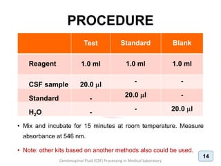
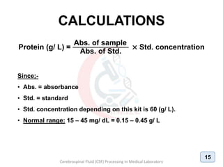
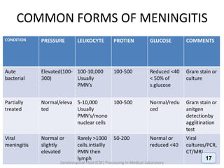
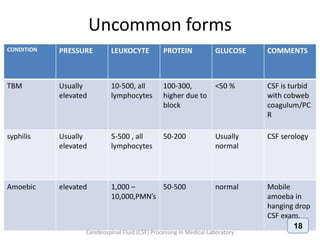
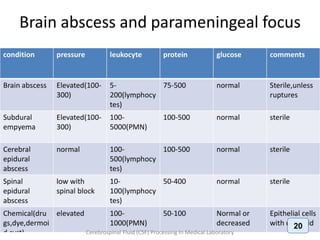
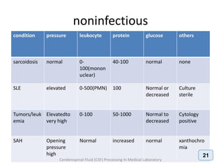
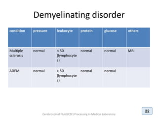
BIOCHEMICAL EXAMINATION
Hussein A. Abid
Laboratory Medicine Specialist
Iraqi Medical Laboratory Association
Scientific Affairs and Cultural Relations Section
Scientific Affairs and Training
Fourth lecture
26/09/2018 (Wednesday)
Dr. Maha Al-Saffar
Laboratory Medicine Specialist
CHEMICAL ANALYSIS
• Routinely performed biochemical tests in CSF
are:
1. Glucose
2. Protein (total and specific)
3. Urea and creatinine
4. Electrolytes (Na+, K+, Mg+2, Cl-)
5. Lactate and lactate dehydrogenase (LDH)
6. Glutamine and acid-base parameters.
Cerebrospinal Fluid (CSF) Processing In Medical Laboratory
2
Remember !!
• Before analysis, the fluid should be centrifuged
to avoid contamination by cellular elements.
• CSF is the most precious biological material.
Often, only small volumes of CSF are available for
analysis due to difficulty in collection; hence
handle this with care.
• The specimen may contain virulent organisms,
so strict safety precautions should be followed.
Cerebrospinal Fluid (CSF) Processing In Medical Laboratory
3
CSF GLUCOSE
• CSF glucose is normally approximately two-
thirds (2/3) of the fasting plasma glucose.
• A glucose level below 40 mg/ dL is significant
and occurs in bacterial and fungal meningitis
and in malignancy.
• Glucose is consumed by leukocytes and tumor
cells.
• It can be elevated in diabetes mellitus (DM).
Cerebrospinal Fluid (CSF) Processing In Medical Laboratory
4
CSF PROTEIN
• Total protein levels in CSF are normally very low, and
albumin makes up approximately two thirds (2/3) of the
total. CSF protein may rise to 500 mg/dl in bacterial
meningitis.
• A more moderate increase (150-200 mg/ dL) occurs
in inflammatory diseases of meninges (meningitis,
encephalitis), intracranial tumors, subarachnoid
hemorrhage, and cerebral infarction.
• A more severe increase occurs in the Guillain-Barré
syndrome and acoustic and spinal schwannoma.
Cerebrospinal Fluid (CSF) Processing In Medical Laboratory
5
CSF PROTEIN
• Multiple sclerosis (MS): CSF protein is normal or mildly
increased.
• Increased IgG in CSF, but not in serum (IgG/ albumin index
normally 10:1). About 90% of MS patients have oligoclonal IgG
bands in the CSF. Oligoclonal bands occur in the CSF only not in
the serum.
• The CSF in MS often contains myelin fragments and myelin basic
protein (MBP).
• MBP can be detected by radioimmunoassay. MBP is not specific
for MS. It can appear in any condition causing brain necrosis,
including infarcts.
Cerebrospinal Fluid (CSF) Processing In Medical Laboratory
6
LACTATE & LDH
• Lactate: the CSF lactate is used mainly to help
differentiate bacterial and fungal meningitis,
which cause increased lactate, from viral
meningitis, which does not.
• Lactate dehydrogenase (LDH): this enzyme
is elevated in bacterial and fungal meningitis,
malignancy, and subarachnoid hemorrhage.
Cerebrospinal Fluid (CSF) Processing In Medical Laboratory
7
PROTEIN TEST
Cerebrospinal Fluid (CSF) Processing In Medical Laboratory
PANDY’S TEST
Principle:
• Proteins are precipitated by a saturated solution of phenol in water.
• The reagent used is phenol (carbolic acid crystals dissolved in water) or,
pyrogallic acid or, cresol, usually termed as Pandy's reagent or Pandy's
solution.
Procedure
• One drop of CSF sample (collected from the patient by lumbar puncture
technique), is added to about 1ml of Pandy's solution. The turbid
appearance signifies the presence of elevated levels of globulin protein in
the CSF and is regarded as positive Pandy's reaction. The CSF from a
normal adult shows no turbidity or precipitates and this is a negative
Pandy's reaction.
Cerebrospinal Fluid (CSF) Processing In Medical Laboratory
9
PANDY’S TEST
Reactions and results
• Proteins in the cerebrospinal fluid, normally albumin and globulin
are present in the ratio of 8 to 1. Increase in protein levels are of
diagnostic value in neurological diseases.
• The normal CSF is clear and transparent fluid. The Pandy's
reaction makes it translucent or opaque.
Positive test
• A bluish white streak of precipitated proteins is seen. The degree
of turbidity depends on the amount of protein in the CSF. It can
vary from faint turbidity (mild to moderate elevation in CSF
proteins) to dense milky precipitate (high protein content in CSF).
Cerebrospinal Fluid (CSF) Processing In Medical Laboratory
10
CSF PROTEIN ASSAY
• Protein present in the CSF is detected by a commercial kits
based on Biuret method or any other method.
• Biuret reagent when interacts with the peptide bonds in the
protein give a blue colored product.
• The intensity of the colour is proportional the amount of protein
in CSF.
• Color intensity is determined by measuring the absorbance by
the colored solution at a wavelength of 546 nm.
• Absorbance is measured by an instrument, spectro-
photometer.
Cerebrospinal Fluid (CSF) Processing In Medical Laboratory
11
SPECTROPHOTOMETER
Most of visible spectrophotometers are composed
of:
• Light source which works with visible wavelengths
(400-700 nm).
• Monochromator filter for choosing desired wavelength.
• Sample holder (cuvette).
• Detector.
• Meter or recorder.
Cerebrospinal Fluid (CSF) Processing In Medical Laboratory
12
Cerebrospinal Fluid (CSF) Processing In Medical Laboratory
Spectrophotometer
PROCEDURE
• Mix and incubate for 15 minutes at room temperature. Measure
absorbance at 546 nm.
• Note: other kits based on another methods also could be used.
Cerebrospinal Fluid (CSF) Processing In Medical Laboratory
14
CALCULATIONS
Protein (g/ L) =
Abs. of sample
Abs. of Std.
× Std. concentration
Since;-
• Abs. = absorbance
• Std. = standard
• Std. concentration depending on this kit is 60 (g/ L).
• Normal range: 15 – 45 mg/ dL = 0.15 – 0.45 g/ L
Cerebrospinal Fluid (CSF) Processing In Medical Laboratory
15
OTHER PARAMETERS
• Other biochemical tests for CSF, will be as
protein test, are also depend on the
commercial kits.
• The biochemical testing results will help in
differentiating the types of meningitis.
Cerebrospinal Fluid (CSF) Processing In Medical Laboratory
16
Cerebrospinal Fluid (CSF) Processing In Medical Laboratory
17
Cerebrospinal Fluid (CSF) Processing In Medical Laboratory
18
Cerebrospinal Fluid (CSF) Processing In Medical Laboratory
19
Cerebrospinal Fluid (CSF) Processing In Medical Laboratory
20
Cerebrospinal Fluid (CSF) Processing In Medical Laboratory
21
Cerebrospinal Fluid (CSF) Processing In Medical Laboratory
22

CSF BIOCHEMICAL EXAMINATION

  • 1.
    BIOCHEMICAL EXAMINATION Hussein A.Abid Laboratory Medicine Specialist Iraqi Medical Laboratory Association Scientific Affairs and Cultural Relations Section Scientific Affairs and Training Fourth lecture 26/09/2018 (Wednesday) Dr. Maha Al-Saffar Laboratory Medicine Specialist
  • 2.
    CHEMICAL ANALYSIS • Routinelyperformed biochemical tests in CSF are: 1. Glucose 2. Protein (total and specific) 3. Urea and creatinine 4. Electrolytes (Na+, K+, Mg+2, Cl-) 5. Lactate and lactate dehydrogenase (LDH) 6. Glutamine and acid-base parameters. Cerebrospinal Fluid (CSF) Processing In Medical Laboratory 2
  • 3.
    Remember !! • Beforeanalysis, the fluid should be centrifuged to avoid contamination by cellular elements. • CSF is the most precious biological material. Often, only small volumes of CSF are available for analysis due to difficulty in collection; hence handle this with care. • The specimen may contain virulent organisms, so strict safety precautions should be followed. Cerebrospinal Fluid (CSF) Processing In Medical Laboratory 3
  • 4.
    CSF GLUCOSE • CSFglucose is normally approximately two- thirds (2/3) of the fasting plasma glucose. • A glucose level below 40 mg/ dL is significant and occurs in bacterial and fungal meningitis and in malignancy. • Glucose is consumed by leukocytes and tumor cells. • It can be elevated in diabetes mellitus (DM). Cerebrospinal Fluid (CSF) Processing In Medical Laboratory 4
  • 5.
    CSF PROTEIN • Totalprotein levels in CSF are normally very low, and albumin makes up approximately two thirds (2/3) of the total. CSF protein may rise to 500 mg/dl in bacterial meningitis. • A more moderate increase (150-200 mg/ dL) occurs in inflammatory diseases of meninges (meningitis, encephalitis), intracranial tumors, subarachnoid hemorrhage, and cerebral infarction. • A more severe increase occurs in the Guillain-Barré syndrome and acoustic and spinal schwannoma. Cerebrospinal Fluid (CSF) Processing In Medical Laboratory 5
  • 6.
    CSF PROTEIN • Multiplesclerosis (MS): CSF protein is normal or mildly increased. • Increased IgG in CSF, but not in serum (IgG/ albumin index normally 10:1). About 90% of MS patients have oligoclonal IgG bands in the CSF. Oligoclonal bands occur in the CSF only not in the serum. • The CSF in MS often contains myelin fragments and myelin basic protein (MBP). • MBP can be detected by radioimmunoassay. MBP is not specific for MS. It can appear in any condition causing brain necrosis, including infarcts. Cerebrospinal Fluid (CSF) Processing In Medical Laboratory 6
  • 7.
    LACTATE & LDH •Lactate: the CSF lactate is used mainly to help differentiate bacterial and fungal meningitis, which cause increased lactate, from viral meningitis, which does not. • Lactate dehydrogenase (LDH): this enzyme is elevated in bacterial and fungal meningitis, malignancy, and subarachnoid hemorrhage. Cerebrospinal Fluid (CSF) Processing In Medical Laboratory 7
  • 8.
    PROTEIN TEST Cerebrospinal Fluid(CSF) Processing In Medical Laboratory
  • 9.
    PANDY’S TEST Principle: • Proteinsare precipitated by a saturated solution of phenol in water. • The reagent used is phenol (carbolic acid crystals dissolved in water) or, pyrogallic acid or, cresol, usually termed as Pandy's reagent or Pandy's solution. Procedure • One drop of CSF sample (collected from the patient by lumbar puncture technique), is added to about 1ml of Pandy's solution. The turbid appearance signifies the presence of elevated levels of globulin protein in the CSF and is regarded as positive Pandy's reaction. The CSF from a normal adult shows no turbidity or precipitates and this is a negative Pandy's reaction. Cerebrospinal Fluid (CSF) Processing In Medical Laboratory 9
  • 10.
    PANDY’S TEST Reactions andresults • Proteins in the cerebrospinal fluid, normally albumin and globulin are present in the ratio of 8 to 1. Increase in protein levels are of diagnostic value in neurological diseases. • The normal CSF is clear and transparent fluid. The Pandy's reaction makes it translucent or opaque. Positive test • A bluish white streak of precipitated proteins is seen. The degree of turbidity depends on the amount of protein in the CSF. It can vary from faint turbidity (mild to moderate elevation in CSF proteins) to dense milky precipitate (high protein content in CSF). Cerebrospinal Fluid (CSF) Processing In Medical Laboratory 10
  • 11.
    CSF PROTEIN ASSAY •Protein present in the CSF is detected by a commercial kits based on Biuret method or any other method. • Biuret reagent when interacts with the peptide bonds in the protein give a blue colored product. • The intensity of the colour is proportional the amount of protein in CSF. • Color intensity is determined by measuring the absorbance by the colored solution at a wavelength of 546 nm. • Absorbance is measured by an instrument, spectro- photometer. Cerebrospinal Fluid (CSF) Processing In Medical Laboratory 11
  • 12.
    SPECTROPHOTOMETER Most of visiblespectrophotometers are composed of: • Light source which works with visible wavelengths (400-700 nm). • Monochromator filter for choosing desired wavelength. • Sample holder (cuvette). • Detector. • Meter or recorder. Cerebrospinal Fluid (CSF) Processing In Medical Laboratory 12
  • 13.
    Cerebrospinal Fluid (CSF)Processing In Medical Laboratory Spectrophotometer
  • 14.
    PROCEDURE • Mix andincubate for 15 minutes at room temperature. Measure absorbance at 546 nm. • Note: other kits based on another methods also could be used. Cerebrospinal Fluid (CSF) Processing In Medical Laboratory 14
  • 15.
    CALCULATIONS Protein (g/ L)= Abs. of sample Abs. of Std. × Std. concentration Since;- • Abs. = absorbance • Std. = standard • Std. concentration depending on this kit is 60 (g/ L). • Normal range: 15 – 45 mg/ dL = 0.15 – 0.45 g/ L Cerebrospinal Fluid (CSF) Processing In Medical Laboratory 15
  • 16.
    OTHER PARAMETERS • Otherbiochemical tests for CSF, will be as protein test, are also depend on the commercial kits. • The biochemical testing results will help in differentiating the types of meningitis. Cerebrospinal Fluid (CSF) Processing In Medical Laboratory 16
  • 17.
    Cerebrospinal Fluid (CSF)Processing In Medical Laboratory 17
  • 18.
    Cerebrospinal Fluid (CSF)Processing In Medical Laboratory 18
  • 19.
    Cerebrospinal Fluid (CSF)Processing In Medical Laboratory 19
  • 20.
    Cerebrospinal Fluid (CSF)Processing In Medical Laboratory 20
  • 21.
    Cerebrospinal Fluid (CSF)Processing In Medical Laboratory 21
  • 22.
    Cerebrospinal Fluid (CSF)Processing In Medical Laboratory 22