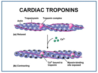
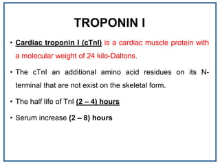
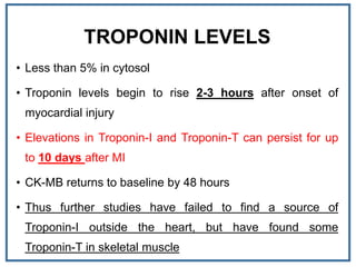
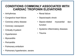
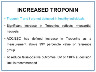
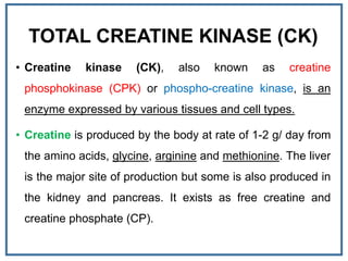
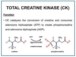
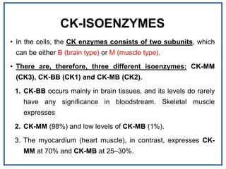
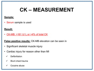
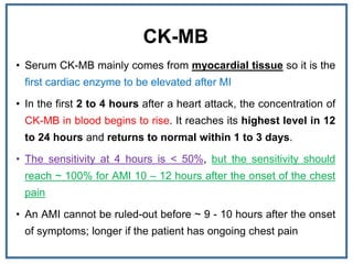
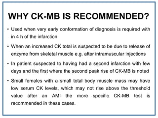
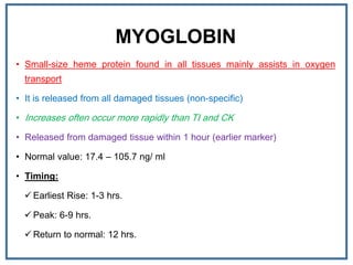
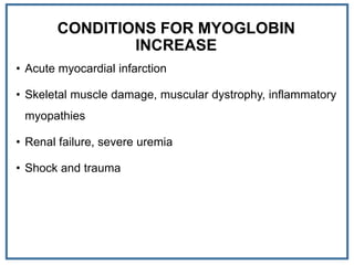
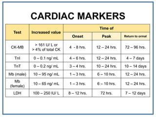
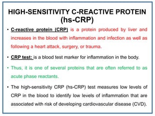
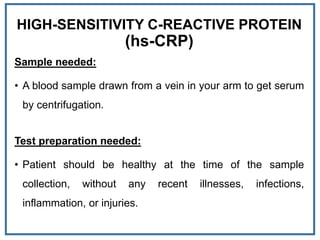
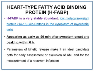
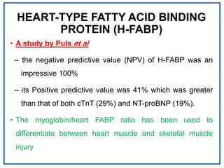
CARDIAC BIOMARKERS - II
Hussein A. Abid
Iraqi Medical Laboratory Association
Scientific Affairs & Cultural Relations
Training and development center
Lecture: 4
Date: 08/08/2018
CARDIAC TROPONINS
• Troponin is a complex of three regulatory proteins is integral to
non-smooth muscle contraction in skeletal as well as cardiac
muscle.
• Troponin is attached to the tropomyosin sitting in the groove
between actin filaments in muscle tissue.
• Troponin has three subunits: TnC, TnT and TnI
 Troponin-C: has calcium binding ability has no diagnostic
value
 Troponin-T: binds the troponin- tropomyosin complex
 Troponin-I: is an inhibitory protein
CARDIAC TROPONINS
CARDIAC TROPONINS
CARDIAC TROPONINS
TROPONIN I
• Cardiac troponin I (cTnI) is a cardiac muscle protein with
a molecular weight of 24 kilo-Daltons.
• The cTnI an additional amino acid residues on its N-
terminal that are not exist on the skeletal form.
• The half life of TnI (2 – 4) hours
• Serum increase (2 – 8) hours
TROPONIN LEVELS
• Less than 5% in cytosol
• Troponin levels begin to rise 2-3 hours after onset of
myocardial injury
• Elevations in Troponin-I and Troponin-T can persist for up
to 10 days after MI
• CK-MB returns to baseline by 48 hours
• Thus further studies have failed to find a source of
Troponin-I outside the heart, but have found some
Troponin-T in skeletal muscle
CONDITIONS COMMONLY ASSOCIATED WITH
CARDIAC TROPONIN ELEVATIONS
• Arrhythmias
• Congestive heart failure
• Coronary artery disease
• Coronary vasospasm
• Critically ill patient
• Hypertension
• Myocarditis
• Pericarditis
• Pulmonary embolism
• Pulmonary hypertension, severe
• Renal failure
• Sepsis/septic shock
• Sepsis-related myocardial dys-
function
• Systemic inflammatory diseases
• Trauma
INCREASED TROPONIN
• Troponin T and I are not detected in healthy individuals
• Significant increase in Troponins reflects myocardial
necrosis
• ACC/ESC has defined increase in Troponins as a
measurement above 99th percentile value of reference
group
• To reduce false-positive outcomes, CV of ≤10% at decision
limit is recommended
TROPONIN ASSAY
• Troponin-T (TnT): (Roche Diagnostics, Germany)
• Troponin-I (TnI): (Siemens Healthcare Diagnostics)
• Troponin T
– 99th percentile limits - 0.01 ng/ mL
– assay ranges - 0.01-25 ng/ mL
• Troponin I
– 99th percentile limits - 0.04 ng/mL
– assay range - 0.04-40 ng/mL
• Reference limits based on the 99th percentile for a healthy population
are 0.01 ng/ mL (Troponin T) and 0.04 ng/ mL (Troponin I)
TOTAL CREATINE KINASE (CK)
• Creatine kinase (CK), also known as creatine
phosphokinase (CPK) or phospho-creatine kinase, is an
enzyme expressed by various tissues and cell types.
• Creatine is produced by the body at rate of 1-2 g/ day from
the amino acids, glycine, arginine and methionine. The liver
is the major site of production but some is also produced in
the kidney and pancreas. It exists as free creatine and
creatine phosphate (CP).
TOTAL CREATINE KINASE (CK)
Function
• CK catalyzes the conversion of creatine and consumes
adenosine triphosphate (ATP) to create phosphocreatine
and adenosine diphosphate (ADP).
TOTAL CREATINE KINASE (CK)
Role of phosphocreatine (PC)
• In tissues and cells that consume ATP rapidly, especially
skeletal muscle, phosphocreatine serves as an energy
reservoir for the rapid buffering and regeneration of ATP in
situ.
• Thus when muscle contracts, ATP is consumed to form
ADP, in this case CK catalyze the re-phosphorylation of
ADP to form ATP using Crp as the phosphorylation
reservoir.
CK-ISOENZYMES
• In the cells, the CK enzymes consists of two subunits, which
can be either B (brain type) or M (muscle type).
• There are, therefore, three different isoenzymes: CK-MM
(CK3), CK-BB (CK1) and CK-MB (CK2).
1. CK-BB occurs mainly in brain tissues, and its levels do rarely
have any significance in bloodstream. Skeletal muscle
expresses
2. CK-MM (98%) and low levels of CK-MB (1%).
3. The myocardium (heart muscle), in contrast, expresses CK-
MM at 70% and CK-MB at 25–30%.
CK – MEASUREMENT
Sample:
• Serum sample is used
Result:
• CK-MB: >161 U/ L or >4% of total CK
False positive results: CK-MB elevation can be seen in
• Significant skeletal muscle injury
• Cardiac injury for reason other than MI
 Defibrillation
 Blunt chest trauma
 Cocaine abuse
INTERPRETATION | CARDIAC DISEASE
Interpretation: (Cardiac disease)
• CK total is elevated rapidly after MI
• In the first 4 to 6 hours after a heart attack, the
concentration of CK in blood begins to rise. It reaches its
highest level in 18 to 24 hours and returns to normal
within 2 to 3 days.
• CK level is directly proportional to the infraction size.
INTERPRETATION | MUSCULAR DISEASE
• Because most of the CPK in the body normally exists in muscle, a rise in the
amount of CPK in the blood indicates that muscle damage has occurred.
Physiological rise: sever muscular exercise, cramps, repeated muscular injections
cause transient CK rise for 2-4 days.
Pathological rise:
 Parasitic infection (trichinosis) caused by Trichinella spiralis
 Convulsions and muscle spasms
 Duchenne's muscular dystrophy: caused by mutation in the gene of dystrophin
protein. In this case high plasma activities are found from birth and before the
onset of clinical signs During the early clinical stages at the disease , very high
activity usually present but at terminal stage of the disease , the level tend to be
fall when the mass of functioning muscle diminish with progression of the disease
CK-MB
• Serum CK-MB mainly comes from myocardial tissue so it is the
first cardiac enzyme to be elevated after MI
• In the first 2 to 4 hours after a heart attack, the concentration of
CK-MB in blood begins to rise. It reaches its highest level in 12
to 24 hours and returns to normal within 1 to 3 days.
• The sensitivity at 4 hours is < 50%, but the sensitivity should
reach ~ 100% for AMI 10 – 12 hours after the onset of the chest
pain
• An AMI cannot be ruled-out before ~ 9 - 10 hours after the onset
of symptoms; longer if the patient has ongoing chest pain
WHY CK-MB IS RECOMMENDED?
• Used when very early conformation of diagnosis is required with
in 4 h of the infarction
• When an increased CK total is suspected to be due to release of
enzyme from skeletal muscle e.g. after intramuscular injections
• In patient suspected to having had a second infarction with few
days and the first where the second peak rise of CK-MB is noted
• Small females with a small total body muscle mass may have
low serum CK levels, which may not rise above the threshold
value after an AMI the more specific CK-MB test is
recommended in these cases.
MYOGLOBIN
• Small-size heme protein found in all tissues mainly assists in oxygen
transport
• It is released from all damaged tissues (non-specific)
• Increases often occur more rapidly than TI and CK
• Released from damaged tissue within 1 hour (earlier marker)
• Normal value: 17.4 – 105.7 ng/ ml
• Timing:
 Earliest Rise: 1-3 hrs.
 Peak: 6-9 hrs.
 Return to normal: 12 hrs.
CONDITIONS FOR MYOGLOBIN
INCREASE
• Acute myocardial infarction
• Skeletal muscle damage, muscular dystrophy, inflammatory
myopathies
• Renal failure, severe uremia
• Shock and trauma
MYOGLOBIN
• Rapid monitor of success of thrombolytic therapy
• Negative predictor of MI
• Due to poor specificity, myoglobin levels do not always
predict myocardial injury
• Not utilized often for AMI/cardiac damage assessment
because of its very rapid metabolism
CARDIAC MARKERS
Time of
Increased valueTest
Return to ormalPeakOnset
72 – 96 hrs.12 – 24 hrs.4 - 8 hrs.
> 161 U/ L or
> 4% of total CK
CK-MB
4 – 7 days12 – 24 hrs.4 – 6 hrs.0 – 0.1 ng/ mLTnI
10 – 14 days10 – 24 hrs.3 – 4 hrs.0 – 0.2 ng/ mLTnT
12 – 24 hrs.6 – 10 hrs.1 – 3 hrs.10 – 95 ng/ mLMb (male)
12 – 24 hrs.6 – 10 hrs.1 – 3 hrs.10 – 65 ng/ mL
Mb
(female)
7 – 12 days72 hrs.8 – 12 hrs.100 – 250 IU/ LLDH
ISCHEMIA MODIFIED ALBUMIN (IMA)
• A novel marker of ischemia, is produced when circulating
serum albumin contacts ischemic heart tissues
• IMA can be measured by the albumin cobalt binding (ACB)
assay that is based on IMA's inability to bind to cobalt
• Mechanism; due to structural change in the amino terminal
end of albumin
• IMA levels rise within 6 hours
• Remains elevated for 12 hours
ISCHEMIA MODIFIED ALBUMIN (IMA)
• IMA levels raised in non-cardiac ischemia. (Is it specific
for myocardial ischemia?)
• Modification to N-terminal end may also be induced by
extracellular hypoxia, acidosis etc.
• FDA in 2010 has approved a multimarker approach for
using the combination of ECG, the cTnI, and the IMA levels
achieving a sensitivity of 95% for ACS.
HIGH-SENSITIVITY C-REACTIVE PROTEIN
(hs-CRP)
• C-reactive protein (CRP) is a protein produced by liver and
increases in the blood with inflammation and infection as well as
following a heart attack, surgery, or trauma.
• CRP test: is a blood test marker for inflammation in the body.
• Thus, it is one of several proteins that are often referred to as
acute phase reactants.
• The high-sensitivity CRP (hs-CRP) test measures low levels of
CRP in the blood to identify low levels of inflammation that are
associated with risk of developing cardiovascular disease (CVD).
HIGH-SENSITIVITY C-REACTIVE PROTEIN
(hs-CRP)
Sample needed:
• A blood sample drawn from a vein in your arm to get serum
by centrifugation.
Test preparation needed:
• Patient should be healthy at the time of the sample
collection, without any recent illnesses, infections,
inflammation, or injuries.
HEART-TYPE FATTY ACID BINDING
PROTEIN (H-FABP)
• H-FABP is a very stable abundant, low molecular-weight
protein (14-15) kilo-Daltons in the cytoplasm of myocardial
cells
• Appearing as early as 90 min after symptom onset and
peaking within 6 h.
• Parameters of kinetic release make it an ideal candidate
both for early assessment or exclusion of AMI and for the
measurement of a recurrent infarction
HEART-TYPE FATTY ACID BINDING
PROTEIN (H-FABP)
• A study by Puls et al
– the negative predictive value (NPV) of H-FABP was an
impressive 100%
– its Positive predictive value was 41% which was greater
than that of both cTnT (29%) and NT-proBNP (19%).
• The myoglobin/heart FABP ratio has been used to
differentiate between heart muscle and skeletal muscle
injury
NATRIURETIC PEPTIDES (NP)
• The NP family includes:
 ANP: α-atrial natriuretic peptide (28 amino-acid), N-
terminal proANP (98 amino-acid)
 BNP: brain natriuretic peptide (32 amino-acid), N-
terminal proBNP (76 amino-acid)
 CNP: C-type natriuretic peptide (22 and 53 amino-acid)
NATRIURETIC PEPTIDES (NP)
• ANP is released primarily in response to atrial wall
stretching and intravascular volume expansion.
• BNP is mainly secreted by the ventricles
• CNP is found predominantly in the brain and also
synthesized by vascular endothelial cells
sCD40 LIGAND
• CD40 ligand is a transmembrane protein related to TNF.
• It has multiple prothrombotic and proatherogenic effects.
• What is usually measured is the soluble form of the
receptor, sCD40 ligand, which has been shown to be a
predictor of events after acute presentation.
• At present, standardized assays, reference interval studies,
nor consistent assay validations are not available.
SERUM AMYLOID PROTEIN A
• Serum amyloid protein A, an acute-phase protein and an
apolipoprotein, has been used with hs-CRP in cross-
sectional studies.
• It can be synergistic with hs-CRP but is much less
commonly used.
• At present, no standardized assays, reference interval
studies, nor consistent assay validations are available.
CYTOKINES
• A variety of stimulatory and inhibitory interleukins (TNF, IL-1, IL-
6, IL-8, IL-12, IL-18) are thought to help mediate the elaboration
of CRP and the development of atherosclerosis and acute
events. These cytokines may stimulate or inhibit leukocytes,
often through T cell–mediated processes and effects on
monocytes, which are indigenous to atherogenesis.
• In some studies, IL-6 is more prognostic than hs-CRP.
• These cytokines often have inhibitors and/or binding proteins
that modulate their effects. At present, standardized assays,
reference intervals studies, and consistent assay validations are
not available.
LIPOPROTEIN-ASSOCIATED PHOSPHOLIPASE A2
• Phospholipase A2 (Lp-PLA2) is a phospholipase associated with LDL that is
thought to be an inflammatory marker.
• It was previously known as platelet-activating factor (PAF) acetyl hydrolase.
• It is synthesized by monocytes and lymphocytes and is thought to cleave
oxidized lipids to produce lipid fragments that are more atherogenic and that
increase endothelial adhesion.
• An FDA-approved assay for this analyte includes obligatory reference intervals.
• It has been shown to be predictive of events in a primary prevention cohort,
even when hsCRP is present in the model, suggesting that it measures
something different from what is measured by the acute-phase reactants
associated with hsCRP.
PREGNANCY-ASSOCIATED PLASMA PROTEIN A
• Pregnancy-associated plasma protein A (PAPP-A) is a
metalloproteinase that is thought to be expressed in plaques that
may be prone to rupture.
• The literature in this regard is mixed at present concerning its
use.
• At present, standardize assays, reference interval studies, and
consistent assay validations are not available.
• Recent data suggest that heparin administration in MI patients is
associated with increased PAPP-A concentrations; this may limit
its prognostic role.
OXIDIZED LDL
• Oxidized LDL has been attributed a key role in the
development of atherosclerosis.
• Several methods have been used to measure it, but they
yield potentially different data.
• Some have correlated malondialdehyde LDL with the
development of atherosclerosis and short-term events.
• Direct identification with antibodies suggests that oxidized
LDL may be released from vessels and may colocalize with
lipoprotein a [Lp(a)] after acute events.
PLACENTAL GROWTH FACTOR
• Placental growth factor is an angiogenic factor related to
vascular endothelial growth factor (VEGF), which
stimulates smooth muscle cells and macrophages.
• It also increases TNF and MCP-1. A novel assay for this
analyte is thought to provide additional prognostic
information on patients who present with ACS.
• At present, standardized assays, reference interval studies,
and consistent assay validations are not available.
MATRIX METALLOPROTEINASES
• Matrix metalloproteinases (MMPs) can degrade the collagen matrix in coronary
artery or myocardium.
• They are integral to remodeling of the coronary artery and/or the heart after
acute events.
• Elaboration of MMP-9, a gelatinase, is thought to be important in plaque
destabilization; thus some have tried to measure it as a prognostic index.
• Other MMPs participate in the elaboration of extracellular matrix in the heart.
• Many MMPs also have inhibitors [tissue inhibitors of metalloproteinase
(TIMPs)] that modulate their effects.
• At present, standardized assays, reference intervals studies, and
• consistent assay validations are not available.
MONOCYTE CHEMOTACTIC PROTEIN
• Monocyte chemotactic protein (MCP-1) is a chemokine that
is thought to be responsible for the recruitment of
monocytes into atherosclerotic plaque.
• It has been reported to be elevated in patients with ACS
and to have long-term predictive value.
• However, at present, standardized assays, reference
interval studies, and consistent assay validations are not
available
TISSUE PLASMINOGEN ACTIVATOR ANTIGEN (t-PA)
AND PLASMINOGEN ACTIVATOR INHIBITOR 1 (PAI-1)
• t-PA is the body’s physiologic fibrinolytic activator.
• PAI-1, its endogenous inhibitor, binds to t-PA.
• Inhibition of fibrinolysis has been suggested to be a reason for
recurrent infarction; the fact that maximal inhibition usually occurs in the
early morning hours provides a potential explanation for the circadian
variability of AMI.
• It may also be the reason why persons with diabetes have such
unstable disease; the growth factor properties of insulin stimulate
increases in PAI-1.
• An accurate assessment of this system includes both tPA and PAI-1,
along with some assessment of bound versus free levels.
SECRETED PLATELET GRANULAR
SUBSTANCES
• Both platelet factor 4 (PF4) and beta thromboglobulin (BTG) are
secreted when platelets aggregate.
• PF4 has a short half-life and is released by heparin.
• BTG is not released by heparin and has a longer halflife.
• Both markers have been used to assess platelet aggregation.
• BTG is by far the more reliable.
• At present, standardized assays, reference interval studies, and
consistent assay validations are not available.
UNBOUND FREE FATTY ACID
• Unbound free fatty acid (uFFA) 39 has also been touted as
a marker of ischemia.
• Most fatty acid is bound, and ischemia is thought to
increase the small unbound fraction.
• Initial studies have reported mixed results.
• At present, standardized assays, reference interval studies,
and consistent assay validations are not available.
NOURIN
• Nourin: is a small protein released rapidly by “stressed
myocytes.”
• It induces changes in a variety of inflammatory cytokines
and attracts neutrophils.
• Preliminary studies have been done to attempt to validate
its use.
• At present, standardized assays, reference interval studies,
and consistent assay validations are not available.
COPEPTIN
• Copeptin, a 30 amino acid glycoprotein constituting the C-terminal portion
of arginine vasopressin, has been shown to be a prognostic biomarker in
hemorrhagic and septic sepsis.
• More recently, data have shown that measurement of copeptin serves as a
rapid and early rule-out biomarker for AMI at presentation in patients with
symptoms suggestive of ACS with a normal cTn value.
• An assay measuring copeptin (CT-proAVP) has been described using the
Brahms Kryptor Immunology Analyzer, Diamond Diagnostics Holliston, MA.
• Additional clinical and analytical validation studies will be necessary,
especially head-to-head comparisons of copeptin versus the new hs-cTn
assays.
NOURIN
• Nourin: is a small protein released rapidly by “stressed
myocytes.”
• It induces changes in a variety of inflammatory cytokines
and attracts neutrophils.
• Preliminary studies have been done to attempt to validate
its use.
• At present, standardized assays, reference interval studies,
and consistent assay validations are not available.

Cardiac biomarkers - II

  • 1.
    CARDIAC BIOMARKERS -II Hussein A. Abid Iraqi Medical Laboratory Association Scientific Affairs & Cultural Relations Training and development center Lecture: 4 Date: 08/08/2018
  • 2.
    CARDIAC TROPONINS • Troponinis a complex of three regulatory proteins is integral to non-smooth muscle contraction in skeletal as well as cardiac muscle. • Troponin is attached to the tropomyosin sitting in the groove between actin filaments in muscle tissue. • Troponin has three subunits: TnC, TnT and TnI  Troponin-C: has calcium binding ability has no diagnostic value  Troponin-T: binds the troponin- tropomyosin complex  Troponin-I: is an inhibitory protein
  • 3.
  • 4.
  • 6.
  • 7.
    TROPONIN I • Cardiactroponin I (cTnI) is a cardiac muscle protein with a molecular weight of 24 kilo-Daltons. • The cTnI an additional amino acid residues on its N- terminal that are not exist on the skeletal form. • The half life of TnI (2 – 4) hours • Serum increase (2 – 8) hours
  • 8.
    TROPONIN LEVELS • Lessthan 5% in cytosol • Troponin levels begin to rise 2-3 hours after onset of myocardial injury • Elevations in Troponin-I and Troponin-T can persist for up to 10 days after MI • CK-MB returns to baseline by 48 hours • Thus further studies have failed to find a source of Troponin-I outside the heart, but have found some Troponin-T in skeletal muscle
  • 9.
    CONDITIONS COMMONLY ASSOCIATEDWITH CARDIAC TROPONIN ELEVATIONS • Arrhythmias • Congestive heart failure • Coronary artery disease • Coronary vasospasm • Critically ill patient • Hypertension • Myocarditis • Pericarditis • Pulmonary embolism • Pulmonary hypertension, severe • Renal failure • Sepsis/septic shock • Sepsis-related myocardial dys- function • Systemic inflammatory diseases • Trauma
  • 10.
    INCREASED TROPONIN • TroponinT and I are not detected in healthy individuals • Significant increase in Troponins reflects myocardial necrosis • ACC/ESC has defined increase in Troponins as a measurement above 99th percentile value of reference group • To reduce false-positive outcomes, CV of ≤10% at decision limit is recommended
  • 11.
    TROPONIN ASSAY • Troponin-T(TnT): (Roche Diagnostics, Germany) • Troponin-I (TnI): (Siemens Healthcare Diagnostics) • Troponin T – 99th percentile limits - 0.01 ng/ mL – assay ranges - 0.01-25 ng/ mL • Troponin I – 99th percentile limits - 0.04 ng/mL – assay range - 0.04-40 ng/mL • Reference limits based on the 99th percentile for a healthy population are 0.01 ng/ mL (Troponin T) and 0.04 ng/ mL (Troponin I)
  • 12.
    TOTAL CREATINE KINASE(CK) • Creatine kinase (CK), also known as creatine phosphokinase (CPK) or phospho-creatine kinase, is an enzyme expressed by various tissues and cell types. • Creatine is produced by the body at rate of 1-2 g/ day from the amino acids, glycine, arginine and methionine. The liver is the major site of production but some is also produced in the kidney and pancreas. It exists as free creatine and creatine phosphate (CP).
  • 13.
    TOTAL CREATINE KINASE(CK) Function • CK catalyzes the conversion of creatine and consumes adenosine triphosphate (ATP) to create phosphocreatine and adenosine diphosphate (ADP).
  • 14.
    TOTAL CREATINE KINASE(CK) Role of phosphocreatine (PC) • In tissues and cells that consume ATP rapidly, especially skeletal muscle, phosphocreatine serves as an energy reservoir for the rapid buffering and regeneration of ATP in situ. • Thus when muscle contracts, ATP is consumed to form ADP, in this case CK catalyze the re-phosphorylation of ADP to form ATP using Crp as the phosphorylation reservoir.
  • 15.
    CK-ISOENZYMES • In thecells, the CK enzymes consists of two subunits, which can be either B (brain type) or M (muscle type). • There are, therefore, three different isoenzymes: CK-MM (CK3), CK-BB (CK1) and CK-MB (CK2). 1. CK-BB occurs mainly in brain tissues, and its levels do rarely have any significance in bloodstream. Skeletal muscle expresses 2. CK-MM (98%) and low levels of CK-MB (1%). 3. The myocardium (heart muscle), in contrast, expresses CK- MM at 70% and CK-MB at 25–30%.
  • 16.
    CK – MEASUREMENT Sample: •Serum sample is used Result: • CK-MB: >161 U/ L or >4% of total CK False positive results: CK-MB elevation can be seen in • Significant skeletal muscle injury • Cardiac injury for reason other than MI  Defibrillation  Blunt chest trauma  Cocaine abuse
  • 17.
    INTERPRETATION | CARDIACDISEASE Interpretation: (Cardiac disease) • CK total is elevated rapidly after MI • In the first 4 to 6 hours after a heart attack, the concentration of CK in blood begins to rise. It reaches its highest level in 18 to 24 hours and returns to normal within 2 to 3 days. • CK level is directly proportional to the infraction size.
  • 18.
    INTERPRETATION | MUSCULARDISEASE • Because most of the CPK in the body normally exists in muscle, a rise in the amount of CPK in the blood indicates that muscle damage has occurred. Physiological rise: sever muscular exercise, cramps, repeated muscular injections cause transient CK rise for 2-4 days. Pathological rise:  Parasitic infection (trichinosis) caused by Trichinella spiralis  Convulsions and muscle spasms  Duchenne's muscular dystrophy: caused by mutation in the gene of dystrophin protein. In this case high plasma activities are found from birth and before the onset of clinical signs During the early clinical stages at the disease , very high activity usually present but at terminal stage of the disease , the level tend to be fall when the mass of functioning muscle diminish with progression of the disease
  • 19.
    CK-MB • Serum CK-MBmainly comes from myocardial tissue so it is the first cardiac enzyme to be elevated after MI • In the first 2 to 4 hours after a heart attack, the concentration of CK-MB in blood begins to rise. It reaches its highest level in 12 to 24 hours and returns to normal within 1 to 3 days. • The sensitivity at 4 hours is < 50%, but the sensitivity should reach ~ 100% for AMI 10 – 12 hours after the onset of the chest pain • An AMI cannot be ruled-out before ~ 9 - 10 hours after the onset of symptoms; longer if the patient has ongoing chest pain
  • 20.
    WHY CK-MB ISRECOMMENDED? • Used when very early conformation of diagnosis is required with in 4 h of the infarction • When an increased CK total is suspected to be due to release of enzyme from skeletal muscle e.g. after intramuscular injections • In patient suspected to having had a second infarction with few days and the first where the second peak rise of CK-MB is noted • Small females with a small total body muscle mass may have low serum CK levels, which may not rise above the threshold value after an AMI the more specific CK-MB test is recommended in these cases.
  • 21.
    MYOGLOBIN • Small-size hemeprotein found in all tissues mainly assists in oxygen transport • It is released from all damaged tissues (non-specific) • Increases often occur more rapidly than TI and CK • Released from damaged tissue within 1 hour (earlier marker) • Normal value: 17.4 – 105.7 ng/ ml • Timing:  Earliest Rise: 1-3 hrs.  Peak: 6-9 hrs.  Return to normal: 12 hrs.
  • 22.
    CONDITIONS FOR MYOGLOBIN INCREASE •Acute myocardial infarction • Skeletal muscle damage, muscular dystrophy, inflammatory myopathies • Renal failure, severe uremia • Shock and trauma
  • 23.
    MYOGLOBIN • Rapid monitorof success of thrombolytic therapy • Negative predictor of MI • Due to poor specificity, myoglobin levels do not always predict myocardial injury • Not utilized often for AMI/cardiac damage assessment because of its very rapid metabolism
  • 24.
    CARDIAC MARKERS Time of IncreasedvalueTest Return to ormalPeakOnset 72 – 96 hrs.12 – 24 hrs.4 - 8 hrs. > 161 U/ L or > 4% of total CK CK-MB 4 – 7 days12 – 24 hrs.4 – 6 hrs.0 – 0.1 ng/ mLTnI 10 – 14 days10 – 24 hrs.3 – 4 hrs.0 – 0.2 ng/ mLTnT 12 – 24 hrs.6 – 10 hrs.1 – 3 hrs.10 – 95 ng/ mLMb (male) 12 – 24 hrs.6 – 10 hrs.1 – 3 hrs.10 – 65 ng/ mL Mb (female) 7 – 12 days72 hrs.8 – 12 hrs.100 – 250 IU/ LLDH
  • 25.
    ISCHEMIA MODIFIED ALBUMIN(IMA) • A novel marker of ischemia, is produced when circulating serum albumin contacts ischemic heart tissues • IMA can be measured by the albumin cobalt binding (ACB) assay that is based on IMA's inability to bind to cobalt • Mechanism; due to structural change in the amino terminal end of albumin • IMA levels rise within 6 hours • Remains elevated for 12 hours
  • 26.
    ISCHEMIA MODIFIED ALBUMIN(IMA) • IMA levels raised in non-cardiac ischemia. (Is it specific for myocardial ischemia?) • Modification to N-terminal end may also be induced by extracellular hypoxia, acidosis etc. • FDA in 2010 has approved a multimarker approach for using the combination of ECG, the cTnI, and the IMA levels achieving a sensitivity of 95% for ACS.
  • 27.
    HIGH-SENSITIVITY C-REACTIVE PROTEIN (hs-CRP) •C-reactive protein (CRP) is a protein produced by liver and increases in the blood with inflammation and infection as well as following a heart attack, surgery, or trauma. • CRP test: is a blood test marker for inflammation in the body. • Thus, it is one of several proteins that are often referred to as acute phase reactants. • The high-sensitivity CRP (hs-CRP) test measures low levels of CRP in the blood to identify low levels of inflammation that are associated with risk of developing cardiovascular disease (CVD).
  • 28.
    HIGH-SENSITIVITY C-REACTIVE PROTEIN (hs-CRP) Sampleneeded: • A blood sample drawn from a vein in your arm to get serum by centrifugation. Test preparation needed: • Patient should be healthy at the time of the sample collection, without any recent illnesses, infections, inflammation, or injuries.
  • 29.
    HEART-TYPE FATTY ACIDBINDING PROTEIN (H-FABP) • H-FABP is a very stable abundant, low molecular-weight protein (14-15) kilo-Daltons in the cytoplasm of myocardial cells • Appearing as early as 90 min after symptom onset and peaking within 6 h. • Parameters of kinetic release make it an ideal candidate both for early assessment or exclusion of AMI and for the measurement of a recurrent infarction
  • 30.
    HEART-TYPE FATTY ACIDBINDING PROTEIN (H-FABP) • A study by Puls et al – the negative predictive value (NPV) of H-FABP was an impressive 100% – its Positive predictive value was 41% which was greater than that of both cTnT (29%) and NT-proBNP (19%). • The myoglobin/heart FABP ratio has been used to differentiate between heart muscle and skeletal muscle injury
  • 31.
    NATRIURETIC PEPTIDES (NP) •The NP family includes:  ANP: α-atrial natriuretic peptide (28 amino-acid), N- terminal proANP (98 amino-acid)  BNP: brain natriuretic peptide (32 amino-acid), N- terminal proBNP (76 amino-acid)  CNP: C-type natriuretic peptide (22 and 53 amino-acid)
  • 32.
    NATRIURETIC PEPTIDES (NP) •ANP is released primarily in response to atrial wall stretching and intravascular volume expansion. • BNP is mainly secreted by the ventricles • CNP is found predominantly in the brain and also synthesized by vascular endothelial cells
  • 34.
    sCD40 LIGAND • CD40ligand is a transmembrane protein related to TNF. • It has multiple prothrombotic and proatherogenic effects. • What is usually measured is the soluble form of the receptor, sCD40 ligand, which has been shown to be a predictor of events after acute presentation. • At present, standardized assays, reference interval studies, nor consistent assay validations are not available.
  • 35.
    SERUM AMYLOID PROTEINA • Serum amyloid protein A, an acute-phase protein and an apolipoprotein, has been used with hs-CRP in cross- sectional studies. • It can be synergistic with hs-CRP but is much less commonly used. • At present, no standardized assays, reference interval studies, nor consistent assay validations are available.
  • 36.
    CYTOKINES • A varietyof stimulatory and inhibitory interleukins (TNF, IL-1, IL- 6, IL-8, IL-12, IL-18) are thought to help mediate the elaboration of CRP and the development of atherosclerosis and acute events. These cytokines may stimulate or inhibit leukocytes, often through T cell–mediated processes and effects on monocytes, which are indigenous to atherogenesis. • In some studies, IL-6 is more prognostic than hs-CRP. • These cytokines often have inhibitors and/or binding proteins that modulate their effects. At present, standardized assays, reference intervals studies, and consistent assay validations are not available.
  • 37.
    LIPOPROTEIN-ASSOCIATED PHOSPHOLIPASE A2 •Phospholipase A2 (Lp-PLA2) is a phospholipase associated with LDL that is thought to be an inflammatory marker. • It was previously known as platelet-activating factor (PAF) acetyl hydrolase. • It is synthesized by monocytes and lymphocytes and is thought to cleave oxidized lipids to produce lipid fragments that are more atherogenic and that increase endothelial adhesion. • An FDA-approved assay for this analyte includes obligatory reference intervals. • It has been shown to be predictive of events in a primary prevention cohort, even when hsCRP is present in the model, suggesting that it measures something different from what is measured by the acute-phase reactants associated with hsCRP.
  • 38.
    PREGNANCY-ASSOCIATED PLASMA PROTEINA • Pregnancy-associated plasma protein A (PAPP-A) is a metalloproteinase that is thought to be expressed in plaques that may be prone to rupture. • The literature in this regard is mixed at present concerning its use. • At present, standardize assays, reference interval studies, and consistent assay validations are not available. • Recent data suggest that heparin administration in MI patients is associated with increased PAPP-A concentrations; this may limit its prognostic role.
  • 39.
    OXIDIZED LDL • OxidizedLDL has been attributed a key role in the development of atherosclerosis. • Several methods have been used to measure it, but they yield potentially different data. • Some have correlated malondialdehyde LDL with the development of atherosclerosis and short-term events. • Direct identification with antibodies suggests that oxidized LDL may be released from vessels and may colocalize with lipoprotein a [Lp(a)] after acute events.
  • 40.
    PLACENTAL GROWTH FACTOR •Placental growth factor is an angiogenic factor related to vascular endothelial growth factor (VEGF), which stimulates smooth muscle cells and macrophages. • It also increases TNF and MCP-1. A novel assay for this analyte is thought to provide additional prognostic information on patients who present with ACS. • At present, standardized assays, reference interval studies, and consistent assay validations are not available.
  • 41.
    MATRIX METALLOPROTEINASES • Matrixmetalloproteinases (MMPs) can degrade the collagen matrix in coronary artery or myocardium. • They are integral to remodeling of the coronary artery and/or the heart after acute events. • Elaboration of MMP-9, a gelatinase, is thought to be important in plaque destabilization; thus some have tried to measure it as a prognostic index. • Other MMPs participate in the elaboration of extracellular matrix in the heart. • Many MMPs also have inhibitors [tissue inhibitors of metalloproteinase (TIMPs)] that modulate their effects. • At present, standardized assays, reference intervals studies, and • consistent assay validations are not available.
  • 42.
    MONOCYTE CHEMOTACTIC PROTEIN •Monocyte chemotactic protein (MCP-1) is a chemokine that is thought to be responsible for the recruitment of monocytes into atherosclerotic plaque. • It has been reported to be elevated in patients with ACS and to have long-term predictive value. • However, at present, standardized assays, reference interval studies, and consistent assay validations are not available
  • 43.
    TISSUE PLASMINOGEN ACTIVATORANTIGEN (t-PA) AND PLASMINOGEN ACTIVATOR INHIBITOR 1 (PAI-1) • t-PA is the body’s physiologic fibrinolytic activator. • PAI-1, its endogenous inhibitor, binds to t-PA. • Inhibition of fibrinolysis has been suggested to be a reason for recurrent infarction; the fact that maximal inhibition usually occurs in the early morning hours provides a potential explanation for the circadian variability of AMI. • It may also be the reason why persons with diabetes have such unstable disease; the growth factor properties of insulin stimulate increases in PAI-1. • An accurate assessment of this system includes both tPA and PAI-1, along with some assessment of bound versus free levels.
  • 44.
    SECRETED PLATELET GRANULAR SUBSTANCES •Both platelet factor 4 (PF4) and beta thromboglobulin (BTG) are secreted when platelets aggregate. • PF4 has a short half-life and is released by heparin. • BTG is not released by heparin and has a longer halflife. • Both markers have been used to assess platelet aggregation. • BTG is by far the more reliable. • At present, standardized assays, reference interval studies, and consistent assay validations are not available.
  • 45.
    UNBOUND FREE FATTYACID • Unbound free fatty acid (uFFA) 39 has also been touted as a marker of ischemia. • Most fatty acid is bound, and ischemia is thought to increase the small unbound fraction. • Initial studies have reported mixed results. • At present, standardized assays, reference interval studies, and consistent assay validations are not available.
  • 46.
    NOURIN • Nourin: isa small protein released rapidly by “stressed myocytes.” • It induces changes in a variety of inflammatory cytokines and attracts neutrophils. • Preliminary studies have been done to attempt to validate its use. • At present, standardized assays, reference interval studies, and consistent assay validations are not available.
  • 47.
    COPEPTIN • Copeptin, a30 amino acid glycoprotein constituting the C-terminal portion of arginine vasopressin, has been shown to be a prognostic biomarker in hemorrhagic and septic sepsis. • More recently, data have shown that measurement of copeptin serves as a rapid and early rule-out biomarker for AMI at presentation in patients with symptoms suggestive of ACS with a normal cTn value. • An assay measuring copeptin (CT-proAVP) has been described using the Brahms Kryptor Immunology Analyzer, Diamond Diagnostics Holliston, MA. • Additional clinical and analytical validation studies will be necessary, especially head-to-head comparisons of copeptin versus the new hs-cTn assays.
  • 48.
    NOURIN • Nourin: isa small protein released rapidly by “stressed myocytes.” • It induces changes in a variety of inflammatory cytokines and attracts neutrophils. • Preliminary studies have been done to attempt to validate its use. • At present, standardized assays, reference interval studies, and consistent assay validations are not available.