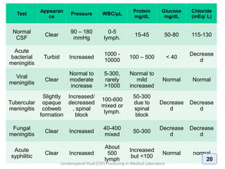
MICROBIOLOGICAL
EXAMINATION – II
Hussein A. Abid
Laboratory Medicine Specialist
Iraqi Medical Laboratory Association
Scientific Affairs and Cultural Relations Section
Scientific Affairs and Training
Third lecture
25/09/2018 (Tuesday)
FIRST DAY PROCESSING
2. Gram staining: a Gram smear is required when
the CSF contains pus cells (neutrophils).
• It should be the first investigation to be performed
and reported when the CSF appears purulent or
cloudy (suggestive of acute pyogenic meningitis).
• A Gram smear may also provide useful information
when a CSF is unsuitable for cell counting or
biochemical testing (e.g. when it is heavily blood
stained or contains clots).
Cerebrospinal Fluid (CSF) Processing In Medical Laboratory
2
FIRST DAY PROCESSING
Making a smear of CSF for Gram staining
1- Mix tube #2 and centrifuge most of it at approximately 1000 g for 5-10
minutes (leave a small amount of uncentrifuged CSF for a cell count
should this be required if there is no third tube).
• Do not centrifuge a purulent fluid. A smear for Gram staining is best
prepared from the uncentrifuged CSF.
2- Transfer the supernatant fluid to another tube (to be used for glucose and
protein tests should these be required).
3- Mix the sediment. Transfer several drops of the sediment to a slide, but
do not make the preparation too thick because this will make it difficult to
decolorize adequately. Allow the preparation to air-dry in a safe place.
4- Alcohol-fix the preparation and stain it by the Gram technique.
Cerebrospinal Fluid (CSF) Processing In Medical Laboratory
3
FIRST DAY PROCESSING
Examining a CSF Gram smear:
• Examine the smear microscopically for pus cells and
bacteria using the 40 and 100 objectives.
• Pus cells: report as many, moderate number, or few.
Pus cells will be found mainly in pyogenic bacterial
meningitis and in amoebic-meningoencephalitis (rare).
• Bacteria: Look in well stained (not too thick) areas for:
 Gram negative intracellular diplococci that could be
N. meningitidis.
Cerebrospinal Fluid (CSF) Processing In Medical Laboratory
4
FIRST DAY PROCESSING
 Gram positive diplococci or short streptococci, that could be
S. pneumoniae. It is often possible to see the capsules as
unstained areas around the bacteria.
 Gram negative rods, possibly H. influenzae, especially if
filamentous or other polymorphic forms are seen.
 Gram negative rods could also be E. coli or other coliforms,
especially when the CSF is from a newborn infant.
• Unevenly stained irregular in size yeast cells (some showing
budding), suggestive of C. neoformans. The large capsule that
surrounds the cell does not stain. It is best seen in an India ink
preparation.
• The smear will usually contain lymphocytes.
Cerebrospinal Fluid (CSF) Processing In Medical Laboratory
5
FIRST DAY PROCESSING
Important:
• Advise the medical officer immediately if the Gram smear
contains bacteria, pus cells, or yeast cells (confirmed as
capsulated in India ink preparation).
Acridine-orange stained smear to detect bacteria in CSF
• When facilities for fluorescence microscopy are available,
examine an acridine-orange (AO) stained smear.
• Bacteria especially when few, are more easily detected in
AO smears. They stain bright orange and cells and
debris stain green or yellow. The organisms can be
detected using the 40 objective.
Cerebrospinal Fluid (CSF) Processing In Medical Laboratory
6
FIRST DAY PROCESSING
• Note: When bacteria and pus cells are seen in the
Gram smear, culture the CSF. There is no need to
perform a cell count or measure the protein or
glucose.
• When the patient has been given antibiotics
(usually as emergency treatment) it will be more
difficult to detect bacteria in the Gram smear and
to isolate pathogens in culture.
• When M. tuberculosis is suspected Ziehl-Neelsen
staining should be performed.
Cerebrospinal Fluid (CSF) Processing In Medical Laboratory
7
FIRST DAY PROCESSING
CELL COUNT:
• A white cell count with an indication whether the
cells are pus cells or lymphocytes, is required
when the CSF appears slightly cloudy or clear or
when the Gram smear does not indicate pyogenic
bacterial meningitis.
• Note: samples that are heavily blood stained or
contain clots are unsuitable for cell counting. Make
a Gram smear and report the presence of pus cells
and bacteria as previously described.
Cerebrospinal Fluid (CSF) Processing In Medical Laboratory
8
FIRST DAY PROCESSING
Method:
1. To identify whether white cells in the CSF. are polymorphonuclear
neutrophils (pus cells) or lymphocytes, dilute the CSF in a fluid which
stains the cells.
2. Isotonic 0.1% toluidine blue is recommended because it stains
lymphocytes and the nuclei of pus cells blue. C. neoformans yeast cells
stain pink. Red cells remain unstained. The motility of trypanosomes is
not affected by the dye.
3. When toluidine blue is unavailable, isotonic methylene blue can be used
which will also stain the nuclei of leucocytes.
4. If preferred, leucocytes can be differentiated by examining a Leishman,
Giemsa or rapid Field’s stained smear (sediment from centrifuged CSF)
after counting the cells.
Cerebrospinal Fluid (CSF) Processing In Medical Laboratory
9
FIRST DAY PROCESSING
1. Mix un-centrifuged CSF, dilute the fluid 1 in 2, i.e. mix 1 drop of CSF with
1 drop of toluidine blue diluting fluid. The drops must be of equal volume,
therefore use Pasteur pipettes of the same bore size for both fluids and
hold the pipettes vertically when dispensing the drops.
2. Assemble a modified Fuchs-Rosenthal ruled counting chamber, making
sure the chamber and cover glass are completely clean. When
unavailable, an improved Neubauer (preferably Bright-Line) chamber can
be used. A Fuchs-Rosenthal chamber is recommended because it has
twice the depth (0.2 mm) and is more suitable for counting WBCs in CSF.
3. Using a fine bore Pasteur pipette or capillary tube, carefully fill the
counting chamber with the well-mixed diluted CSF. The fluid must not
overflow into the channels on each side of the chamber.
Cerebrospinal Fluid (CSF) Processing In Medical Laboratory
10
FIRST DAY PROCESSING
4. Wait about 2 minutes for the cells to settle. Count the cells
microscopically.
5. Focus the cells and rulings using the 10 objective with the
condenser iris closed sufficiently to give good contrast. Before
starting the count, use the 40 objective to check that the cells are
white cells and not red cells (unstained smaller cells without a
nucleus) and note whether the white cells are mainly
polymorphonuclear neutrophils (with lobed nucleus) or
lymphocytes.
• If a mixture of both, estimate approximately the percentage of
each type of cell.
Cerebrospinal Fluid (CSF) Processing In Medical Laboratory
11
FIRST DAY PROCESSING
• When yeast cells are seen, examine an India ink
preparation.
• Note: When red cells are seen, mention this in the
report. When many red cells are present, the CSF is
unsuitable for WBC cell counting.
6.Count the cells in 4 large squares (Improved Neubaur).
• Note: When the cells are too many to count, dilute the
CSF 1 in 10 (1 drop CSF mixed with 9 drops of diluting
fluid), refill the chamber and count the cells.
Cerebrospinal Fluid (CSF) Processing In Medical Laboratory
12
FIRST DAY PROCESSING
• Multiply the cells counted by 2. Report the number of cells per liter (L) of
CSF.
• Example (Using 1 in 2 CSF dilution and Fuchs- Rosenthal chamber), If
240 cells are counted in 5 squares:
240 × 2 = 480, report as 480 × 106 cells/ L
• Formerly, 480 × 106 cells/ L would have been reported as 480 cells/ mm3
or 480 cells/ L.
• When using an Improved Neubauer chamber: To count the cells in 4
large squares, multiply the cells counted by 5. Report the number of
cells per liter of CSF.
• Example, If 64 cells are counted in 4 squares:
64 × 5 = 320, report as 320 × 106 cells/ L
Cerebrospinal Fluid (CSF) Processing In Medical Laboratory
13
FIRST DAY PROCESSING
Calculation factors (1 in 10 CSF dilution)
• Fuchs-Rosenthal chamber: Multiply cells
counted in 5 squares by 10. Report number of
cells per liter of CSF.
• Normal CSF contains up to 5 × 10⁶ cells/
liter (higher in neonates).
• When no WBCs are seen, report the count as:
5 cells × 106 cells/ L
Cerebrospinal Fluid (CSF) Processing In Medical Laboratory
14
FIRST DAY PROCESSING
Immunological diagnosis of acute bacterial meningitis
• Direct antigen testing of CSF may provide a rapid
diagnosis of acute bacterial meningitis, particularly when
the patient has been treated with antimicrobials and
bacteria cannot be detected in a Gram smear or by
culture.
• Tests are available to detect N. meningitidis groups A, B,
C, Y and W135 (Group B reagent cross-reacts with E. coli
K1 antigen), H. influenzae type b, S. pneumoniae, and S.
agalactiae.
Cerebrospinal Fluid (CSF) Processing In Medical Laboratory
15
FIRST DAY PROCESSING
3.CSF culture: culture the CSF when bacteria are seen in
the Gram smear and, or, cells are present, or the protein
concentration is raised.
• Use CSF sample when the CSF is clear or slightly cloudy,
centrifuge the sample in a sterile capped tube for about
15 minutes, and use the sediment to inoculate the culture
media.
• Important: CSF must be cultured as soon as possible
after collection. When a delay is unavoidable, the fluid
should be kept at 35-37 °C (not refrigerated).
Cerebrospinal Fluid (CSF) Processing In Medical Laboratory
16
FIRST DAY PROCESSING
Chocolate (heated blood) agar and blood agar:
• Inoculate the specimen on chocolate agar and blood agar.
When Gram positive diplococci are seen in the Gram smear,
add an optochin disc to the blood agar plate to assist in the
identification of S. pneumoniae.
• Incubate both plates in a carbon dioxide enriched
atmosphere at 35-37 °C for up to 48 hours, checking for
growth after overnight incubation.
• When patient is a newborn infant: inoculate the specimen
also on MacConkey agar. Incubate aerobically at 35-37 °C
overnight.
Cerebrospinal Fluid (CSF) Processing In Medical Laboratory
17
SECOND DAY
Examine and report the cultures
1- Chocolate agar and blood agar cultures: look especially for
colonies that could be:
 Neisseria meningitidis (growing on chocolate agar and blood
agar, oxidase positive)
 Streptococcus pneumoniae (sensitive to optochin)
 Haemophilus influenzae (growing only on chocolate agar)
 Cryptococcus neoformans (Gram stain the colonies).
 Streptococcus agalactiae.
 Listeria monocytogenes.
Cerebrospinal Fluid (CSF) Processing In Medical Laboratory
18
SECOND DAY
Examine and report the cultures
2- MacConkey agar culture: look especially for colonies could be;
 Escherichia coli or other coliform.
 Other bacteria that cause neonatal meningitis.
3- MacConkey agar culture: antimicrobial susceptibility testing (AST)
 Test isolates of S. pneumoniae for susceptibility to chloramphenicol
and penicillin (use 1 g oxacillin disc).
 Test H. influenzae for beta-lactamase production and susceptibility to
chloramphenicol (using chocolate agar). Perform susceptibility testing
on Gram negative rods as indicated.
Cerebrospinal Fluid (CSF) Processing In Medical Laboratory
19
Test
Appearan
ce
Pressure WBC/μL
Protein
mg/dL
Glucose
mg/dL
Chloride
(mEq/ L)
Normal
CSF
Clear
90 – 180
mmHg
0-5
lymph.
15-45 50-80 115-130
Acute
bacterial
meningitis
Turbid Increased
1000 -
10000
100 – 500 < 40
Decrease
d
Viral
meningitis
Clear
Normal to
moderate
increase
5-300,
rarely
>1000
Normal to
mild
increased
Normal Normal
Tubercular
meningitis
Slightly
opaque
cobweb
formation
Increased/
decreased
, spinal
block
100-600
mixed or
lymph.
50-300
due to
spinal
block
Decrease
d
Decrease
d
Fungal
meningitis
Clear Increased
40-400
mixed
50-300
Decrease
d
Decrease
d
Acute
syphilitic
Clear Increased
About
500
lymph
Increased
but <100
Normal normal
Cerebrospinal Fluid (CSF) Processing In Medical Laboratory
20
IMPORTANT NOTES
Interfering factors
• Patient on antibiotic therapy.
• Improper sample collection.
Result reporting
• Results of the microscopy and all positive cultures of CSF are
reported immediately to the treating physician. Negative bacterial
results are sent out 72 hours after the CSF is received.
Turn around time (TAT)
• Gram stain result is reported within 30 minutes of specimen receipt
• Positive Culture results: 3-5 days
• Negative Culture results: 2-3 days
Cerebrospinal Fluid (CSF) Processing In Medical Laboratory
21
IMPORTANT NOTES
Additional information
• Several antigen detection methods are available for the
direct detection of the polysaccharide capsular antigen
of H. influenzae, N. meningitidis, S. pneumoniae and
Group B streptococci in CSF which showed specificity
and sensitivity of about 90-97%.
• Direct detection of Cryptococcus antigen in CSF is also
available which replaced India ink in many laboratories.
• The routine culture for CSF does not include all
organisms mentioned in the above table.
Cerebrospinal Fluid (CSF) Processing In Medical Laboratory
22

CSF MICROBIOLOGICAL EXAMINATION – II

  • 1.
    MICROBIOLOGICAL EXAMINATION – II HusseinA. Abid Laboratory Medicine Specialist Iraqi Medical Laboratory Association Scientific Affairs and Cultural Relations Section Scientific Affairs and Training Third lecture 25/09/2018 (Tuesday)
  • 2.
    FIRST DAY PROCESSING 2.Gram staining: a Gram smear is required when the CSF contains pus cells (neutrophils). • It should be the first investigation to be performed and reported when the CSF appears purulent or cloudy (suggestive of acute pyogenic meningitis). • A Gram smear may also provide useful information when a CSF is unsuitable for cell counting or biochemical testing (e.g. when it is heavily blood stained or contains clots). Cerebrospinal Fluid (CSF) Processing In Medical Laboratory 2
  • 3.
    FIRST DAY PROCESSING Makinga smear of CSF for Gram staining 1- Mix tube #2 and centrifuge most of it at approximately 1000 g for 5-10 minutes (leave a small amount of uncentrifuged CSF for a cell count should this be required if there is no third tube). • Do not centrifuge a purulent fluid. A smear for Gram staining is best prepared from the uncentrifuged CSF. 2- Transfer the supernatant fluid to another tube (to be used for glucose and protein tests should these be required). 3- Mix the sediment. Transfer several drops of the sediment to a slide, but do not make the preparation too thick because this will make it difficult to decolorize adequately. Allow the preparation to air-dry in a safe place. 4- Alcohol-fix the preparation and stain it by the Gram technique. Cerebrospinal Fluid (CSF) Processing In Medical Laboratory 3
  • 4.
    FIRST DAY PROCESSING Examininga CSF Gram smear: • Examine the smear microscopically for pus cells and bacteria using the 40 and 100 objectives. • Pus cells: report as many, moderate number, or few. Pus cells will be found mainly in pyogenic bacterial meningitis and in amoebic-meningoencephalitis (rare). • Bacteria: Look in well stained (not too thick) areas for:  Gram negative intracellular diplococci that could be N. meningitidis. Cerebrospinal Fluid (CSF) Processing In Medical Laboratory 4
  • 5.
    FIRST DAY PROCESSING Gram positive diplococci or short streptococci, that could be S. pneumoniae. It is often possible to see the capsules as unstained areas around the bacteria.  Gram negative rods, possibly H. influenzae, especially if filamentous or other polymorphic forms are seen.  Gram negative rods could also be E. coli or other coliforms, especially when the CSF is from a newborn infant. • Unevenly stained irregular in size yeast cells (some showing budding), suggestive of C. neoformans. The large capsule that surrounds the cell does not stain. It is best seen in an India ink preparation. • The smear will usually contain lymphocytes. Cerebrospinal Fluid (CSF) Processing In Medical Laboratory 5
  • 6.
    FIRST DAY PROCESSING Important: •Advise the medical officer immediately if the Gram smear contains bacteria, pus cells, or yeast cells (confirmed as capsulated in India ink preparation). Acridine-orange stained smear to detect bacteria in CSF • When facilities for fluorescence microscopy are available, examine an acridine-orange (AO) stained smear. • Bacteria especially when few, are more easily detected in AO smears. They stain bright orange and cells and debris stain green or yellow. The organisms can be detected using the 40 objective. Cerebrospinal Fluid (CSF) Processing In Medical Laboratory 6
  • 7.
    FIRST DAY PROCESSING •Note: When bacteria and pus cells are seen in the Gram smear, culture the CSF. There is no need to perform a cell count or measure the protein or glucose. • When the patient has been given antibiotics (usually as emergency treatment) it will be more difficult to detect bacteria in the Gram smear and to isolate pathogens in culture. • When M. tuberculosis is suspected Ziehl-Neelsen staining should be performed. Cerebrospinal Fluid (CSF) Processing In Medical Laboratory 7
  • 8.
    FIRST DAY PROCESSING CELLCOUNT: • A white cell count with an indication whether the cells are pus cells or lymphocytes, is required when the CSF appears slightly cloudy or clear or when the Gram smear does not indicate pyogenic bacterial meningitis. • Note: samples that are heavily blood stained or contain clots are unsuitable for cell counting. Make a Gram smear and report the presence of pus cells and bacteria as previously described. Cerebrospinal Fluid (CSF) Processing In Medical Laboratory 8
  • 9.
    FIRST DAY PROCESSING Method: 1.To identify whether white cells in the CSF. are polymorphonuclear neutrophils (pus cells) or lymphocytes, dilute the CSF in a fluid which stains the cells. 2. Isotonic 0.1% toluidine blue is recommended because it stains lymphocytes and the nuclei of pus cells blue. C. neoformans yeast cells stain pink. Red cells remain unstained. The motility of trypanosomes is not affected by the dye. 3. When toluidine blue is unavailable, isotonic methylene blue can be used which will also stain the nuclei of leucocytes. 4. If preferred, leucocytes can be differentiated by examining a Leishman, Giemsa or rapid Field’s stained smear (sediment from centrifuged CSF) after counting the cells. Cerebrospinal Fluid (CSF) Processing In Medical Laboratory 9
  • 10.
    FIRST DAY PROCESSING 1.Mix un-centrifuged CSF, dilute the fluid 1 in 2, i.e. mix 1 drop of CSF with 1 drop of toluidine blue diluting fluid. The drops must be of equal volume, therefore use Pasteur pipettes of the same bore size for both fluids and hold the pipettes vertically when dispensing the drops. 2. Assemble a modified Fuchs-Rosenthal ruled counting chamber, making sure the chamber and cover glass are completely clean. When unavailable, an improved Neubauer (preferably Bright-Line) chamber can be used. A Fuchs-Rosenthal chamber is recommended because it has twice the depth (0.2 mm) and is more suitable for counting WBCs in CSF. 3. Using a fine bore Pasteur pipette or capillary tube, carefully fill the counting chamber with the well-mixed diluted CSF. The fluid must not overflow into the channels on each side of the chamber. Cerebrospinal Fluid (CSF) Processing In Medical Laboratory 10
  • 11.
    FIRST DAY PROCESSING 4.Wait about 2 minutes for the cells to settle. Count the cells microscopically. 5. Focus the cells and rulings using the 10 objective with the condenser iris closed sufficiently to give good contrast. Before starting the count, use the 40 objective to check that the cells are white cells and not red cells (unstained smaller cells without a nucleus) and note whether the white cells are mainly polymorphonuclear neutrophils (with lobed nucleus) or lymphocytes. • If a mixture of both, estimate approximately the percentage of each type of cell. Cerebrospinal Fluid (CSF) Processing In Medical Laboratory 11
  • 12.
    FIRST DAY PROCESSING •When yeast cells are seen, examine an India ink preparation. • Note: When red cells are seen, mention this in the report. When many red cells are present, the CSF is unsuitable for WBC cell counting. 6.Count the cells in 4 large squares (Improved Neubaur). • Note: When the cells are too many to count, dilute the CSF 1 in 10 (1 drop CSF mixed with 9 drops of diluting fluid), refill the chamber and count the cells. Cerebrospinal Fluid (CSF) Processing In Medical Laboratory 12
  • 13.
    FIRST DAY PROCESSING •Multiply the cells counted by 2. Report the number of cells per liter (L) of CSF. • Example (Using 1 in 2 CSF dilution and Fuchs- Rosenthal chamber), If 240 cells are counted in 5 squares: 240 × 2 = 480, report as 480 × 106 cells/ L • Formerly, 480 × 106 cells/ L would have been reported as 480 cells/ mm3 or 480 cells/ L. • When using an Improved Neubauer chamber: To count the cells in 4 large squares, multiply the cells counted by 5. Report the number of cells per liter of CSF. • Example, If 64 cells are counted in 4 squares: 64 × 5 = 320, report as 320 × 106 cells/ L Cerebrospinal Fluid (CSF) Processing In Medical Laboratory 13
  • 14.
    FIRST DAY PROCESSING Calculationfactors (1 in 10 CSF dilution) • Fuchs-Rosenthal chamber: Multiply cells counted in 5 squares by 10. Report number of cells per liter of CSF. • Normal CSF contains up to 5 × 10⁶ cells/ liter (higher in neonates). • When no WBCs are seen, report the count as: 5 cells × 106 cells/ L Cerebrospinal Fluid (CSF) Processing In Medical Laboratory 14
  • 15.
    FIRST DAY PROCESSING Immunologicaldiagnosis of acute bacterial meningitis • Direct antigen testing of CSF may provide a rapid diagnosis of acute bacterial meningitis, particularly when the patient has been treated with antimicrobials and bacteria cannot be detected in a Gram smear or by culture. • Tests are available to detect N. meningitidis groups A, B, C, Y and W135 (Group B reagent cross-reacts with E. coli K1 antigen), H. influenzae type b, S. pneumoniae, and S. agalactiae. Cerebrospinal Fluid (CSF) Processing In Medical Laboratory 15
  • 16.
    FIRST DAY PROCESSING 3.CSFculture: culture the CSF when bacteria are seen in the Gram smear and, or, cells are present, or the protein concentration is raised. • Use CSF sample when the CSF is clear or slightly cloudy, centrifuge the sample in a sterile capped tube for about 15 minutes, and use the sediment to inoculate the culture media. • Important: CSF must be cultured as soon as possible after collection. When a delay is unavoidable, the fluid should be kept at 35-37 °C (not refrigerated). Cerebrospinal Fluid (CSF) Processing In Medical Laboratory 16
  • 17.
    FIRST DAY PROCESSING Chocolate(heated blood) agar and blood agar: • Inoculate the specimen on chocolate agar and blood agar. When Gram positive diplococci are seen in the Gram smear, add an optochin disc to the blood agar plate to assist in the identification of S. pneumoniae. • Incubate both plates in a carbon dioxide enriched atmosphere at 35-37 °C for up to 48 hours, checking for growth after overnight incubation. • When patient is a newborn infant: inoculate the specimen also on MacConkey agar. Incubate aerobically at 35-37 °C overnight. Cerebrospinal Fluid (CSF) Processing In Medical Laboratory 17
  • 18.
    SECOND DAY Examine andreport the cultures 1- Chocolate agar and blood agar cultures: look especially for colonies that could be:  Neisseria meningitidis (growing on chocolate agar and blood agar, oxidase positive)  Streptococcus pneumoniae (sensitive to optochin)  Haemophilus influenzae (growing only on chocolate agar)  Cryptococcus neoformans (Gram stain the colonies).  Streptococcus agalactiae.  Listeria monocytogenes. Cerebrospinal Fluid (CSF) Processing In Medical Laboratory 18
  • 19.
    SECOND DAY Examine andreport the cultures 2- MacConkey agar culture: look especially for colonies could be;  Escherichia coli or other coliform.  Other bacteria that cause neonatal meningitis. 3- MacConkey agar culture: antimicrobial susceptibility testing (AST)  Test isolates of S. pneumoniae for susceptibility to chloramphenicol and penicillin (use 1 g oxacillin disc).  Test H. influenzae for beta-lactamase production and susceptibility to chloramphenicol (using chocolate agar). Perform susceptibility testing on Gram negative rods as indicated. Cerebrospinal Fluid (CSF) Processing In Medical Laboratory 19
  • 20.
    Test Appearan ce Pressure WBC/μL Protein mg/dL Glucose mg/dL Chloride (mEq/ L) Normal CSF Clear 90– 180 mmHg 0-5 lymph. 15-45 50-80 115-130 Acute bacterial meningitis Turbid Increased 1000 - 10000 100 – 500 < 40 Decrease d Viral meningitis Clear Normal to moderate increase 5-300, rarely >1000 Normal to mild increased Normal Normal Tubercular meningitis Slightly opaque cobweb formation Increased/ decreased , spinal block 100-600 mixed or lymph. 50-300 due to spinal block Decrease d Decrease d Fungal meningitis Clear Increased 40-400 mixed 50-300 Decrease d Decrease d Acute syphilitic Clear Increased About 500 lymph Increased but <100 Normal normal Cerebrospinal Fluid (CSF) Processing In Medical Laboratory 20
  • 21.
    IMPORTANT NOTES Interfering factors •Patient on antibiotic therapy. • Improper sample collection. Result reporting • Results of the microscopy and all positive cultures of CSF are reported immediately to the treating physician. Negative bacterial results are sent out 72 hours after the CSF is received. Turn around time (TAT) • Gram stain result is reported within 30 minutes of specimen receipt • Positive Culture results: 3-5 days • Negative Culture results: 2-3 days Cerebrospinal Fluid (CSF) Processing In Medical Laboratory 21
  • 22.
    IMPORTANT NOTES Additional information •Several antigen detection methods are available for the direct detection of the polysaccharide capsular antigen of H. influenzae, N. meningitidis, S. pneumoniae and Group B streptococci in CSF which showed specificity and sensitivity of about 90-97%. • Direct detection of Cryptococcus antigen in CSF is also available which replaced India ink in many laboratories. • The routine culture for CSF does not include all organisms mentioned in the above table. Cerebrospinal Fluid (CSF) Processing In Medical Laboratory 22