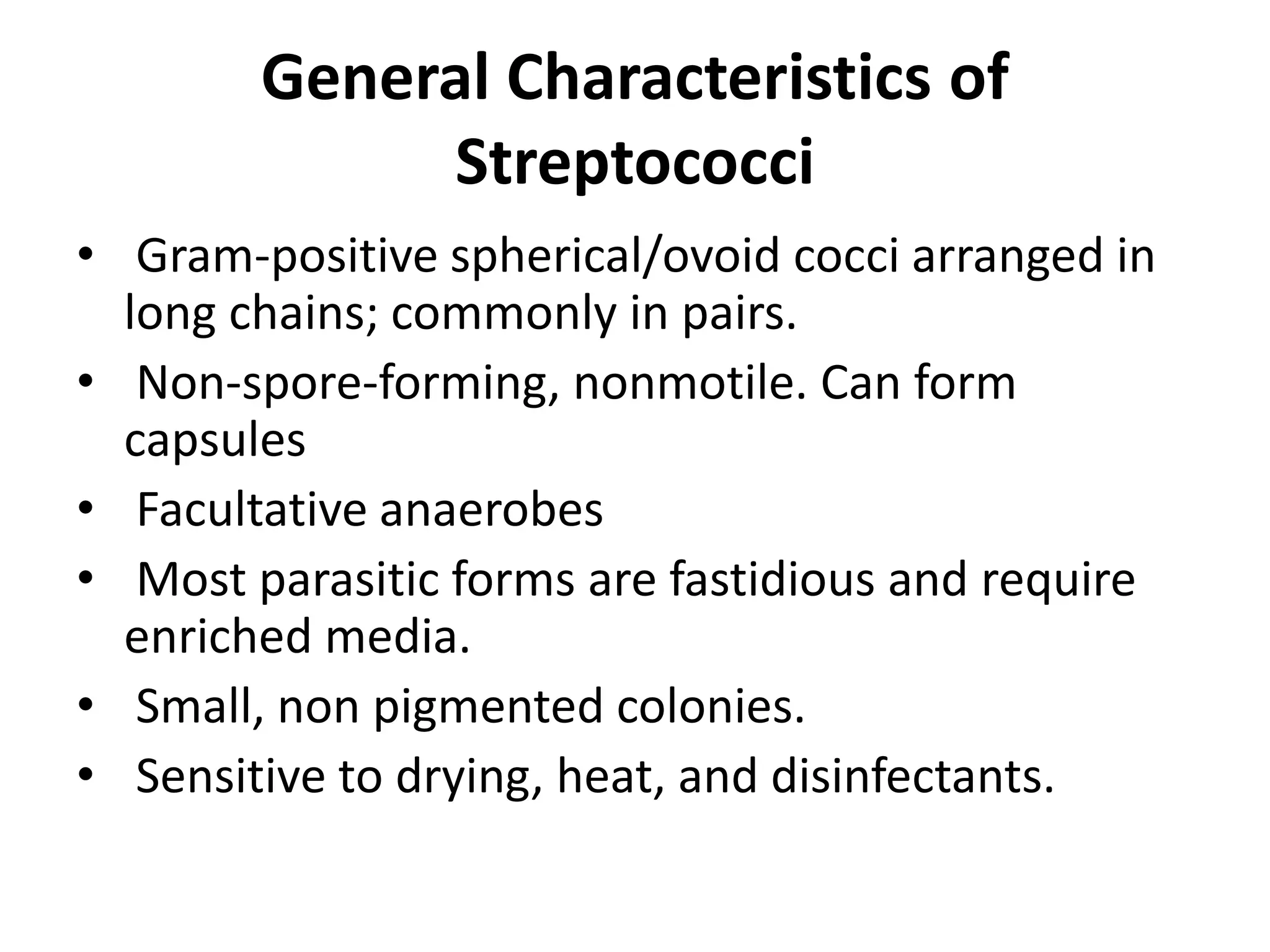
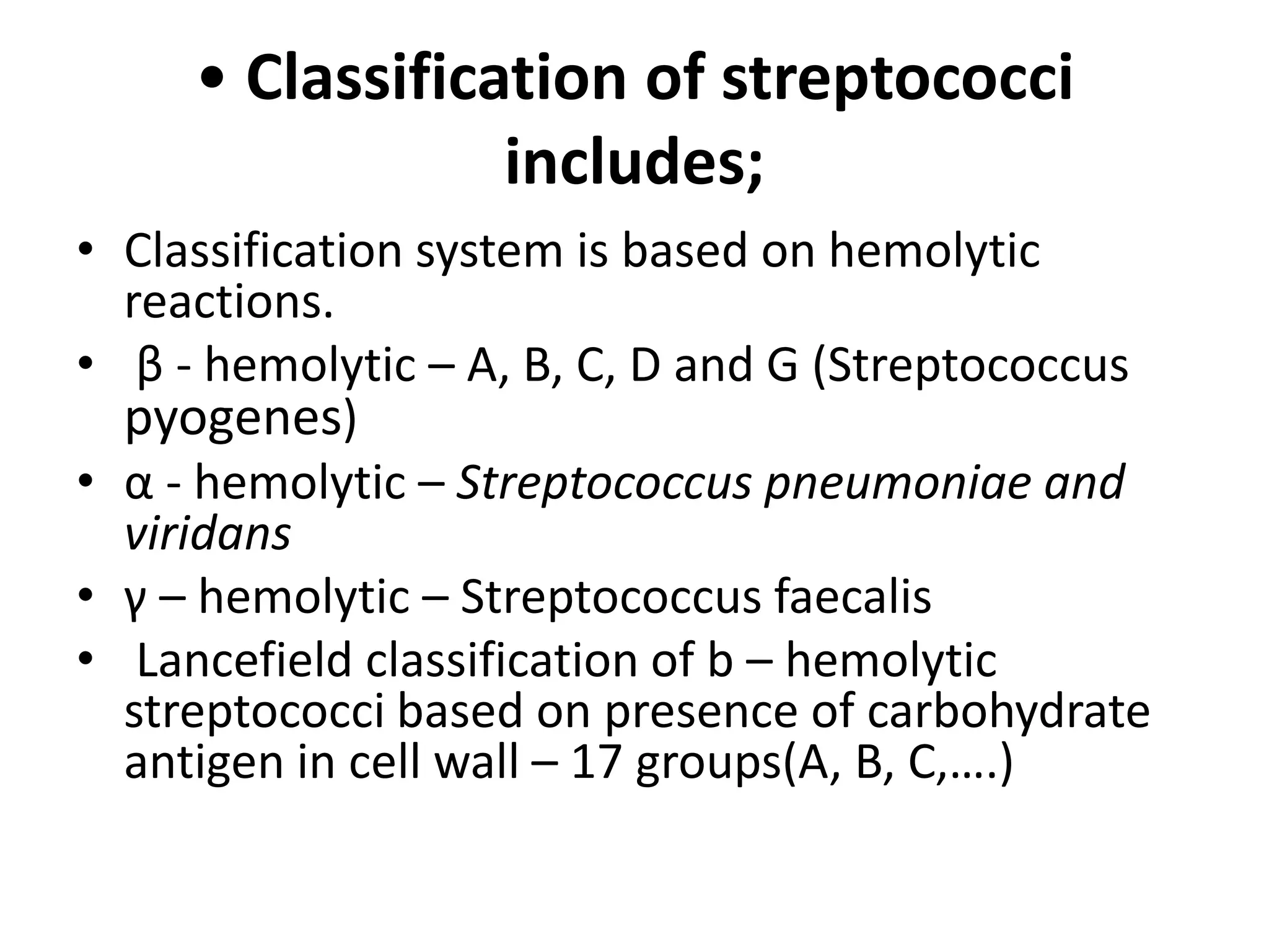
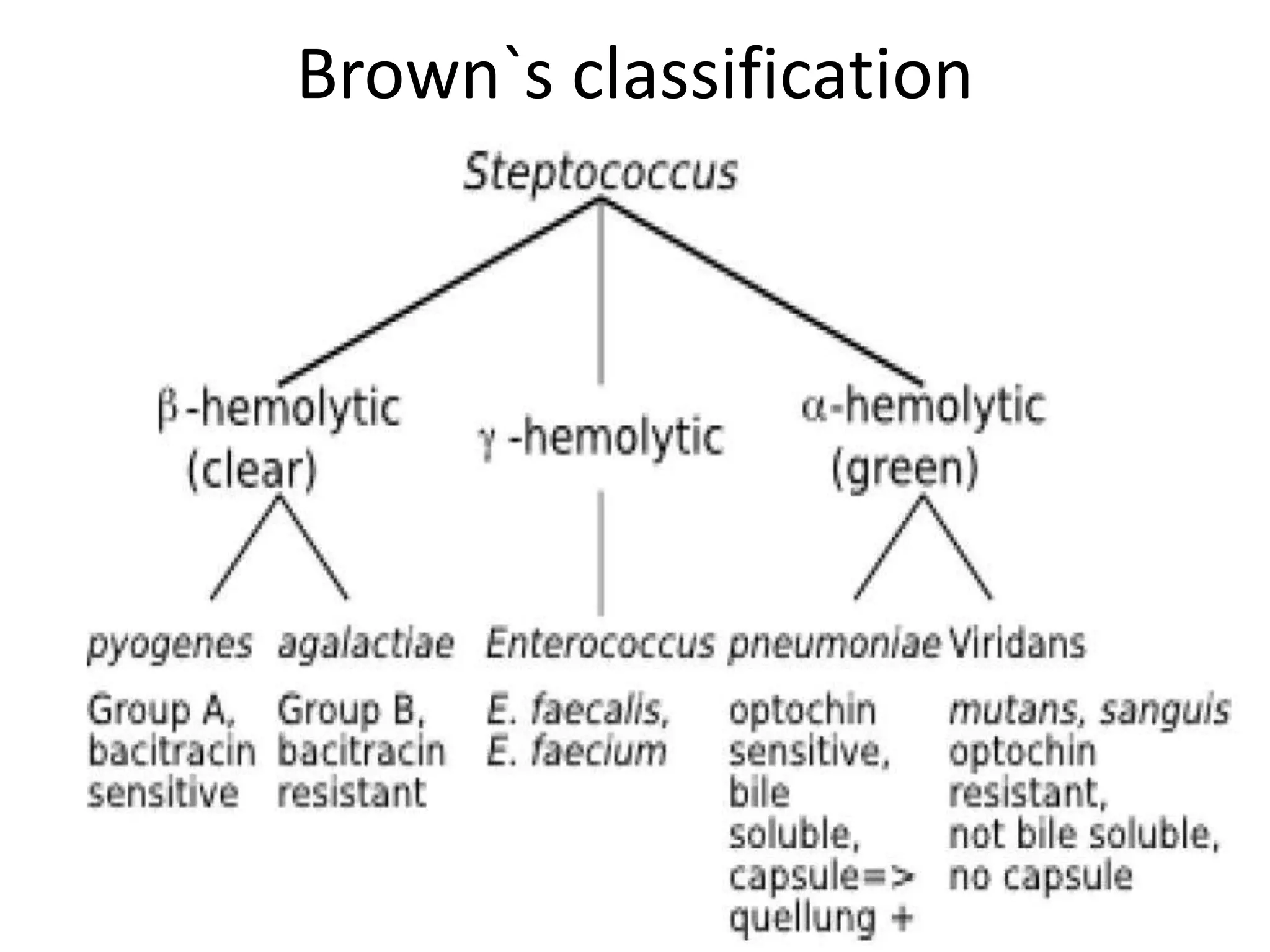
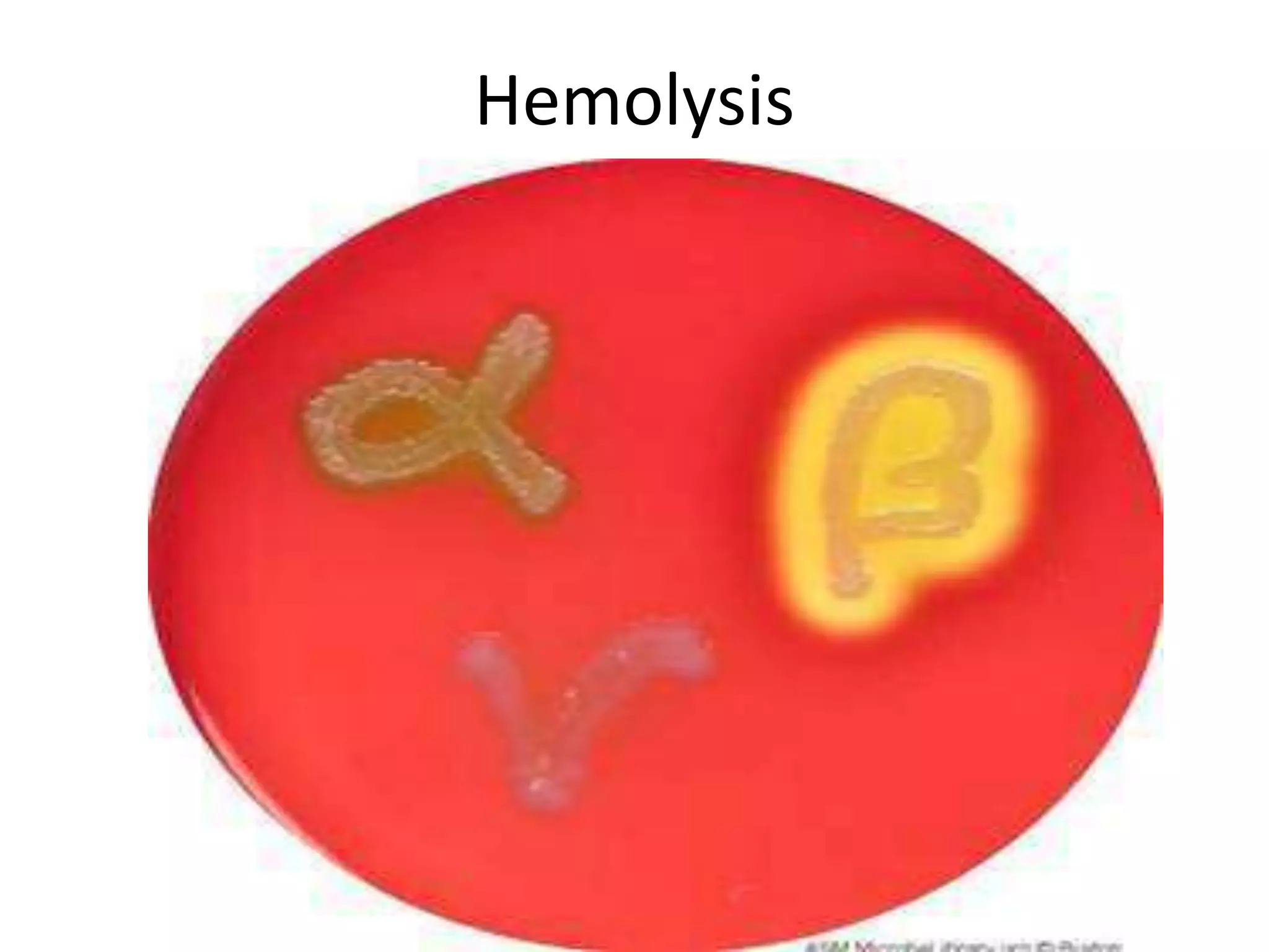
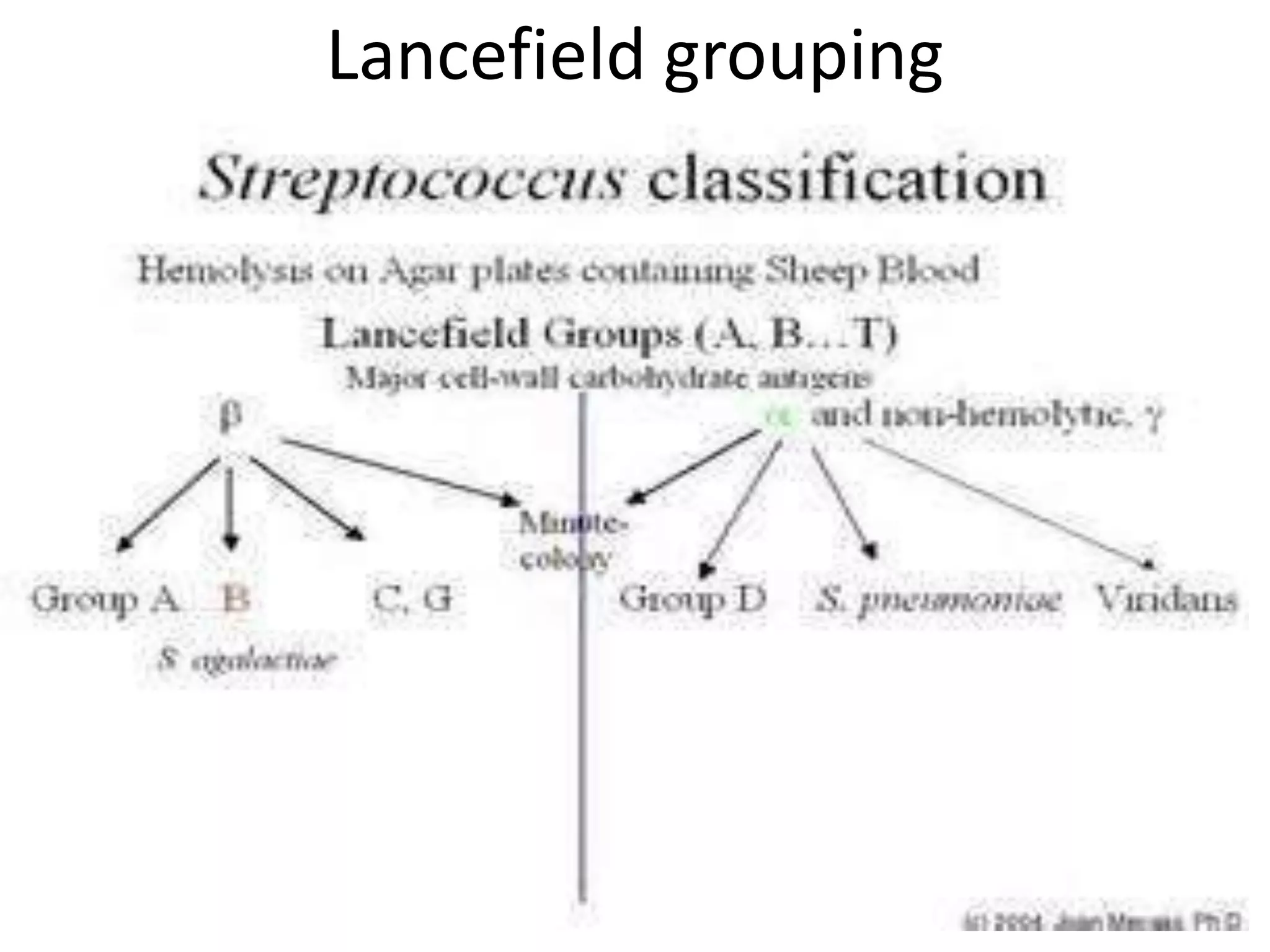
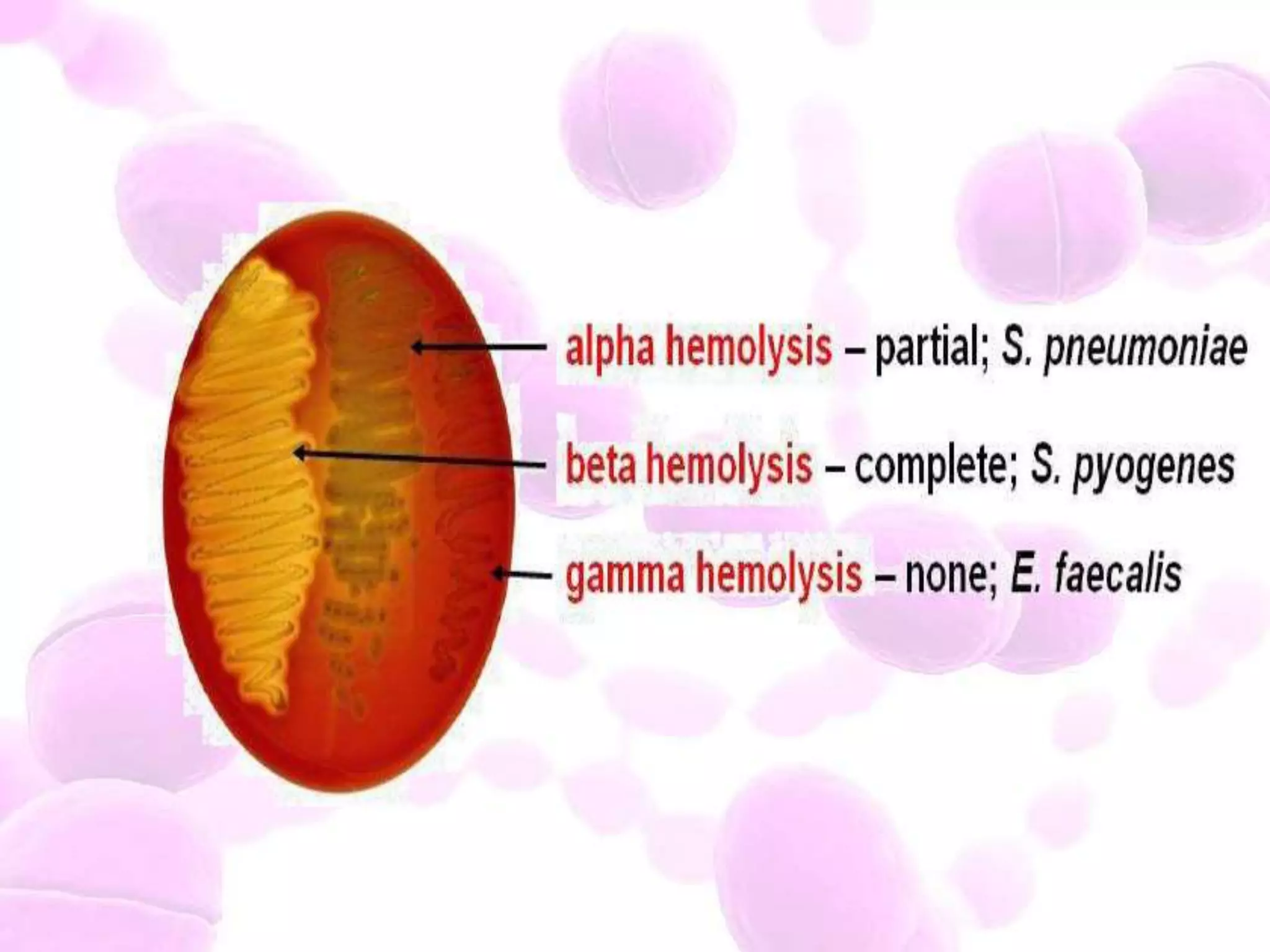
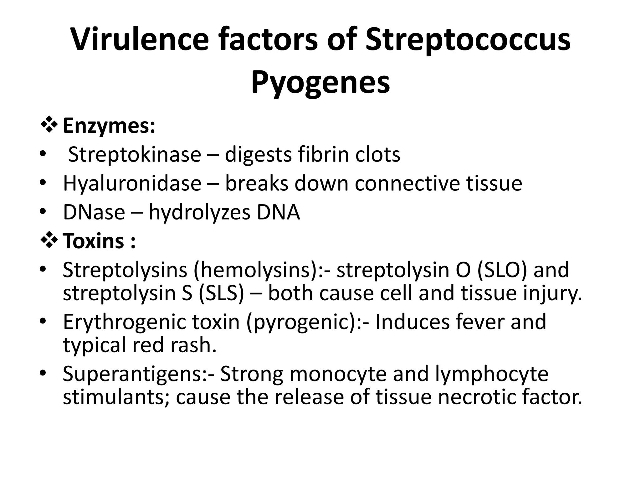
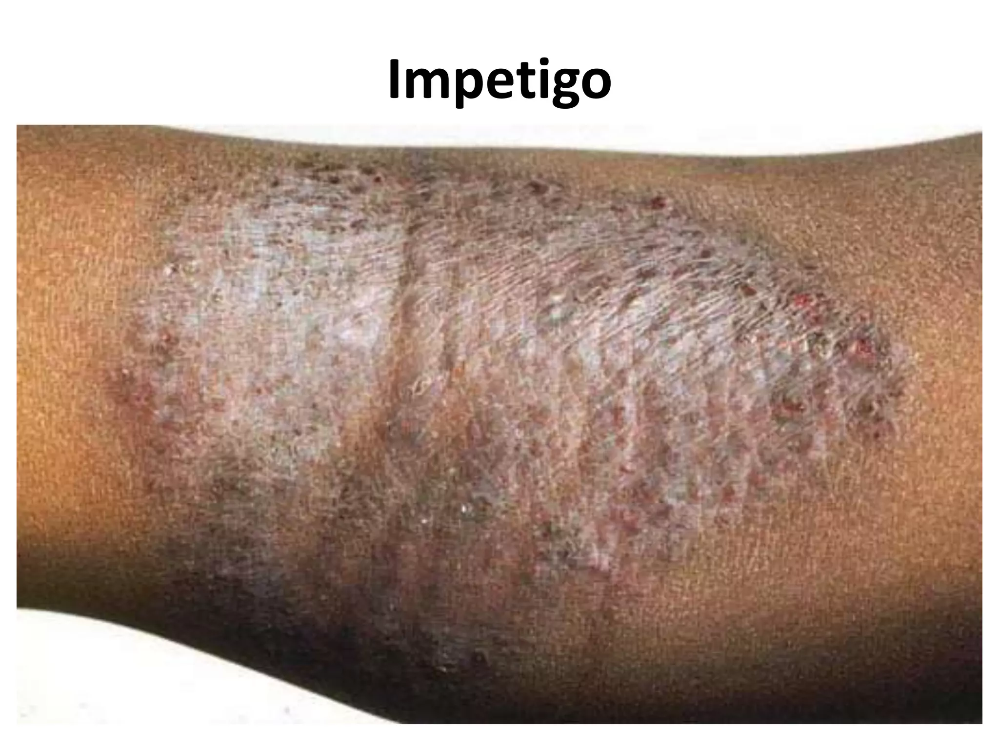
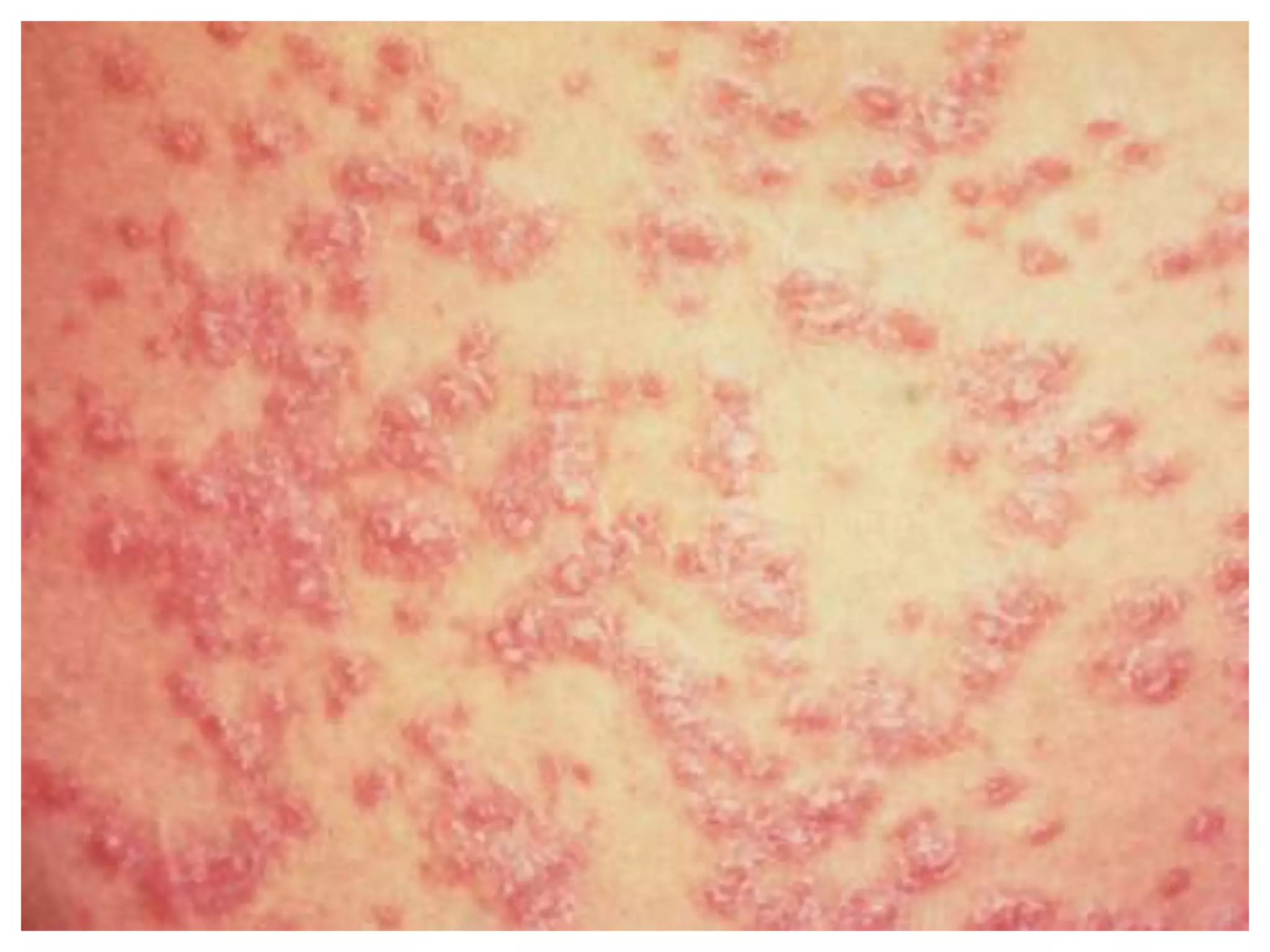
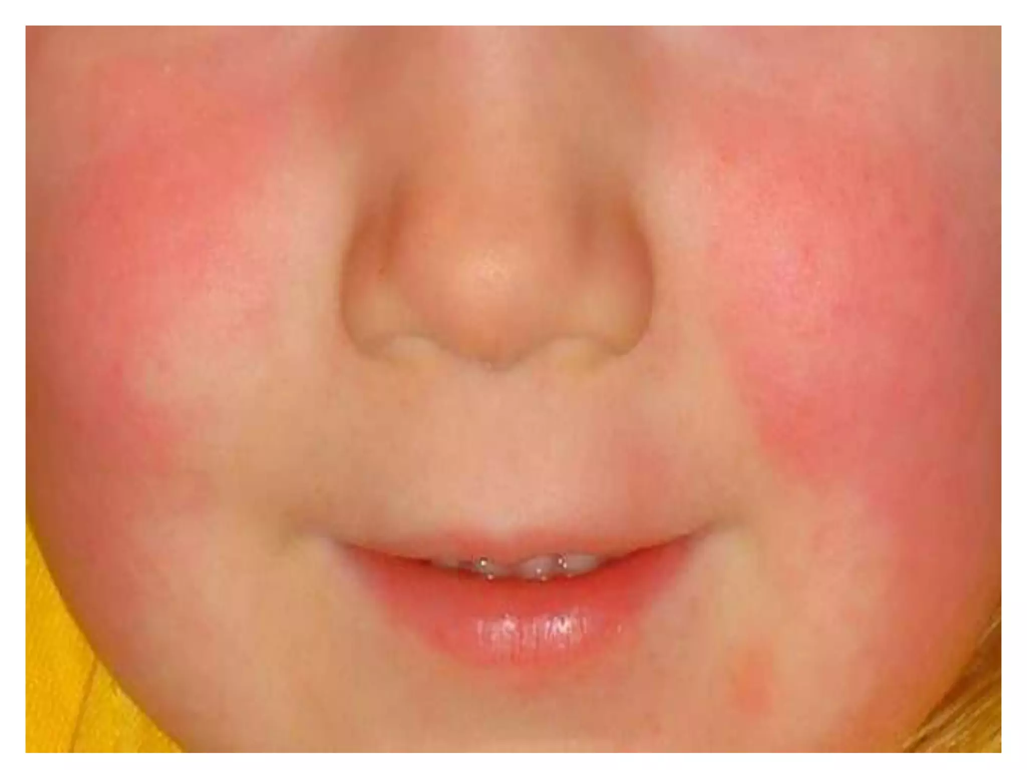
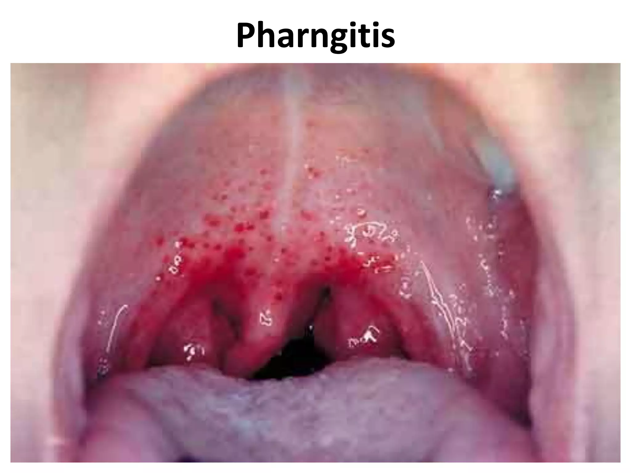
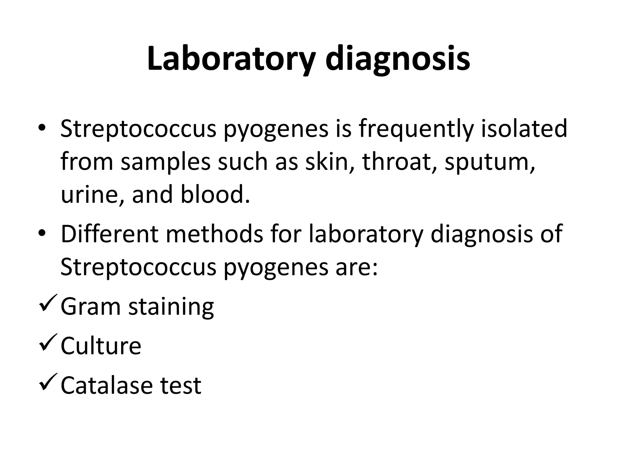
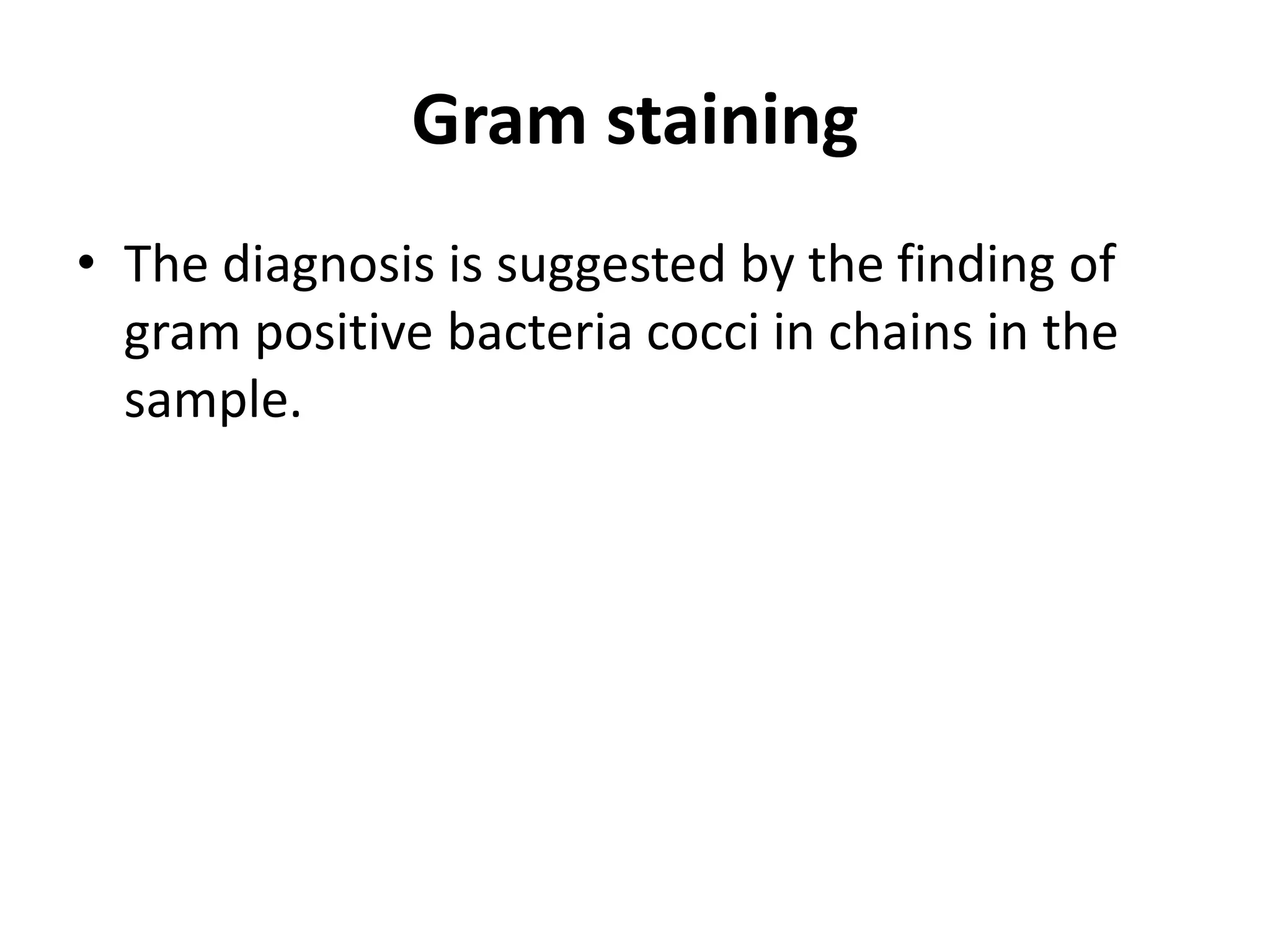
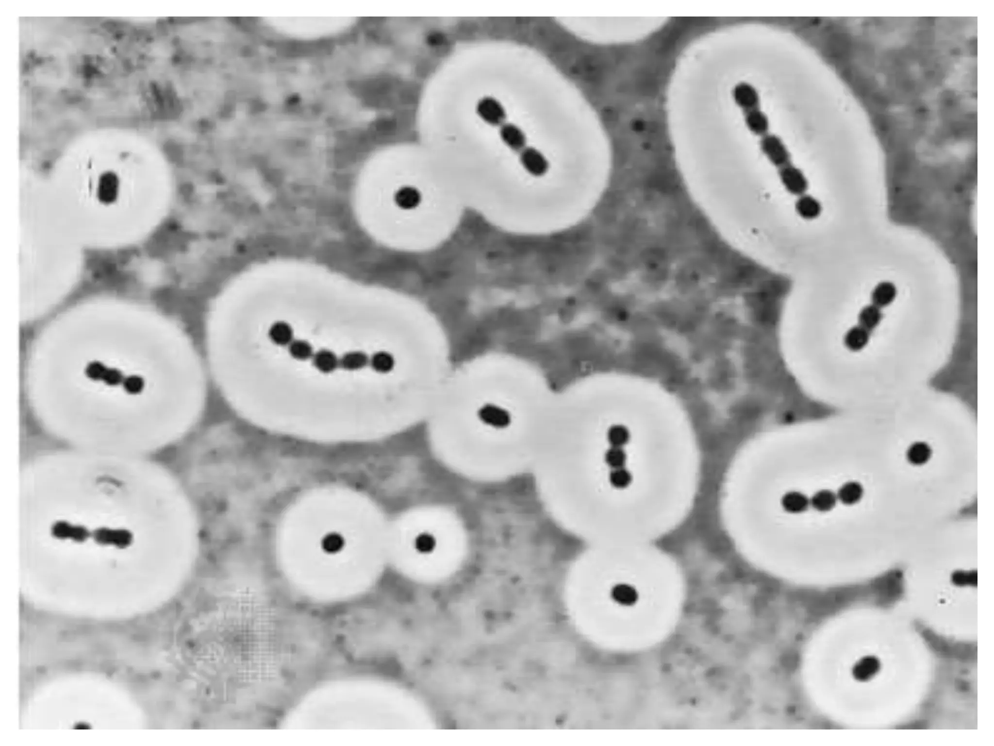
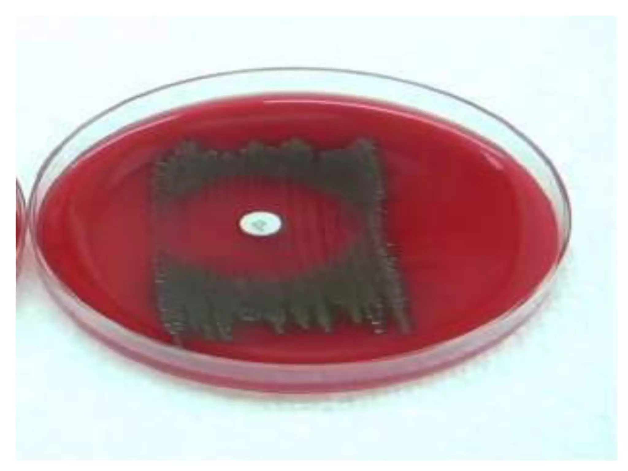
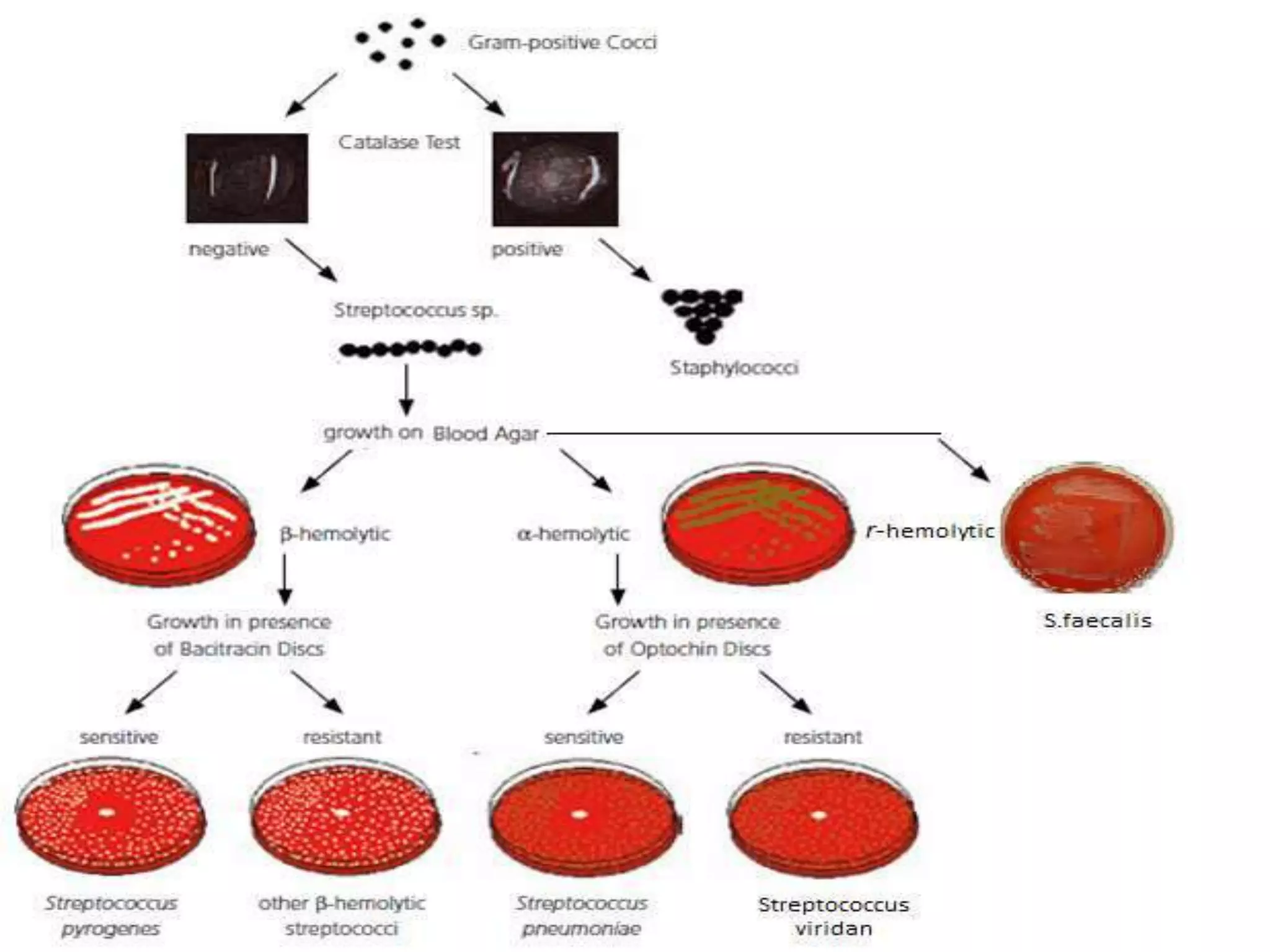
Mr. Byiringiro Pacifique, STD
018/SI/5037
STREPTOCOCCUS
• Streptococci is Gram-positive spherical/ovoid
• cocci arranged in long chains; commonly in
pairs.
• Streptococci are classified according to their
ability to break down blood in fresh blood
agar plates. Some streptococci have no effect
on blood ( non-haemolytic streptococci).
General Characteristics of
Streptococci
• Gram-positive spherical/ovoid cocci arranged in
long chains; commonly in pairs.
• Non-spore-forming, nonmotile. Can form
capsules
• Facultative anaerobes
• Most parasitic forms are fastidious and require
enriched media.
• Small, non pigmented colonies.
• Sensitive to drying, heat, and disinfectants.
Gram positive cocci in chains
• Classification of streptococci
includes;
• Classification system is based on hemolytic
reactions.
• β - hemolytic – A, B, C, D and G (Streptococcus
pyogenes)
• α - hemolytic – Streptococcus pneumoniae and
viridans
• γ – hemolytic – Streptococcus faecalis
• Lancefield classification of b – hemolytic
streptococci based on presence of carbohydrate
antigen in cell wall – 17 groups(A, B, C,….)
Classification of Streptococci
• Brown`s classification
• Lancefield grouping
• Griffith typing
Brown`s classification
Hemolysis
Lancefield grouping
Human Streptococcal Pathogens
• Streptococcus Pyogenes
• Streptococcus Viridans
• Streptococcus Pneumoniae
• Streptoccus Faecalis
Streptococcus Pyogenes
• Most serious streptococcal pathogen. It is a
parasite and inhabits throat, nasopharynx,
occasionally skin.
Morphology
• Gram-positive
• Spherical/ovoid cocci arranged in long chains.
• Nonmotile.
• Can form capsules.
Cultural characteristics
• Facultative anaerobes.
• Best growth achieved at pH 7.4-7.6 and
temperature 37⁰C.
• Required enriched media for growth.
• Colonies are around 1mm in diameter,
surrounded by a zone of clear hemolysis, semi
transparent and vary in appearance.
Biochemical properties
• They Produces many virulence factors. Means
that there is production of enzymes and
toxins.
Virulence factors of Streptococcus
Pyogenes
Enzymes:
• Streptokinase – digests fibrin clots
• Hyaluronidase – breaks down connective tissue
• DNase – hydrolyzes DNA
Toxins :
• Streptolysins (hemolysins):- streptolysin O (SLO) and
streptolysin S (SLS) – both cause cell and tissue injury.
• Erythrogenic toxin (pyrogenic):- Induces fever and
typical red rash.
• Superantigens:- Strong monocyte and lymphocyte
stimulants; cause the release of tissue necrotic factor.
Pathogenesis
• Humans are the only reservoir.
• In apparent carriers.(visible carriers of toxins and
enzyme)
• Transmission is through contact, droplets, food,
fomites
• Portal of entry is generally through skin or pharynx.
• Children are predominant group affected for cutaneous
and throat infections
• Systemic infections and progressive diseases may occur
if untreated.
Streptococcus pyogenes diseases
• Impetigo (pyoderma)- It is a skin infection with
superficial lesions that break and form highly
contagious crust.
• Throat infections
• Rheumatic fever – It has subclinical pharyngitis
in children; carditis with extensive heart valve
damage possible, arthritis, chorea, fever.
• Acute glomerulonephritis – It is the
inflammation of the nephrons.
Impetigo
Pharngitis
Laboratory diagnosis
• Streptococcus pyogenes is frequently isolated
from samples such as skin, throat, sputum,
urine, and blood.
• Different methods for laboratory diagnosis of
Streptococcus pyogenes are:
Gram staining
Culture
Catalase test
Gram staining
• The diagnosis is suggested by the finding of
gram positive bacteria cocci in chains in the
sample.
Culture
• The organism is cultured on blood agar with
an added bacitracin antibiotic disk to show
beta-hemolytic colonies and sensitivity (zone
of inhibition around the disk) for the
antibiotic.
Catalase test
• Transfer a small amount of bacterial colony to
a surface of clean, dry glass slide using a loop
or sterile wooden stick. Place a drop of 3%
H2O2 on to the slide and mix. A positive result
is the rapid evolution of oxygen (within 5-10
sec.) as evidenced by bubbling. A negative
result is no bubbles or only a few scattered
bubbles. Streptococcus Pyogenes are catalase
negative.
Treatment
• Streptococcus Pyogenes infections are treated
with penicillin
• Erythromycin is recommended for patients
who are allergic to penicillin.
Prevention
• Long-term penicillin prophylaxis for people
with a history of rheumatic fever or recurrent
strep throat.
• Streptococcus Pyogenes infections are best
prevented through effective hand hygiene.
STREPTOCOCCUS PNEUMONIAE
• These are commonly seen in nasopharynx of
healthy persons. It will not cause any illness
itself unless a viral infection or other factors
provokes.
Morphology
• They are gram positive cocci.
• Measures 0.5 – 1.25 μm
• Non motile and non sporing
• Capsulated
• Small, oval-shaped cells arranged in pairs and
short chains.
Cultural characteristics
• Culture requires blood or chocolate agar.
• Growth improved by 5-10% CO2.
• Facultative anaerobes.
• Best growth achieved temperature 25 - 40⁰C.
• Colonies are surrounded by greenish
hemolysis under aerobic conditions and clear
under anaerobic conditions.
Biochemical properties
• No significant toxin is produced in pneumococci.
• Presence of some of the extra cellular products
• – Haemolysin
• – Immunoglobilin A1 protease
• – Neuraminidase
• – Hyaluronidase
• Lack catalase and peroxidases.
• Ferment glucose, lactose and sucrose with
production of acid.
Pathogenesis
• 5-50% of all people carry it as normal flora in
the nasopharynx.
• Young children, elderly, immune
compromised, those with other lung diseases
or viral infections, are at risk.
• Pneumococci multiply and induce an
inflammatory response.
• Gains access to middle ear by way of
eustachian tube.
Pneumococci diseases
• Pneumonia : infection of lung parenchyma.
• Otitis media : infection of middle ear
Laboratory diagnosis
• Streptococcus Pneumococci is frequently isolated
from samples such as sputum, blood, wound, CSF.
• Different methods for laboratory diagnosis of S.
Pneumococci are:
Gram staining
Quellung test
Culture
Catalase test
Gram staining
• positive bacteria cocci arranged in pairs and
short chains in the sample.
Quellung test
• The characteristic capsule can be best
detected using the Quellung test. In this test,
application of antiserum followed by staining
with India ink causes the capsule to appear
like a halo around the organism. The capsule is
also visible in smears stained with methylene
blue.
Culture
• The organism is cultured on blood agar or
chocolate agar with incubation in an
environment of carbon dioxide. The organism
is cultured on blood agar with an added
optochin disk to show alphahemolytic
colonies.
Catalase test
• Streptococcus pneumococci is catalase
negative.
Treatment and Prevention
• Traditionally treated with penicillin G or V.
• Broad spectrum antibiotics are recommended
for patients who are allergic to penicillin.
• Two vaccines available for high risk
individuals:
– Capsular antigen vaccine for older adults and
other high risk individuals – effective 5 years.
– Conjugate vaccine for children 2 to 23 months.
Streptococcus Viridans
• It is alpha haemolytic streptococci.
• It is oval in shape and found in short chains.
• It has six species groups (viridans group);
 Streptococcus mutans, Streptococcus oralis, Streptococcus
salivarus,
 Streptococcus sanguis, Streptococcus milleri, Streptococcus
mitis.
• Found in gums and teeth, oral cavity, and also in
nasopharynx, genital tract, skin.
• Not very invasive; dental or surgical procedures facilitate
entrance.
• It causes dental caries, sepsis and endocarditis.
• Persons with preexisting heart conditions should receive
prophylactic antibiotics before surgery or dental
procedures.
Streptococcus Faecalis
• It is also known as enterococci.
• It is a Gram-positive
• Oval cocci in pairs or short chains.
• Identified in MaConkey agar. Colonies are magenta in
color and pin point.
• It can grow in the range of 10 to 45°C and survive at
temperatures of 60°C for 30 min.
• It is non-motile, facultative anaerobic microbe.
• It ferments glucose and does not produce a catalase.
• It is associated with urinary tract infections, biliary tract
infections, septicemia, endocarditis and intra
abdominal abscess.
• Aminogycosides are drug of choice.
References
1. Ananthanarayan R. & Paniker C. J. (2009).
Textbook of Microbiology. Hyderabad:
Universities Press (India) Private Limited.
2. Harvey Richard A. et all. Lippincott’s Illustrated
Reviews Microbiology, Wolters Kluwer (India) Pvt.
Ltd.,2nd edition.
3. Chakraborty P. (2009). A Textbook of
Microbiology. Kolkata: New Central Book Agency
(P) Ltd.
4. Murray et all. Medical microbiology, Elsevier
Saunders, 7th edition.
5.Dr.T.V.Rao MD in 2012 about Group A
streptococcus pyogenes.
6.Dr.M.Malathi Postgraduate final year
Department of Microbiology Chengalpattu
Medical College about classifications of
streptococcus.
7.Mrs. Jincy Ealias M.Sc. (N) Asst. Professor
8.Mr. Binu Babu MBA, M.Sc. (N) Asst. Professor
Presentation of streptococcus

Presentation of streptococcus

  • 1.
  • 2.
    STREPTOCOCCUS • Streptococci isGram-positive spherical/ovoid • cocci arranged in long chains; commonly in pairs. • Streptococci are classified according to their ability to break down blood in fresh blood agar plates. Some streptococci have no effect on blood ( non-haemolytic streptococci).
  • 3.
    General Characteristics of Streptococci •Gram-positive spherical/ovoid cocci arranged in long chains; commonly in pairs. • Non-spore-forming, nonmotile. Can form capsules • Facultative anaerobes • Most parasitic forms are fastidious and require enriched media. • Small, non pigmented colonies. • Sensitive to drying, heat, and disinfectants.
  • 4.
  • 5.
    • Classification ofstreptococci includes; • Classification system is based on hemolytic reactions. • β - hemolytic – A, B, C, D and G (Streptococcus pyogenes) • α - hemolytic – Streptococcus pneumoniae and viridans • γ – hemolytic – Streptococcus faecalis • Lancefield classification of b – hemolytic streptococci based on presence of carbohydrate antigen in cell wall – 17 groups(A, B, C,….)
  • 6.
    Classification of Streptococci •Brown`s classification • Lancefield grouping • Griffith typing
  • 7.
  • 8.
  • 9.
  • 11.
    Human Streptococcal Pathogens •Streptococcus Pyogenes • Streptococcus Viridans • Streptococcus Pneumoniae • Streptoccus Faecalis
  • 12.
    Streptococcus Pyogenes • Mostserious streptococcal pathogen. It is a parasite and inhabits throat, nasopharynx, occasionally skin.
  • 13.
    Morphology • Gram-positive • Spherical/ovoidcocci arranged in long chains. • Nonmotile. • Can form capsules.
  • 14.
    Cultural characteristics • Facultativeanaerobes. • Best growth achieved at pH 7.4-7.6 and temperature 37⁰C. • Required enriched media for growth. • Colonies are around 1mm in diameter, surrounded by a zone of clear hemolysis, semi transparent and vary in appearance.
  • 16.
    Biochemical properties • TheyProduces many virulence factors. Means that there is production of enzymes and toxins.
  • 17.
    Virulence factors ofStreptococcus Pyogenes Enzymes: • Streptokinase – digests fibrin clots • Hyaluronidase – breaks down connective tissue • DNase – hydrolyzes DNA Toxins : • Streptolysins (hemolysins):- streptolysin O (SLO) and streptolysin S (SLS) – both cause cell and tissue injury. • Erythrogenic toxin (pyrogenic):- Induces fever and typical red rash. • Superantigens:- Strong monocyte and lymphocyte stimulants; cause the release of tissue necrotic factor.
  • 18.
    Pathogenesis • Humans arethe only reservoir. • In apparent carriers.(visible carriers of toxins and enzyme) • Transmission is through contact, droplets, food, fomites • Portal of entry is generally through skin or pharynx. • Children are predominant group affected for cutaneous and throat infections • Systemic infections and progressive diseases may occur if untreated.
  • 19.
    Streptococcus pyogenes diseases •Impetigo (pyoderma)- It is a skin infection with superficial lesions that break and form highly contagious crust. • Throat infections • Rheumatic fever – It has subclinical pharyngitis in children; carditis with extensive heart valve damage possible, arthritis, chorea, fever. • Acute glomerulonephritis – It is the inflammation of the nephrons.
  • 20.
  • 23.
  • 24.
    Laboratory diagnosis • Streptococcuspyogenes is frequently isolated from samples such as skin, throat, sputum, urine, and blood. • Different methods for laboratory diagnosis of Streptococcus pyogenes are: Gram staining Culture Catalase test
  • 25.
    Gram staining • Thediagnosis is suggested by the finding of gram positive bacteria cocci in chains in the sample.
  • 26.
    Culture • The organismis cultured on blood agar with an added bacitracin antibiotic disk to show beta-hemolytic colonies and sensitivity (zone of inhibition around the disk) for the antibiotic.
  • 27.
    Catalase test • Transfera small amount of bacterial colony to a surface of clean, dry glass slide using a loop or sterile wooden stick. Place a drop of 3% H2O2 on to the slide and mix. A positive result is the rapid evolution of oxygen (within 5-10 sec.) as evidenced by bubbling. A negative result is no bubbles or only a few scattered bubbles. Streptococcus Pyogenes are catalase negative.
  • 29.
    Treatment • Streptococcus Pyogenesinfections are treated with penicillin • Erythromycin is recommended for patients who are allergic to penicillin.
  • 30.
    Prevention • Long-term penicillinprophylaxis for people with a history of rheumatic fever or recurrent strep throat. • Streptococcus Pyogenes infections are best prevented through effective hand hygiene.
  • 31.
    STREPTOCOCCUS PNEUMONIAE • Theseare commonly seen in nasopharynx of healthy persons. It will not cause any illness itself unless a viral infection or other factors provokes.
  • 32.
    Morphology • They aregram positive cocci. • Measures 0.5 – 1.25 μm • Non motile and non sporing • Capsulated • Small, oval-shaped cells arranged in pairs and short chains.
  • 34.
    Cultural characteristics • Culturerequires blood or chocolate agar. • Growth improved by 5-10% CO2. • Facultative anaerobes. • Best growth achieved temperature 25 - 40⁰C. • Colonies are surrounded by greenish hemolysis under aerobic conditions and clear under anaerobic conditions.
  • 36.
    Biochemical properties • Nosignificant toxin is produced in pneumococci. • Presence of some of the extra cellular products • – Haemolysin • – Immunoglobilin A1 protease • – Neuraminidase • – Hyaluronidase • Lack catalase and peroxidases. • Ferment glucose, lactose and sucrose with production of acid.
  • 37.
    Pathogenesis • 5-50% ofall people carry it as normal flora in the nasopharynx. • Young children, elderly, immune compromised, those with other lung diseases or viral infections, are at risk. • Pneumococci multiply and induce an inflammatory response. • Gains access to middle ear by way of eustachian tube.
  • 38.
    Pneumococci diseases • Pneumonia: infection of lung parenchyma. • Otitis media : infection of middle ear
  • 39.
    Laboratory diagnosis • StreptococcusPneumococci is frequently isolated from samples such as sputum, blood, wound, CSF. • Different methods for laboratory diagnosis of S. Pneumococci are: Gram staining Quellung test Culture Catalase test
  • 40.
    Gram staining • positivebacteria cocci arranged in pairs and short chains in the sample.
  • 41.
    Quellung test • Thecharacteristic capsule can be best detected using the Quellung test. In this test, application of antiserum followed by staining with India ink causes the capsule to appear like a halo around the organism. The capsule is also visible in smears stained with methylene blue.
  • 43.
    Culture • The organismis cultured on blood agar or chocolate agar with incubation in an environment of carbon dioxide. The organism is cultured on blood agar with an added optochin disk to show alphahemolytic colonies.
  • 45.
    Catalase test • Streptococcuspneumococci is catalase negative.
  • 46.
    Treatment and Prevention •Traditionally treated with penicillin G or V. • Broad spectrum antibiotics are recommended for patients who are allergic to penicillin. • Two vaccines available for high risk individuals: – Capsular antigen vaccine for older adults and other high risk individuals – effective 5 years. – Conjugate vaccine for children 2 to 23 months.
  • 47.
    Streptococcus Viridans • Itis alpha haemolytic streptococci. • It is oval in shape and found in short chains. • It has six species groups (viridans group);  Streptococcus mutans, Streptococcus oralis, Streptococcus salivarus,  Streptococcus sanguis, Streptococcus milleri, Streptococcus mitis. • Found in gums and teeth, oral cavity, and also in nasopharynx, genital tract, skin. • Not very invasive; dental or surgical procedures facilitate entrance. • It causes dental caries, sepsis and endocarditis. • Persons with preexisting heart conditions should receive prophylactic antibiotics before surgery or dental procedures.
  • 48.
    Streptococcus Faecalis • Itis also known as enterococci. • It is a Gram-positive • Oval cocci in pairs or short chains. • Identified in MaConkey agar. Colonies are magenta in color and pin point. • It can grow in the range of 10 to 45°C and survive at temperatures of 60°C for 30 min. • It is non-motile, facultative anaerobic microbe. • It ferments glucose and does not produce a catalase. • It is associated with urinary tract infections, biliary tract infections, septicemia, endocarditis and intra abdominal abscess. • Aminogycosides are drug of choice.
  • 50.
    References 1. Ananthanarayan R.& Paniker C. J. (2009). Textbook of Microbiology. Hyderabad: Universities Press (India) Private Limited. 2. Harvey Richard A. et all. Lippincott’s Illustrated Reviews Microbiology, Wolters Kluwer (India) Pvt. Ltd.,2nd edition. 3. Chakraborty P. (2009). A Textbook of Microbiology. Kolkata: New Central Book Agency (P) Ltd. 4. Murray et all. Medical microbiology, Elsevier Saunders, 7th edition.
  • 51.
    5.Dr.T.V.Rao MD in2012 about Group A streptococcus pyogenes. 6.Dr.M.Malathi Postgraduate final year Department of Microbiology Chengalpattu Medical College about classifications of streptococcus. 7.Mrs. Jincy Ealias M.Sc. (N) Asst. Professor 8.Mr. Binu Babu MBA, M.Sc. (N) Asst. Professor