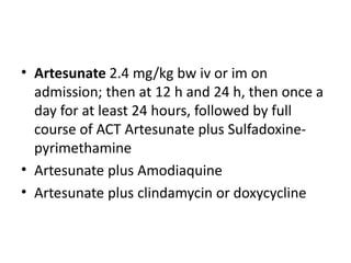
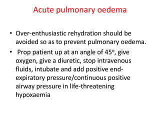
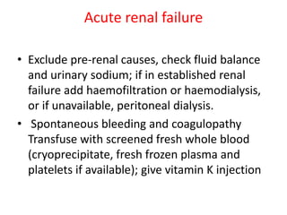
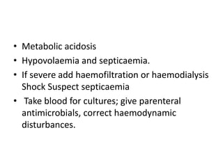
COMPLICATED FALCIPARUM
MALARIA
Introduction
• Malaria is the most important parasitic
disease of man.
• Approximately 5% of the world’s population is
infected, and it causes over 1 million deaths
each year.
• The disease is a protozoan infection of red
blood cells transmitted by the bite of a blood-
feeding female anopheline mosquito.
epidemiology
Mosquito vector
• Malaria is transmitted by some species of
anopheline mosquitoes.
• The optimum conditions for transmission are
high humidity and an ambient temperature
between 20 and 30°C.
LIFE CYCLE
parasite
pathophysiology
• The pathophysiology of malaria results from
destruction of erythrocytes,the liberation of
parasite and erythrocyte material into the
circulation, and the host reaction to these events.
• P. falciparum malaria-infected erythrocytes
sequester in the microcirculation of vital organs,
interfering with microcirculatory flow and host
tissue metabolism.
Toxicity and cytokines
• A glycolipid material with many of the
properties of bacterial endotoxin is released
on meront rupture.
• The products of malaria parasites, and the
crude malaria pigment which is released at
schizont rupture, induce activation of the
cytokine cascade .
• Initially (TNF), (IL)-1, and (γIFN) are produced and
these in turn induce release of ‘pro-infl ammatory’
cytokines including IL-6, IL-8,IL-12, IL-18.
• Cytokines are responsible for many of the
symptoms and signs of the infection, fever and
malaise.
• Cytokines are involved in placental dysfunction,
suppression of erythropoiesis and inhibition of
gluconeogenesis.
• TNF played a causal role in coma and cerebral
dysfunction.
• Cytokines upregulate the endothelial expression of
vascular ligands for P. falciparum-infected
erythrocytes, notably ICAM-1, and thus promote
cytoadherence.
• They may also be important mediators of parasite
killing by activating leukocytes, and , to release toxic
oxygen species, nitric oxide, and by generating
parasiticidal lipid peroxides, and causing fever.
• Thus, whereas high concentrations of cytokines
appear to be harmful, lower levels probably benefit
the host.
Sequestration
• Erythrocytes containing mature forms of P.
falciparum adhere to microvascular endothelium
(‘cytoadherence’) and disappear from the
circulation.
• This process is known as sequestration.
• Sequestration is thought to be central to the
pathophysiology of falciparum malaria.
• Sequestration occurs predominantly in the venules
of vital organs .
• It is not distributed uniformly throughout the body.
• greatest in the brain, particularly the white matter,
prominent in the heart, eyes, liver, kidneys,
intestines and adipose tissue, and least in the skin.
• Cytoadherence and the related phenomena of
rosetting and autoagglutination lead to
microcirculatory obstruction in falciparum malaria.
• The consequences of microcirculatory obstruction
are
• activation of the vascular endothelium
• endothelial dysfunction
• reduced oxygen and substrate supply
• which leads to anaerobic glycolysis, lactic acidosis
and cellular dysfunction
Rosetting
• Erythrocytes containing mature parasites adhere
to uninfected erythrocytes.
• This process leads to the formation of ‘rosettes’
• Rosetting is mediated by attachment of specific
domains of PfEMP1 to the complement receptor
CR1, heparan sulphate,blood group A antigen.
• Rosetting might encourage cytoadherence by
reducing flow (shear rate), which would enhance
anaerobic glycolysis,reduce pH and facilitate
adherence of infected erythrocytes to venular
endothelium.
• Rosetting tends to start in venules, and this could
certainly reduce flow.
• .
Clinical features
The classical attack lasts 6-10 hours.
• a cold stage (sensation of cold, shivering)
• a hot stage (fever, headaches, vomiting;
seizures in young children) , fever up to 104 F
a sweating stage (sweats, return to
normal temperature, tiredness)
Paroxysms coincide synchronus rupture
schizont
• Tertian Malaria , where paroxysms of malaria is
repeated after 48 hrs or fever occurs every third
day. It is feature of P.falciparum, P.ovale and
P.vivax
• Quartan Malaria , where paroxysms occurs after
every 72 hour or fever occurs every fourth day. It
is seen in P.malariae
Renal failure
• The renal injury in severe malaria results from
acute tubular necrosis.
• Acute tubular necrosis results from renal
microvascular obstruction and cellular injury
consequent upon sequestration in the kidney and
the filtration of nephrotoxins such as free
haemoglobin, myoglobin and other cellular
material.
• Massive haemolysis compounds the insult in
blackwater fever complicating malaria, and
haemoglobinuria may itself lead to renal
impairment.
• It always recovers fully in survivors.
• Significant glomerulonephritis is very rare.
Pulmonary edema
• Pulmonary oedema in malaria results from a
sudden increase in pulmonary capillary
permeability. ( Non cardiogenic pulmonary
oedema)
• presence of sequestered PRBC and host
leukocytes in pulmonary capillaries may have a
role in causing pulmonary capillary endothelial
cell dysfunction.
Anaemia
It results from
• the obligatory destruction of red cells containing parasites
at merogony,
• the shortened survival of red cells from which parasites
have been extracted by the spleen, and
• the accelerated destruction of non-parasitized red cells
that parallels disease severity.
• compounded by bone marrow dyserythropoeisis.
• The cause of the dyserythropoiesis is thought to
be related to intramedullary cytokine
production.
• Loss of unparasitized erythrocytes accounts for
approximately 90% of the acute anaemia
resulting from a single uncomplicated infection.
Coagulopathy and thrombocytopenia
In acute malaria, coagulation cascade activity is
accelerated with
• accelerated fibrinogen turnover,
• consumption of antithrombin III,
• reduced factor XIII, and
• increased concentrations of fibrin
degradation products.
• In severe infections the antithrombin
III,protein S and protein C are further reduced
and prothrombin and partial thromboplastin
times may be prolonged.
• Thrombocytopenia is common to all the four
human malarias and is caused by increased
splenic clearance
• Plasma concentrations of macrophage colony
stimulating factor are high, which stimulate
macrophage activity,and may increase platelet
destruction
Black water fever
• Blackwater fever is a condition in which there
is massive intravascular haemolysis and the
passage of ‘Coca-Cola’-coloured urine.
Blackwater (black urine) occurs in three circumstances:
• (1) when patients with G6PD defi ciency take oxidant
drugs (e.g. primaquine,sulphones or sulphonamides)
irrespective of whether they have malaria or not;
• (2) occasionally when patients with G6PD deficiency have
malaria and receive quinine treatment;
• (3) in some patients with severe falciparum malaria who
have normal erythrocyte G6PD levels irrespective of the
treatment given and
Liver dysfunction
• Jaundice is common in adults with severe
malaria.
• Other features are reduced clotting factor
synthesis, reduced metabolic clearance of the
antimalarial drugs, and a failure of
gluconeogenesis which contributes to lactic
acidosis and hypoglycaemia.
• Jaundice in malaria appears to have
haemolytic,hepatic, and cholestatic
components.
• Cholestatic jaundice may persist well into the
recovery period.
• There is no residual liver damage following
malaria
Acidosis
• Acidosis is a major cause of death in severe
falciparum malaria, both in adults and
children.
• Mainly a lactic acidosis, although ketoacidosis
may predominate in children, and the acidosis
of renal failure is common in adults
• Acid-base assessment or venous lactate
concentrations on or 4 h after admission to
hospital are very good indicators of prognosis in
severe malaria.
• Lactic acidosis results from several discrete
processes: the tissue anaerobic glycolysis
consequent upon microvascular obstruction;
• a failure of hepatic and renal lactate clearance;
and the production of lactate by the parasite
Hypoglycemia
Causes:
• an increased peripheral requirement for glucose
consequent upon anaerobic glycolysis (the Pasteur effect),
• the increased metabolic demands of the febrile illness,and
• the obligatory demands of the parasites, which use glucose
as their major fuel ; and
• a failure of hepatic gluconeogenesis and glycogenolysis
(reduced supply).
• In patients treated with quinine, this is
compounded by quinine-stimulated pancreatic β-
cell insulin secretion.
• Hyperinsulinaemia is balanced by a reduced tissue
sensitivity to insulin, which returns to normal as
the patient improves.
• This probably explains why quinine-induced
hypoglycaemia tends to occur after the first24 h of
treatment, whereas malaria-related hypoglycaemia
is present when the patient with severe malaria
first presents.
Placental dysfunction
• Pregnancy increases susceptibility to malaria. This is
caused by a suppression of systemic and placental cell-
mediated immune responses.
• There is intense sequestration of P. falciparum infected
erythrocytes in the placenta, local activation of pro-infl
ammatory cytokine production, and maternal anaemia.
• This leads to cellular infiltration and thickening of the
syncytiotrophoblast and placental insufficiency with
consequent fetal growth retardation.
Bacterial infections
• Patients with severe malaria are vulnerable to
bacterial infections , particularly of the lungs and
urinary tract (following catheterization).
• Postpartum sepsis is also common.
• Spontaneous bacterial septicaemia may also
occur in severe malaria
Relapse
• Both P. vivax and P. ovale have a tendency to
relapse after resolution of the primary infection.
• Relapse, which results from maturation of
persistent hypnozoites in the liver, must be
distinguished from recrudescence of the primary
infection because of incomplete treatment.
• P. falciparum is the usual cause of
recrudescent infections and these tend to
arise 2–4 weeks following treatment.
• Relapses occur weeks or months (or even
years) after the primary infection
• The symptoms of a relapse start more abruptly
than in the primary infection as the infection is
more synchronous.
• They may begin with a sudden chill or rigor.
• Primaquine given for 14 days will eradicate
hypnozoites and prevent relapse in over 80% of
patients
Malaria in pregnancy
• Anaemia and LBW in primigravida.
• If a pregnant woman does develop severe
malaria, fetal loss is common, and the
maternal mortality is very high.
• The mortality of cerebral malaria in pregnancy
is approximately 50%, compared with 15–20%
in non-pregnant adults.
Cerebral malaria
• Unarousable coma (i.e. there is a non-purposeful
response or no response to a painful stimulus) in
falciparum malaria.
• the onset of coma may be sudden, often
following a generalized seizure,
• or gradual, with initial drowsiness, confusion,
disorientation, delirium or agitation, followed by
unconsciousness.
• Extreme agitation is a poor prognostic sign in
falciparum malaria.
• On examination, the patient is febrile and
unrousable.
• There may be some passive resistance to neck fl
exion.
• Sustained hyperventilation is a poor prognostic
sign as it indicates metabolic acidosis if the chest is
clear, or pneumonia or pulmonary oedema if it is
not.
• On examination of the nervous system, the gaze is
usually normal or divergent (but there is no
evidence of extraocular muscle paresis).
• Retinal whitening, retinal haemorrhages, focal
whitening of vessels, papilloedema and cotton wool
spots are seen in fundus.
• Papilloedema is unusual and is a sign of poor
prognosis, as is retinal oedema.
• The haemorrhages are often flame or boat shaped,
and may have a pale centre resembling Roth spots.
• They rarely affect the macula
• Patients may exhibit phasic increases in tone with
extensor posturing of the decorticate (arm flexed,
legs extended), or more usually decerebrate (arms
and legs extended) types.
• The back may arch as in opisthotonus,with
sustained, usually upward and lateral, ocular
deviation.
• The posturing is commonly associated with noisy
hyperventilation.
TREATMENT
• Artesunate 2.4 mg/kg bw iv or im on
admission; then at 12 h and 24 h, then once a
day for at least 24 hours, followed by full
course of ACT Artesunate plus Sulfadoxine-
pyrimethamine
• Artesunate plus Amodiaquine
• Artesunate plus clindamycin or doxycycline
• OR Artemether 3.2 mg/kg bw i.m. given on
admission then 1.6 mg/kg bw per day for at
least 24 hours, followed by full course of ACT
Artemether plus Lumefantrine
Cerebral malaria
• Coma –
• Maintain airway, place patient on his or her
side, exclude other treatable causes of coma
(e.g. hypoglycaemia, bacterial meningitis)
• intubate if necessary
• Hyperpyrexia Administer tepid sponging,
fanning, cooling blanket and antipyretic drugs.
• Convulsions Maintain airways; treat promptly
with intravenous or rectal diazepam.
• Hypoglycaemia (blood glucose concentration
of<2.2 mmol/l; <40 mg/100ml) Check blood
glucose, correct hypoglycaemia and maintain
with glucose-containing infusion
• Severe anaemia (haemoglobin <5 g/100ml or
packed cell volume <15%) Transfuse with
screened fresh whole blood
Acute pulmonary oedema
• Over-enthusiastic rehydration should be
avoided so as to prevent pulmonary oedema.
• Prop patient up at an angle of 45o, give
oxygen, give a diuretic, stop intravenous
fluids, intubate and add positive end-
expiratory pressure/continuous positive
airway pressure in life-threatening
hypoxaemia
Acute renal failure
• Exclude pre-renal causes, check fluid balance
and urinary sodium; if in established renal
failure add haemofiltration or haemodialysis,
or if unavailable, peritoneal dialysis.
• Spontaneous bleeding and coagulopathy
Transfuse with screened fresh whole blood
(cryoprecipitate, fresh frozen plasma and
platelets if available); give vitamin K injection
• Metabolic acidosis
• Hypovolaemia and septicaemia.
• If severe add haemofiltration or haemodialysis
Shock Suspect septicaemia
• Take blood for cultures; give parenteral
antimicrobials, correct haemodynamic
disturbances.
• Quinine 20 mg salt/kg bw on admission (iv
infusion over 4 hrs, then 10 mg/kg bw every 8
h; infusion rate should not exceed 5 mg
salt/kg bw per hour; course for 3 days. 7 days
for malaria acquired in SE Asia[4] Doxycyclined
100mgs BID for 7 days OR Clindamycin 20mg
base/kg/day divided in three doses for 7
days[4] in pregnancy.
• Blood transfusion:
• In high transmission settings, blood transfusion is recommended for
children with a haemoglobin level of <5 g/100 ml (haematocrit
<15%).
• In low-transmission settings, a threshold of 20% (haemoglobin 7
g/100ml) is recommended.
• These general recommendations still need to be tailored to the
individual, as the pathological consequences of rapid development
of anaemia.
THANK YOU

Malaria part 1

  • 1.
  • 2.
    Introduction • Malaria isthe most important parasitic disease of man. • Approximately 5% of the world’s population is infected, and it causes over 1 million deaths each year. • The disease is a protozoan infection of red blood cells transmitted by the bite of a blood- feeding female anopheline mosquito.
  • 3.
  • 6.
    Mosquito vector • Malariais transmitted by some species of anopheline mosquitoes. • The optimum conditions for transmission are high humidity and an ambient temperature between 20 and 30°C.
  • 7.
  • 8.
  • 10.
    pathophysiology • The pathophysiologyof malaria results from destruction of erythrocytes,the liberation of parasite and erythrocyte material into the circulation, and the host reaction to these events. • P. falciparum malaria-infected erythrocytes sequester in the microcirculation of vital organs, interfering with microcirculatory flow and host tissue metabolism.
  • 11.
    Toxicity and cytokines •A glycolipid material with many of the properties of bacterial endotoxin is released on meront rupture. • The products of malaria parasites, and the crude malaria pigment which is released at schizont rupture, induce activation of the cytokine cascade .
  • 12.
    • Initially (TNF),(IL)-1, and (γIFN) are produced and these in turn induce release of ‘pro-infl ammatory’ cytokines including IL-6, IL-8,IL-12, IL-18. • Cytokines are responsible for many of the symptoms and signs of the infection, fever and malaise. • Cytokines are involved in placental dysfunction, suppression of erythropoiesis and inhibition of gluconeogenesis. • TNF played a causal role in coma and cerebral dysfunction.
  • 13.
    • Cytokines upregulatethe endothelial expression of vascular ligands for P. falciparum-infected erythrocytes, notably ICAM-1, and thus promote cytoadherence. • They may also be important mediators of parasite killing by activating leukocytes, and , to release toxic oxygen species, nitric oxide, and by generating parasiticidal lipid peroxides, and causing fever. • Thus, whereas high concentrations of cytokines appear to be harmful, lower levels probably benefit the host.
  • 14.
    Sequestration • Erythrocytes containingmature forms of P. falciparum adhere to microvascular endothelium (‘cytoadherence’) and disappear from the circulation. • This process is known as sequestration. • Sequestration is thought to be central to the pathophysiology of falciparum malaria.
  • 16.
    • Sequestration occurspredominantly in the venules of vital organs . • It is not distributed uniformly throughout the body. • greatest in the brain, particularly the white matter, prominent in the heart, eyes, liver, kidneys, intestines and adipose tissue, and least in the skin.
  • 17.
    • Cytoadherence andthe related phenomena of rosetting and autoagglutination lead to microcirculatory obstruction in falciparum malaria. • The consequences of microcirculatory obstruction are • activation of the vascular endothelium • endothelial dysfunction • reduced oxygen and substrate supply • which leads to anaerobic glycolysis, lactic acidosis and cellular dysfunction
  • 18.
    Rosetting • Erythrocytes containingmature parasites adhere to uninfected erythrocytes. • This process leads to the formation of ‘rosettes’ • Rosetting is mediated by attachment of specific domains of PfEMP1 to the complement receptor CR1, heparan sulphate,blood group A antigen.
  • 20.
    • Rosetting mightencourage cytoadherence by reducing flow (shear rate), which would enhance anaerobic glycolysis,reduce pH and facilitate adherence of infected erythrocytes to venular endothelium. • Rosetting tends to start in venules, and this could certainly reduce flow. • .
  • 21.
    Clinical features The classicalattack lasts 6-10 hours. • a cold stage (sensation of cold, shivering) • a hot stage (fever, headaches, vomiting; seizures in young children) , fever up to 104 F a sweating stage (sweats, return to normal temperature, tiredness) Paroxysms coincide synchronus rupture schizont
  • 22.
    • Tertian Malaria, where paroxysms of malaria is repeated after 48 hrs or fever occurs every third day. It is feature of P.falciparum, P.ovale and P.vivax • Quartan Malaria , where paroxysms occurs after every 72 hour or fever occurs every fourth day. It is seen in P.malariae
  • 23.
    Renal failure • Therenal injury in severe malaria results from acute tubular necrosis. • Acute tubular necrosis results from renal microvascular obstruction and cellular injury consequent upon sequestration in the kidney and the filtration of nephrotoxins such as free haemoglobin, myoglobin and other cellular material.
  • 24.
    • Massive haemolysiscompounds the insult in blackwater fever complicating malaria, and haemoglobinuria may itself lead to renal impairment. • It always recovers fully in survivors. • Significant glomerulonephritis is very rare.
  • 25.
    Pulmonary edema • Pulmonaryoedema in malaria results from a sudden increase in pulmonary capillary permeability. ( Non cardiogenic pulmonary oedema) • presence of sequestered PRBC and host leukocytes in pulmonary capillaries may have a role in causing pulmonary capillary endothelial cell dysfunction.
  • 26.
    Anaemia It results from •the obligatory destruction of red cells containing parasites at merogony, • the shortened survival of red cells from which parasites have been extracted by the spleen, and • the accelerated destruction of non-parasitized red cells that parallels disease severity. • compounded by bone marrow dyserythropoeisis.
  • 27.
    • The causeof the dyserythropoiesis is thought to be related to intramedullary cytokine production. • Loss of unparasitized erythrocytes accounts for approximately 90% of the acute anaemia resulting from a single uncomplicated infection.
  • 28.
    Coagulopathy and thrombocytopenia Inacute malaria, coagulation cascade activity is accelerated with • accelerated fibrinogen turnover, • consumption of antithrombin III, • reduced factor XIII, and • increased concentrations of fibrin degradation products.
  • 29.
    • In severeinfections the antithrombin III,protein S and protein C are further reduced and prothrombin and partial thromboplastin times may be prolonged. • Thrombocytopenia is common to all the four human malarias and is caused by increased splenic clearance
  • 30.
    • Plasma concentrationsof macrophage colony stimulating factor are high, which stimulate macrophage activity,and may increase platelet destruction
  • 31.
    Black water fever •Blackwater fever is a condition in which there is massive intravascular haemolysis and the passage of ‘Coca-Cola’-coloured urine.
  • 32.
    Blackwater (black urine)occurs in three circumstances: • (1) when patients with G6PD defi ciency take oxidant drugs (e.g. primaquine,sulphones or sulphonamides) irrespective of whether they have malaria or not; • (2) occasionally when patients with G6PD deficiency have malaria and receive quinine treatment; • (3) in some patients with severe falciparum malaria who have normal erythrocyte G6PD levels irrespective of the treatment given and
  • 33.
    Liver dysfunction • Jaundiceis common in adults with severe malaria. • Other features are reduced clotting factor synthesis, reduced metabolic clearance of the antimalarial drugs, and a failure of gluconeogenesis which contributes to lactic acidosis and hypoglycaemia.
  • 34.
    • Jaundice inmalaria appears to have haemolytic,hepatic, and cholestatic components. • Cholestatic jaundice may persist well into the recovery period. • There is no residual liver damage following malaria
  • 35.
    Acidosis • Acidosis isa major cause of death in severe falciparum malaria, both in adults and children. • Mainly a lactic acidosis, although ketoacidosis may predominate in children, and the acidosis of renal failure is common in adults
  • 36.
    • Acid-base assessmentor venous lactate concentrations on or 4 h after admission to hospital are very good indicators of prognosis in severe malaria. • Lactic acidosis results from several discrete processes: the tissue anaerobic glycolysis consequent upon microvascular obstruction; • a failure of hepatic and renal lactate clearance; and the production of lactate by the parasite
  • 37.
    Hypoglycemia Causes: • an increasedperipheral requirement for glucose consequent upon anaerobic glycolysis (the Pasteur effect), • the increased metabolic demands of the febrile illness,and • the obligatory demands of the parasites, which use glucose as their major fuel ; and • a failure of hepatic gluconeogenesis and glycogenolysis (reduced supply).
  • 38.
    • In patientstreated with quinine, this is compounded by quinine-stimulated pancreatic β- cell insulin secretion. • Hyperinsulinaemia is balanced by a reduced tissue sensitivity to insulin, which returns to normal as the patient improves. • This probably explains why quinine-induced hypoglycaemia tends to occur after the first24 h of treatment, whereas malaria-related hypoglycaemia is present when the patient with severe malaria first presents.
  • 39.
    Placental dysfunction • Pregnancyincreases susceptibility to malaria. This is caused by a suppression of systemic and placental cell- mediated immune responses. • There is intense sequestration of P. falciparum infected erythrocytes in the placenta, local activation of pro-infl ammatory cytokine production, and maternal anaemia. • This leads to cellular infiltration and thickening of the syncytiotrophoblast and placental insufficiency with consequent fetal growth retardation.
  • 40.
    Bacterial infections • Patientswith severe malaria are vulnerable to bacterial infections , particularly of the lungs and urinary tract (following catheterization). • Postpartum sepsis is also common. • Spontaneous bacterial septicaemia may also occur in severe malaria
  • 41.
    Relapse • Both P.vivax and P. ovale have a tendency to relapse after resolution of the primary infection. • Relapse, which results from maturation of persistent hypnozoites in the liver, must be distinguished from recrudescence of the primary infection because of incomplete treatment.
  • 42.
    • P. falciparumis the usual cause of recrudescent infections and these tend to arise 2–4 weeks following treatment. • Relapses occur weeks or months (or even years) after the primary infection
  • 43.
    • The symptomsof a relapse start more abruptly than in the primary infection as the infection is more synchronous. • They may begin with a sudden chill or rigor. • Primaquine given for 14 days will eradicate hypnozoites and prevent relapse in over 80% of patients
  • 44.
    Malaria in pregnancy •Anaemia and LBW in primigravida. • If a pregnant woman does develop severe malaria, fetal loss is common, and the maternal mortality is very high. • The mortality of cerebral malaria in pregnancy is approximately 50%, compared with 15–20% in non-pregnant adults.
  • 45.
    Cerebral malaria • Unarousablecoma (i.e. there is a non-purposeful response or no response to a painful stimulus) in falciparum malaria. • the onset of coma may be sudden, often following a generalized seizure, • or gradual, with initial drowsiness, confusion, disorientation, delirium or agitation, followed by unconsciousness.
  • 46.
    • Extreme agitationis a poor prognostic sign in falciparum malaria. • On examination, the patient is febrile and unrousable. • There may be some passive resistance to neck fl exion. • Sustained hyperventilation is a poor prognostic sign as it indicates metabolic acidosis if the chest is clear, or pneumonia or pulmonary oedema if it is not.
  • 47.
    • On examinationof the nervous system, the gaze is usually normal or divergent (but there is no evidence of extraocular muscle paresis). • Retinal whitening, retinal haemorrhages, focal whitening of vessels, papilloedema and cotton wool spots are seen in fundus. • Papilloedema is unusual and is a sign of poor prognosis, as is retinal oedema. • The haemorrhages are often flame or boat shaped, and may have a pale centre resembling Roth spots. • They rarely affect the macula
  • 48.
    • Patients mayexhibit phasic increases in tone with extensor posturing of the decorticate (arm flexed, legs extended), or more usually decerebrate (arms and legs extended) types. • The back may arch as in opisthotonus,with sustained, usually upward and lateral, ocular deviation. • The posturing is commonly associated with noisy hyperventilation.
  • 49.
  • 50.
    • Artesunate 2.4mg/kg bw iv or im on admission; then at 12 h and 24 h, then once a day for at least 24 hours, followed by full course of ACT Artesunate plus Sulfadoxine- pyrimethamine • Artesunate plus Amodiaquine • Artesunate plus clindamycin or doxycycline
  • 51.
    • OR Artemether3.2 mg/kg bw i.m. given on admission then 1.6 mg/kg bw per day for at least 24 hours, followed by full course of ACT Artemether plus Lumefantrine
  • 52.
    Cerebral malaria • Coma– • Maintain airway, place patient on his or her side, exclude other treatable causes of coma (e.g. hypoglycaemia, bacterial meningitis) • intubate if necessary
  • 53.
    • Hyperpyrexia Administertepid sponging, fanning, cooling blanket and antipyretic drugs. • Convulsions Maintain airways; treat promptly with intravenous or rectal diazepam.
  • 54.
    • Hypoglycaemia (bloodglucose concentration of<2.2 mmol/l; <40 mg/100ml) Check blood glucose, correct hypoglycaemia and maintain with glucose-containing infusion • Severe anaemia (haemoglobin <5 g/100ml or packed cell volume <15%) Transfuse with screened fresh whole blood
  • 55.
    Acute pulmonary oedema •Over-enthusiastic rehydration should be avoided so as to prevent pulmonary oedema. • Prop patient up at an angle of 45o, give oxygen, give a diuretic, stop intravenous fluids, intubate and add positive end- expiratory pressure/continuous positive airway pressure in life-threatening hypoxaemia
  • 56.
    Acute renal failure •Exclude pre-renal causes, check fluid balance and urinary sodium; if in established renal failure add haemofiltration or haemodialysis, or if unavailable, peritoneal dialysis. • Spontaneous bleeding and coagulopathy Transfuse with screened fresh whole blood (cryoprecipitate, fresh frozen plasma and platelets if available); give vitamin K injection
  • 57.
    • Metabolic acidosis •Hypovolaemia and septicaemia. • If severe add haemofiltration or haemodialysis Shock Suspect septicaemia • Take blood for cultures; give parenteral antimicrobials, correct haemodynamic disturbances.
  • 58.
    • Quinine 20mg salt/kg bw on admission (iv infusion over 4 hrs, then 10 mg/kg bw every 8 h; infusion rate should not exceed 5 mg salt/kg bw per hour; course for 3 days. 7 days for malaria acquired in SE Asia[4] Doxycyclined 100mgs BID for 7 days OR Clindamycin 20mg base/kg/day divided in three doses for 7 days[4] in pregnancy.
  • 59.
    • Blood transfusion: •In high transmission settings, blood transfusion is recommended for children with a haemoglobin level of <5 g/100 ml (haematocrit <15%). • In low-transmission settings, a threshold of 20% (haemoglobin 7 g/100ml) is recommended. • These general recommendations still need to be tailored to the individual, as the pathological consequences of rapid development of anaemia.
  • 60.