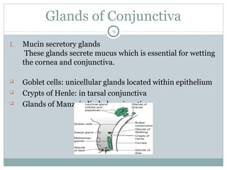
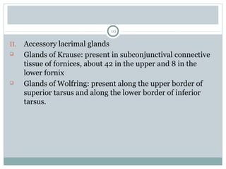
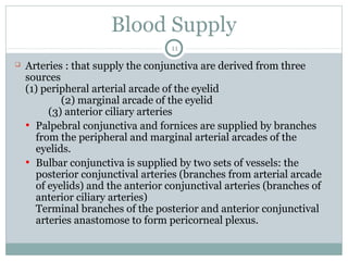
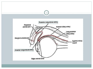
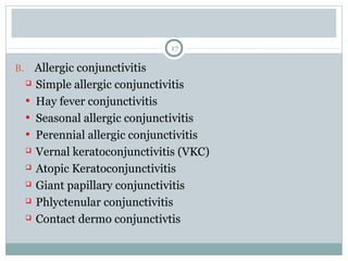
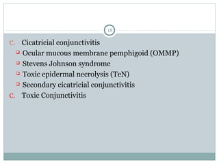
Anatomy of Conjunctiva
& Classification of
Conjunctivitis
1
Contents
I. Anatomy of conjunctiva
 Parts of conjunctiva
 Structure of conjunctiva
 Glands of conjunctiva
 Blood supply
 Nerve supply
I. Types of conjunctivitis
2
Parts of Conjunctiva
I. Palpebral Conjunctiva
 Marginal Conjunctiva extends from
the lid margin to about 2mm on the
back of the lid up to a shallow
groove, the sulcus subtarsalis.
 Tarsal Conjunctiva
• Thin, transparent and highly
vascular.
• In the upper lid – firmly adherent
to whole tarsal plate.
• In the lower lid – adherent only to
half width of tarsus.
• Tarsal glands seen through it as
yellow streaks.
3
 Orbital part, lies loose between the tarsal plate and fornix.
II.Bulbar Conjunctiva
 Thin, transparent
 Separated from the anterior sclera by episcleral tissue and
tenon’s capsule
 3mm ridge around cornea – Limbal Conjunctiva
 In the area of the limbus, the conjunctiva, tenon’s capsule
and episcleral tissue are fused into a dense tissue, adherent
to corneoscleral junction
4
III. Conjunctival Fornix
 Joins the bulbar conjunctiva with the palpebral
conjunctiva
 Subdivided into superior, inferior, medial and lateral
fornices.
 Plica Semilunaris : Pinkish crescentic fold of conjunctiva, persent in
the medial canthus. It is a vestigeal structure in humans and
represents the nictitating membrane of lower animals.
 Caruncle : Small, ovoid, pinkish mass, in the inner canthus, just
medial to the plica semilunaris. It is a piece of modified skin and
hence is covered with stratified squamous epithelium, sebacious
glands, etc.
5
Structure of Conjunctiva
I. Epithelium
 2-5 layered, non-keratinized epithelium. Also contains
goblet cells.
 The layer of epithelial cells varies from region to region
• Marginal conjunctiva has 5 layered stratified squamous
type epithelium.
• Tarsal conjunctiva has 2 layered epithelium: superficial
cylinderical and deep cuboidal cells.
• Fornix & bulbar conjunctiva have 3
layered epithelium: superficial
cylinderical, middle polyhedral and
deep cuboidal cells.
6
7
• Limbal Conjunctiva has again many layered (5 to 6) stratified
squamous epithelium.
II. Adenoid Layer
 Also called lymphoid layer
 Consists of fine connective tissue reticulum in the meshes
of which lie lymphocytes.
 It is not present since birth but develops after 3-4 months
of life. For this reason conjunctival inflammation in an
infant does not produce follicular reaction.
8
III. Fibrous Layer
 Consists of a meshwork of collagenous and elastic fibres.
 This layer contains vessels and nerves of conjunctiva
 It blends with the underlying Tenon’s capsule in the
region of the bulbar conjunctiva
Glands of Conjunctiva
9
I. Mucin secretory glands
These glands secrete mucus which is essential for wetting
the cornea and conjunctiva.
 Goblet cells: unicellular glands located within epithelium
 Crypts of Henle: in tarsal conjunctiva
 Glands of Manz: in limbal conjunctiva
10
II. Accessory lacrimal glands
 Glands of Krause: present in subconjunctival connective
tissue of fornices, about 42 in the upper and 8 in the
lower fornix
 Glands of Wolfring: present along the upper border of
superior tarsus and along the lower border of inferior
tarsus.
Blood Supply
11
 Arteries : that supply the conjunctiva are derived from three
sources
(1) peripheral arterial arcade of the eyelid
(2) marginal arcade of the eyelid
(3) anterior ciliary arteries
• Palpebral conjunctiva and fornices are supplied by branches
from the peripheral and marginal arterial arcades of the
eyelids.
• Bulbar conjunctiva is supplied by two sets of vessels: the
posterior conjunctival arteries (branches from arterial arcade
of eyelids) and the anterior conjunctival arteries (branches of
anterior ciliary arteries)
Terminal branches of the posterior and anterior conjunctival
arteries anastomose to form pericorneal plexus.
12
13
 Veins : from the conjunctiva drain into the venous plexus of
eyelids and some around the cornea into the anterior ciliary
veins.
 Lymphatics : are arranged in two layers – superficial and
deep
Lymphatics from the lateral side drain into pre auricular
lymph nodes and those from medial side into the
submandibular lymph nodes.
Nerve Supply
14
 A circumcorneal zone of conjunctiva is supplied by
branches from long ciliary nerves, which supply the cornea.
 Rest of the conjunctiva is supplied by the branches from
lacrimal, infratrochlear, supratrochlear, supraorbital and
frontal nerves.
Types of Conjunctivitis
15
 Inflammations of the conjunctiva (conjunctivitis) is defined
as conjunctival hyperaemia, associated with a discharge,
which maybe watery, mucoid, mucopurulent or purulent.
 Types: Common types include
A. Infective Conjunctivitis
 Bacterial Conjunctivitis
• Acute bacterial conjunctivitis
• Hyperacute bacterial conjunctivitis
• Chronic bacterial conjunctivitis
• Angular bacterial conjunctivitis
16
 Chlamydial Conjunctivitis
• Trachoma
• Adult inclusion conjunctivitis
• Neonatal chlamydial conjunctivitis
 Viral Conjunctivitis
• Adenovirus conjunctivitis
• Enterovirus conjunctivitis
• Molluscum contagiosum conjunctivitis
• Herpes Simplex conjunctivitis
 Ophthalmia neonatorum
 Granulomatous conjunctivitis
17
B. Allergic conjunctivitis
 Simple allergic conjunctivitis
• Hay fever conjunctivitis
• Seasonal allergic conjunctivitis
• Perennial allergic conjunctivitis
 Vernal keratoconjunctivitis (VKC)
 Atopic Keratoconjunctivitis
 Giant papillary conjunctivitis
 Phlyctenular conjunctivitis
 Contact dermo conjunctivtis
18
C. Cicatricial conjunctivitis
 Ocular mucous membrane pemphigoid (OMMP)
 Stevens Johnson syndrome
 Toxic epidermal necrolysis (TeN)
 Secondary cicatricial conjunctivitis
C. Toxic Conjunctivitis
Thank You
19

Conjuctiva

  • 1.
    Anatomy of Conjunctiva &Classification of Conjunctivitis 1
  • 2.
    Contents I. Anatomy ofconjunctiva  Parts of conjunctiva  Structure of conjunctiva  Glands of conjunctiva  Blood supply  Nerve supply I. Types of conjunctivitis 2
  • 3.
    Parts of Conjunctiva I.Palpebral Conjunctiva  Marginal Conjunctiva extends from the lid margin to about 2mm on the back of the lid up to a shallow groove, the sulcus subtarsalis.  Tarsal Conjunctiva • Thin, transparent and highly vascular. • In the upper lid – firmly adherent to whole tarsal plate. • In the lower lid – adherent only to half width of tarsus. • Tarsal glands seen through it as yellow streaks. 3
  • 4.
     Orbital part,lies loose between the tarsal plate and fornix. II.Bulbar Conjunctiva  Thin, transparent  Separated from the anterior sclera by episcleral tissue and tenon’s capsule  3mm ridge around cornea – Limbal Conjunctiva  In the area of the limbus, the conjunctiva, tenon’s capsule and episcleral tissue are fused into a dense tissue, adherent to corneoscleral junction 4
  • 5.
    III. Conjunctival Fornix Joins the bulbar conjunctiva with the palpebral conjunctiva  Subdivided into superior, inferior, medial and lateral fornices.  Plica Semilunaris : Pinkish crescentic fold of conjunctiva, persent in the medial canthus. It is a vestigeal structure in humans and represents the nictitating membrane of lower animals.  Caruncle : Small, ovoid, pinkish mass, in the inner canthus, just medial to the plica semilunaris. It is a piece of modified skin and hence is covered with stratified squamous epithelium, sebacious glands, etc. 5
  • 6.
    Structure of Conjunctiva I.Epithelium  2-5 layered, non-keratinized epithelium. Also contains goblet cells.  The layer of epithelial cells varies from region to region • Marginal conjunctiva has 5 layered stratified squamous type epithelium. • Tarsal conjunctiva has 2 layered epithelium: superficial cylinderical and deep cuboidal cells. • Fornix & bulbar conjunctiva have 3 layered epithelium: superficial cylinderical, middle polyhedral and deep cuboidal cells. 6
  • 7.
    7 • Limbal Conjunctivahas again many layered (5 to 6) stratified squamous epithelium. II. Adenoid Layer  Also called lymphoid layer  Consists of fine connective tissue reticulum in the meshes of which lie lymphocytes.  It is not present since birth but develops after 3-4 months of life. For this reason conjunctival inflammation in an infant does not produce follicular reaction.
  • 8.
    8 III. Fibrous Layer Consists of a meshwork of collagenous and elastic fibres.  This layer contains vessels and nerves of conjunctiva  It blends with the underlying Tenon’s capsule in the region of the bulbar conjunctiva
  • 9.
    Glands of Conjunctiva 9 I.Mucin secretory glands These glands secrete mucus which is essential for wetting the cornea and conjunctiva.  Goblet cells: unicellular glands located within epithelium  Crypts of Henle: in tarsal conjunctiva  Glands of Manz: in limbal conjunctiva
  • 10.
    10 II. Accessory lacrimalglands  Glands of Krause: present in subconjunctival connective tissue of fornices, about 42 in the upper and 8 in the lower fornix  Glands of Wolfring: present along the upper border of superior tarsus and along the lower border of inferior tarsus.
  • 11.
    Blood Supply 11  Arteries: that supply the conjunctiva are derived from three sources (1) peripheral arterial arcade of the eyelid (2) marginal arcade of the eyelid (3) anterior ciliary arteries • Palpebral conjunctiva and fornices are supplied by branches from the peripheral and marginal arterial arcades of the eyelids. • Bulbar conjunctiva is supplied by two sets of vessels: the posterior conjunctival arteries (branches from arterial arcade of eyelids) and the anterior conjunctival arteries (branches of anterior ciliary arteries) Terminal branches of the posterior and anterior conjunctival arteries anastomose to form pericorneal plexus.
  • 12.
  • 13.
    13  Veins :from the conjunctiva drain into the venous plexus of eyelids and some around the cornea into the anterior ciliary veins.  Lymphatics : are arranged in two layers – superficial and deep Lymphatics from the lateral side drain into pre auricular lymph nodes and those from medial side into the submandibular lymph nodes.
  • 14.
    Nerve Supply 14  Acircumcorneal zone of conjunctiva is supplied by branches from long ciliary nerves, which supply the cornea.  Rest of the conjunctiva is supplied by the branches from lacrimal, infratrochlear, supratrochlear, supraorbital and frontal nerves.
  • 15.
    Types of Conjunctivitis 15 Inflammations of the conjunctiva (conjunctivitis) is defined as conjunctival hyperaemia, associated with a discharge, which maybe watery, mucoid, mucopurulent or purulent.  Types: Common types include A. Infective Conjunctivitis  Bacterial Conjunctivitis • Acute bacterial conjunctivitis • Hyperacute bacterial conjunctivitis • Chronic bacterial conjunctivitis • Angular bacterial conjunctivitis
  • 16.
    16  Chlamydial Conjunctivitis •Trachoma • Adult inclusion conjunctivitis • Neonatal chlamydial conjunctivitis  Viral Conjunctivitis • Adenovirus conjunctivitis • Enterovirus conjunctivitis • Molluscum contagiosum conjunctivitis • Herpes Simplex conjunctivitis  Ophthalmia neonatorum  Granulomatous conjunctivitis
  • 17.
    17 B. Allergic conjunctivitis Simple allergic conjunctivitis • Hay fever conjunctivitis • Seasonal allergic conjunctivitis • Perennial allergic conjunctivitis  Vernal keratoconjunctivitis (VKC)  Atopic Keratoconjunctivitis  Giant papillary conjunctivitis  Phlyctenular conjunctivitis  Contact dermo conjunctivtis
  • 18.
    18 C. Cicatricial conjunctivitis Ocular mucous membrane pemphigoid (OMMP)  Stevens Johnson syndrome  Toxic epidermal necrolysis (TeN)  Secondary cicatricial conjunctivitis C. Toxic Conjunctivitis
  • 19.