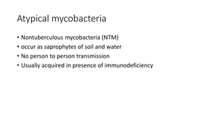
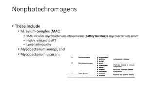
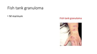
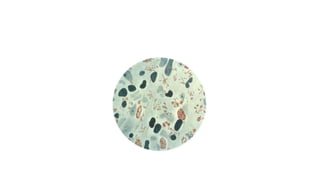
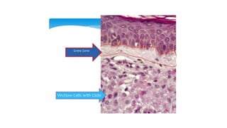
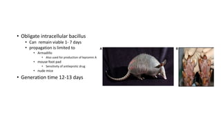
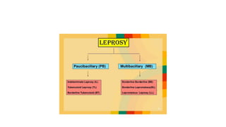
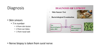
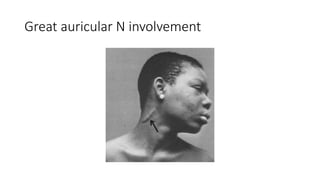
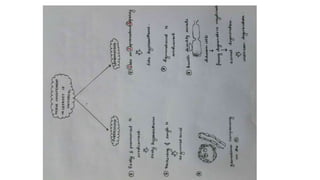
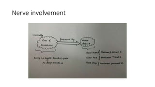
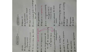
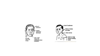
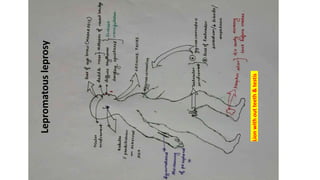
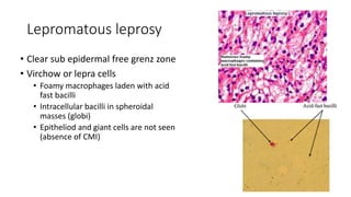
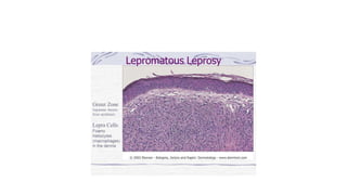
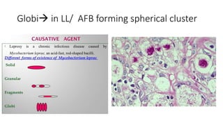
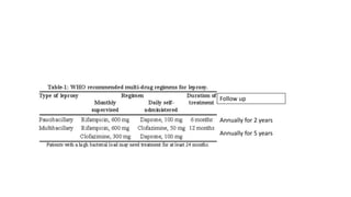
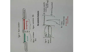
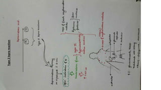
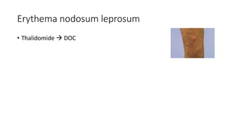
Mycobacteria
Atypical mycobacteria
• Nontuberculous mycobacteria (NTM)
• occur as saprophytes of soil and water
• No person to person transmission
• Usually acquired in presence of immunodeficiency
Runyons classification
Photochromogens
• M. kansasii
• Pulmonary disease
• Mycobacterium marinum,
• Swimming pool granuloma
• Fish tank granuloma
• development of superficial granulomatous lesions in the skin
• Mycobacterium simiae
Scotochromogens
• Mycobacterium scrofulaceum,
• lymphadenopathy
• Mycobacterium gordonae, and
• Mycobacterium szulga
Nonphotochromogens
• These include
• M. avium complex (MAC)
• MAC includes mycobacterium intracellulare (battey bacillus)& mycobacterium avium
• Highly resistant to ATT
• Lymphadenopathy
• Mycobacterium xenopi, and
• Mycobacterium ulcerans
Rapid Growers
• Among this group itself involve
• photochromogens,
• M phlei M smegmatis
• scotochromogens, or
• nonphotochromogens species
• M fortuitum
• M chelonie
Post
trauma/injection
abscess
M ulcerans
•  burulis disease
• Only mycobacteria producing toxin
• Saprophytic mycobacteria are non pathogenic
• M phlei
• M smegmatis
• M gordonae
• M paratuberculosis
Pulmonary disease
• MAC is the commonest cause of pulmonary disease d/t non
tuberculous mycobacteria in developed countries
• M kansasii produces pulmonary ds indistinguishable from TB
Fish tank granuloma
• M marinum
Disseminated ds
• In AIDS related
• Caused by MAC
Mycobacterium leprae
• Acid fast (5 % H2SO4)
• Less acid fast than M tuberculosis (20 % H2S04)
M leprae
• Less acid fast than M tuberculae (hence 5 % H2SO4 or 1% HCl is used)
• First bacillus to be associated with human disease
• Modified fite stain
• In tissue sections
• Phenolic glycolipoprotein 1 plays
important role in pathogenesis
• LAM B1 external to cell wall in
serodiagnosis
• Lepra bacilli has affinity for RE cells & schwann cells
• Prefer cooler areas of body
• Skin / peripheral N / anterior chamber of eye /URT/ testis
• sparing warmer areas
• Axila groin scalp midline of back
• Schwaan cells are first cell to be involved in leprosy
• Virchows cells are
undifferentiated macrophages
• Abundant in lepromatous
leprosy
• Obligate intracellular bacillus
• Can remain viable 1- 7 days
• propagation is limited to
• Armadillo
• Also used for production of lepromin A
• mouse foot pad
• Sensitivity of antileprotic drug
• nude mice
• Generation time 12-13 days
Cardinal signs of leprosy
• Hypopigmented or erythematous skin lesions with definite loss of
sensation
• Involvement of peripheral nerves  definite thickening
• Skin smear  AFB
• Highly infectious with low pathogenicity
• Attack rate among house hold contacts is 4.4 -12 %
• Clinical infection Source of infection
• Active leprosy
• Multibacillary >>sub
• Portal of entry  skin and respiratory tract
• Chief mode of exit  nasal mucosa of untreated LL patients
• Modes of transmiision
• Droplets >>contact>breast milk>transplacental>insect vectors
• The Ridley Jopling classification
• Indeterminate
• Tuberculoid
• Borderline:
• borderline-tuberculoid,
• borderline-borderline,
• borderline-lepromatous
• Lepromatous
Indian classification (by indian leprosy
association)
• In TT is the most common form of leprosy
WHO Classification  treatment
Paucibacillary
• 1-5 lesion
• No nerve or only one nerve
• Skin smears –ve at all sites
• BI <2
• Involves IL BT TT
Multibacillary
• > 6 lesions
• > 1 nerve irrespective of skin lesions
• Skin smears + at all sites
• BI > 2
• Involves BB BL LL
Diagnosis
• Skin smears
• 7 in number
• 4 from skin lesion
• 2 from ear lobes
• 1 from nasal swb
• Nerve biopsy is taken from sural nerve
Morphological index response to Rx/drug
resistance/ disease activity/
• Percentage of solid staining bacilli in a stained smear to total no of
bacilli
Live bacilli  solid stained
Dead bacilli Fragmented
Bacteriological index  density of bacilli in
stained smear
• Bacilli are absent in lungs , ovaries and CNS
• Most common cranial nerve to get involve is VII
• Goll & burdech tract  proprioception
• Not involved in leprosy
• Other organs involved in leprosy are
• Eyes
• Iritis
• Corneal beading
• Testes
• Muscles
• Myopathy , wasting
Mouse foot pad culture
• Detect drug resistance
• Evaluating potency of antileprosy treatment
• Detecting viability of bacilli during Rx
Histamine test
• Very reliable method to detect at an early stage
• Peripheral N damage d/t leprosy  flare response is lost if peripheral N is
damaged
Lepromin test  to detect CMI / Test for prognosis
Early or Fernandez reaction
• after 48 hours of injection
• Erythema & induration >10 mm
is +ve
• Delayed hypersensitivity
reaction to soluble constituents
Late or Mitsuda reaction
• After 3 weeks
• Nodule > 5 mm is +ve
• indicates CMI to bacillary
component of Ag of lepra bacilli
Mitsuda lepromin / Dharmendra lepromin antigen intradermally
BCG vaccination can covert it from
negative to positive
Uses of lepromin test
• To classify lesions into tuberculoid or lepromatous
• To assess prognosis
• If positive  good prognosis
• Not used for diagnosis
• In the first 6 mth of life most children are lepromin negative
Nerve commonly involved
• Ulnar N  elbow (posteriorly)
• Median N  at wrist
• Dorsal cutaneous N of ulnar @ wrist
• Common peroneal  around fibular neck
• Superficial peroneal in front of ankle
• Posterior tibial  below medial malleoli
Great auricular N involvement
Nerve involvement
Types
Indeterminate leprosy
• Bacteriologically negative
• One or two vague hypopigmented macules
• Definite sensory impairement
Tuberculoid leprosy (most common type of
leprosy in INDIA and Africa
• CMI ↑↑
• Lepromin +ve
• Bacillary load ↓↓
• Skin lesions anaesthetic early
• One or few sharply defined
annular asymmetric macules or
plaques with a tendency towards
central clearing , elevated border
Tuberculoid leprosy histopathology
• Noncaseating granuloma with
epitheliod and giant cells
Grenz zone is involved in TL
Borderline Tuberculoid
• SATellite lesions
• Nerve Abscess (mainly ulnar nerve abscess)
• Rx with I & D
• SANTA BanTa
SATellite lesions
Borderline leprosy (BL)
• Inverted saucer shaped ulcers
• All kinds of bizzare lesions in single
patient distributed asymmetrically
• Epitheliod cells +
Lepromatous leprosy
• CMI ↓↓
• lepromin test negative
• Bacillary load ↑↑
• Temperature and pain lost first >>
Hypoaesthesia is a late sign
• Ulnar N most common >>low median
>posterior tibial>
• Radial N is involved last  serious
• Acral,distal,symmetric anaesthesia
common
• M.leprae PGL-1(phenolic glycolipid)
ab +
Lion with out teeth
• Leonine facies
• Atrophy of anterior nasal spine  collapse of nose
• Alveolar resorption Loosening of teeth
• Diffuse dermal infiltration of face
• Loss of eye brow
Lepromatousleprosy
Lionwithoutteeth&testis
Lepromatous leprosy
• Clear sub epidermal free grenz zone
• Virchow or lepra cells
• Foamy macrophages laden with acid
fast bacilli
• Intracellular bacilli in spheroidal
masses (globi)
• Epitheliod and giant cells are not seen
(absence of CMI)
Globi in LL/ AFB forming spherical cluster
Lucio phenomenon
• On lower extremities
• High parasitism in endothelial
cell  endothelial proliferation
 thrombus formation
• Characterised by arteritis and
ulcers on legs is limited to
patients with diffuse non
nodular lepromatous leprosy
• Untreated patients
• Mexico and carribean only
Histoid leprosy
• Type of LL
• Shiny cutaneous and subcutaneous
nodules
• Caused by drug resistant lepra bacilli
• In b/w skin appears normal
• Pure neuritic (indian classification)
• Asymmetric nerve involvement with no skin lesion and usually of tuberculoid
origin
• Painless trophic ulcers
Treatment
• Chaulmoogra oil
• First effective antileprosy Rx
• Form Myanmar
• MDT is used since 1982
• Not C/I in HIV infected patients and is safe in pregnancy
• Rifampicin  highly bactericidal and most effective against M leprae
• Dapsone is bacteriostatic
Blister packets for MDT
• Paucibacillary
• Under surveillance for
atleast 2years
• Multibacillary
• Under surveillance for
5vyeras
Treatment of single lesion leprosy
• ROM regimen
• Rmp
• Ofloxacin
• Minocycline
• ROM -6 (Monthly for 6 months) - Paucibacillary
• ROM -12 (Monthly for 12 months) – Multibacillary
Follow up
Annually for 2 years
Annually for 5 years
• Rifampicin is the most active (bactericidal & rapidly acting) for
leprosy
• In pure neuritic type
• Corticosteroids are also given along with multi drug therapy
Lepra reactions
Type 1 (reversal)
• Borderline type of leprosy (BT,BB,BL)
• In 1st month or yr after Rx initiation
• Sudden increase in effective CMI in
reponse to rapid killing of bacilli
• Type IV
• a/c tenderness and swelling at the site
of skin or nerve lesion
• a/c neuritis  wrist drop
• Rx
• Mild  NSAIDS
• Severe  high dose glucocorticoid
Type 2 (erythema nodosum leprosum)
• In lepromatoum leprosy and also in BL
• With in 2 years of RX
• d/t activation of t helper cells
• Type III HS (arthus reaction)
• Tender inflamed subcutaneous nodules
• Rx
• Glucocorticoids
• Thalidomide (DOC)
• Cloazimine
• Aspirin
Nosystemicfeatures
Type2leprareaction
Erythema nodosum leprosum
• Thalidomide  DOC
Downgrading reaction
• In untreated patients and in 3rd trimester of pregnancy
• Clinically mimic reversal reaction
Dapsone
• Bacteriostatic
• 1-2 mg/kg
• Half life >24 hours
• s/e
• Hemolutic anemia
• Methaemoglobinemia
• c./I
• Hb <7
• G6PD deficiency
clofazimine
• Dye with leprostatic and anti inflammatory property
• s/e
• Reddish brown discolourarion of exposed parts
• Hair and body secretions
• Dry & itchy skin
New drugs
• Rifapentine
• Most effective

Mycobacteria

  • 1.
  • 2.
  • 3.
    Atypical mycobacteria • Nontuberculousmycobacteria (NTM) • occur as saprophytes of soil and water • No person to person transmission • Usually acquired in presence of immunodeficiency
  • 4.
  • 7.
    Photochromogens • M. kansasii •Pulmonary disease • Mycobacterium marinum, • Swimming pool granuloma • Fish tank granuloma • development of superficial granulomatous lesions in the skin • Mycobacterium simiae
  • 8.
    Scotochromogens • Mycobacterium scrofulaceum, •lymphadenopathy • Mycobacterium gordonae, and • Mycobacterium szulga
  • 9.
    Nonphotochromogens • These include •M. avium complex (MAC) • MAC includes mycobacterium intracellulare (battey bacillus)& mycobacterium avium • Highly resistant to ATT • Lymphadenopathy • Mycobacterium xenopi, and • Mycobacterium ulcerans
  • 10.
    Rapid Growers • Amongthis group itself involve • photochromogens, • M phlei M smegmatis • scotochromogens, or • nonphotochromogens species • M fortuitum • M chelonie Post trauma/injection abscess
  • 11.
    M ulcerans • burulis disease • Only mycobacteria producing toxin
  • 12.
    • Saprophytic mycobacteriaare non pathogenic • M phlei • M smegmatis • M gordonae • M paratuberculosis
  • 13.
    Pulmonary disease • MACis the commonest cause of pulmonary disease d/t non tuberculous mycobacteria in developed countries • M kansasii produces pulmonary ds indistinguishable from TB
  • 14.
  • 15.
    Disseminated ds • InAIDS related • Caused by MAC
  • 16.
  • 17.
    Mycobacterium leprae • Acidfast (5 % H2SO4) • Less acid fast than M tuberculosis (20 % H2S04)
  • 18.
    M leprae • Lessacid fast than M tuberculae (hence 5 % H2SO4 or 1% HCl is used) • First bacillus to be associated with human disease • Modified fite stain • In tissue sections
  • 20.
    • Phenolic glycolipoprotein1 plays important role in pathogenesis • LAM B1 external to cell wall in serodiagnosis
  • 21.
    • Lepra bacillihas affinity for RE cells & schwann cells • Prefer cooler areas of body • Skin / peripheral N / anterior chamber of eye /URT/ testis • sparing warmer areas • Axila groin scalp midline of back
  • 22.
    • Schwaan cellsare first cell to be involved in leprosy
  • 23.
    • Virchows cellsare undifferentiated macrophages • Abundant in lepromatous leprosy
  • 25.
    • Obligate intracellularbacillus • Can remain viable 1- 7 days • propagation is limited to • Armadillo • Also used for production of lepromin A • mouse foot pad • Sensitivity of antileprotic drug • nude mice • Generation time 12-13 days
  • 26.
    Cardinal signs ofleprosy • Hypopigmented or erythematous skin lesions with definite loss of sensation • Involvement of peripheral nerves  definite thickening • Skin smear  AFB
  • 27.
    • Highly infectiouswith low pathogenicity • Attack rate among house hold contacts is 4.4 -12 % • Clinical infection Source of infection • Active leprosy • Multibacillary >>sub • Portal of entry  skin and respiratory tract • Chief mode of exit  nasal mucosa of untreated LL patients • Modes of transmiision • Droplets >>contact>breast milk>transplacental>insect vectors
  • 28.
    • The RidleyJopling classification • Indeterminate • Tuberculoid • Borderline: • borderline-tuberculoid, • borderline-borderline, • borderline-lepromatous • Lepromatous
  • 29.
    Indian classification (byindian leprosy association)
  • 30.
    • In TTis the most common form of leprosy
  • 31.
    WHO Classification treatment Paucibacillary • 1-5 lesion • No nerve or only one nerve • Skin smears –ve at all sites • BI <2 • Involves IL BT TT Multibacillary • > 6 lesions • > 1 nerve irrespective of skin lesions • Skin smears + at all sites • BI > 2 • Involves BB BL LL
  • 33.
    Diagnosis • Skin smears •7 in number • 4 from skin lesion • 2 from ear lobes • 1 from nasal swb • Nerve biopsy is taken from sural nerve
  • 36.
    Morphological index responseto Rx/drug resistance/ disease activity/ • Percentage of solid staining bacilli in a stained smear to total no of bacilli Live bacilli  solid stained Dead bacilli Fragmented
  • 37.
    Bacteriological index density of bacilli in stained smear
  • 38.
    • Bacilli areabsent in lungs , ovaries and CNS • Most common cranial nerve to get involve is VII • Goll & burdech tract  proprioception • Not involved in leprosy • Other organs involved in leprosy are • Eyes • Iritis • Corneal beading • Testes • Muscles • Myopathy , wasting
  • 39.
    Mouse foot padculture • Detect drug resistance • Evaluating potency of antileprosy treatment • Detecting viability of bacilli during Rx
  • 40.
    Histamine test • Veryreliable method to detect at an early stage • Peripheral N damage d/t leprosy  flare response is lost if peripheral N is damaged
  • 41.
    Lepromin test to detect CMI / Test for prognosis Early or Fernandez reaction • after 48 hours of injection • Erythema & induration >10 mm is +ve • Delayed hypersensitivity reaction to soluble constituents Late or Mitsuda reaction • After 3 weeks • Nodule > 5 mm is +ve • indicates CMI to bacillary component of Ag of lepra bacilli Mitsuda lepromin / Dharmendra lepromin antigen intradermally BCG vaccination can covert it from negative to positive
  • 42.
    Uses of lepromintest • To classify lesions into tuberculoid or lepromatous • To assess prognosis • If positive  good prognosis • Not used for diagnosis • In the first 6 mth of life most children are lepromin negative
  • 43.
    Nerve commonly involved •Ulnar N  elbow (posteriorly) • Median N  at wrist • Dorsal cutaneous N of ulnar @ wrist • Common peroneal  around fibular neck • Superficial peroneal in front of ankle • Posterior tibial  below medial malleoli
  • 44.
    Great auricular Ninvolvement
  • 46.
  • 47.
  • 49.
    Indeterminate leprosy • Bacteriologicallynegative • One or two vague hypopigmented macules • Definite sensory impairement
  • 50.
    Tuberculoid leprosy (mostcommon type of leprosy in INDIA and Africa • CMI ↑↑ • Lepromin +ve • Bacillary load ↓↓ • Skin lesions anaesthetic early • One or few sharply defined annular asymmetric macules or plaques with a tendency towards central clearing , elevated border
  • 51.
    Tuberculoid leprosy histopathology •Noncaseating granuloma with epitheliod and giant cells
  • 52.
    Grenz zone isinvolved in TL
  • 53.
    Borderline Tuberculoid • SATellitelesions • Nerve Abscess (mainly ulnar nerve abscess) • Rx with I & D • SANTA BanTa SATellite lesions
  • 54.
    Borderline leprosy (BL) •Inverted saucer shaped ulcers • All kinds of bizzare lesions in single patient distributed asymmetrically • Epitheliod cells +
  • 55.
    Lepromatous leprosy • CMI↓↓ • lepromin test negative • Bacillary load ↑↑ • Temperature and pain lost first >> Hypoaesthesia is a late sign • Ulnar N most common >>low median >posterior tibial> • Radial N is involved last  serious • Acral,distal,symmetric anaesthesia common • M.leprae PGL-1(phenolic glycolipid) ab +
  • 56.
    Lion with outteeth • Leonine facies • Atrophy of anterior nasal spine  collapse of nose • Alveolar resorption Loosening of teeth • Diffuse dermal infiltration of face • Loss of eye brow
  • 58.
  • 59.
    Lepromatous leprosy • Clearsub epidermal free grenz zone • Virchow or lepra cells • Foamy macrophages laden with acid fast bacilli • Intracellular bacilli in spheroidal masses (globi) • Epitheliod and giant cells are not seen (absence of CMI)
  • 61.
    Globi in LL/AFB forming spherical cluster
  • 62.
    Lucio phenomenon • Onlower extremities • High parasitism in endothelial cell  endothelial proliferation  thrombus formation • Characterised by arteritis and ulcers on legs is limited to patients with diffuse non nodular lepromatous leprosy • Untreated patients • Mexico and carribean only
  • 63.
    Histoid leprosy • Typeof LL • Shiny cutaneous and subcutaneous nodules • Caused by drug resistant lepra bacilli • In b/w skin appears normal
  • 64.
    • Pure neuritic(indian classification) • Asymmetric nerve involvement with no skin lesion and usually of tuberculoid origin • Painless trophic ulcers
  • 65.
    Treatment • Chaulmoogra oil •First effective antileprosy Rx • Form Myanmar • MDT is used since 1982 • Not C/I in HIV infected patients and is safe in pregnancy • Rifampicin  highly bactericidal and most effective against M leprae • Dapsone is bacteriostatic
  • 66.
  • 67.
    • Paucibacillary • Undersurveillance for atleast 2years • Multibacillary • Under surveillance for 5vyeras
  • 68.
    Treatment of singlelesion leprosy • ROM regimen • Rmp • Ofloxacin • Minocycline • ROM -6 (Monthly for 6 months) - Paucibacillary • ROM -12 (Monthly for 12 months) – Multibacillary
  • 69.
    Follow up Annually for2 years Annually for 5 years
  • 70.
    • Rifampicin isthe most active (bactericidal & rapidly acting) for leprosy
  • 71.
    • In pureneuritic type • Corticosteroids are also given along with multi drug therapy
  • 72.
    Lepra reactions Type 1(reversal) • Borderline type of leprosy (BT,BB,BL) • In 1st month or yr after Rx initiation • Sudden increase in effective CMI in reponse to rapid killing of bacilli • Type IV • a/c tenderness and swelling at the site of skin or nerve lesion • a/c neuritis  wrist drop • Rx • Mild  NSAIDS • Severe  high dose glucocorticoid Type 2 (erythema nodosum leprosum) • In lepromatoum leprosy and also in BL • With in 2 years of RX • d/t activation of t helper cells • Type III HS (arthus reaction) • Tender inflamed subcutaneous nodules • Rx • Glucocorticoids • Thalidomide (DOC) • Cloazimine • Aspirin
  • 73.
  • 74.
  • 75.
    Erythema nodosum leprosum •Thalidomide  DOC
  • 76.
    Downgrading reaction • Inuntreated patients and in 3rd trimester of pregnancy • Clinically mimic reversal reaction
  • 77.
    Dapsone • Bacteriostatic • 1-2mg/kg • Half life >24 hours • s/e • Hemolutic anemia • Methaemoglobinemia • c./I • Hb <7 • G6PD deficiency
  • 78.
    clofazimine • Dye withleprostatic and anti inflammatory property • s/e • Reddish brown discolourarion of exposed parts • Hair and body secretions • Dry & itchy skin
  • 79.