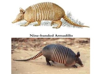
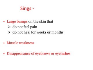
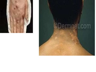
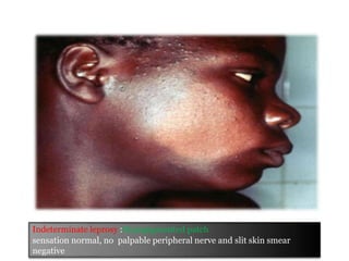
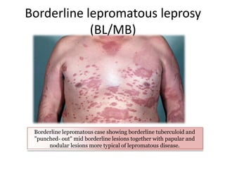
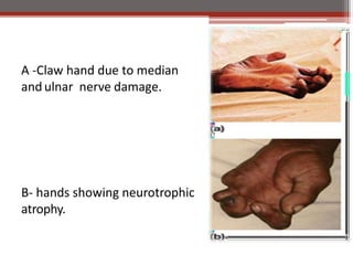
This document contains an MCQ discussion on leprosy. It begins with 13 multiple choice questions about leprosy, including topics like who discovered the causative bacteria, the generation time of Mycobacterium leprae, clinical presentations of leprosy, types of leprosy, treatment, and more. The document then provides explanations and key facts about leprosy, including that it is caused by Mycobacterium leprae, clinical features, types according to skin smear and clinical classification, signs and symptoms, pathogenesis, diagnosis including skin smear microscopy, and treatment with multidrug therapy.
4. 2. What is the generation time of
Mycobacterium laprae
a. 20 minutes
b. 20 hours
c. 12-13 days
d. 20 weeks
5. 3. A 40-year-old woman who has lived in southwest Louisiana all
her life presents to the emergency room. She has strange-
looking, raised areas on her face, arms, and legs. She also
complains that she is losing feeling in her fingers and toes. She
says that when she cuts or burns herself, she does not feel it
and that makes the injury even greater because she does not
know to pull away from the injuring source. A Gram stain of
scrapings from the raised areas on her skin shows thin bacterial
rods that do not take up the stain. However, an acid fast stain
of the same material shows numerous bacilli. What disease is
the woman most likely to have?
a. Borderline tuberculoid leprosy
b. Lepromatous leprosy
c. Scrofula
d. Tuberculoid leprosy
6. • Lady Windermere syndrome is caused by M. avium-
complex, usually in elderly female nonsmokers, resulting in
a very serious lung infection with this organism.
• Scrofula is a term usually reserved for tuberculosis of the
neck, but the disease is really a cervical lymphadenopathy
caused by Mycobacterium tuberculosis.
• The woman obviously has leprosy due to the presence of
acid-fast staining bacilli in the skin scrapings and the loss of
feeling in her fingers and toes. This is caused by the
bacterium infecting and destroying nervous tissue in these
areas.
• Tuberculoid leprosy (also called paucibacillary Hansen
disease) is the mild form of the disease with few organisms
observed in skin scrapings.
• Lepromatous leprosy is the fulminant form of the disease
with numerous bacilli seen in skin scrapings.
7. 4. Single skin lesion is seen in which type
of Leprosy:
a. LL
b. TT
c. BL
d. BT
8. 5. The most infectious type of leprosy
is:
a. Lepromatous leprosy
b. Tuberculoid leprosy
c. Borderline-tubercoloid leprosy
d. None of the above
9. 6. Lepromatous leprosy is observed in
patients with:
a. Deficient cell mediated immunity
b. Adequate cell mediated immunity
c. Adequate humoral immunity
d. None of the above
10. 7. Which type of hypersensitivity reaction
is involved in lepromin test?
a. Type I
b. Type II
c. Type III
d. Type IV
11. 8. The following test is not used
for diagnosis of leprosy :
a. Lepromin test
b. Slit skin smear
c. Fine needle aspiration cytology
d. Skin biopsy
12. 9. The characteristic finding in a case
of leprosy is:
a. Culture test is positive in 2-3 months in
LJ media
b. Long contact with tuberculoid leprosy
can transmit the disease
c. CMI is seen in Lepromatous leprosy
d. Macule lesion heals spontaneous
13. 10. Exacerbation of lesions in patients of
borderline leprosy is seen in :
a. ENL (erythema nodosum leprosum)
b. Lepra reaction type 1
c. Jarisch-Herxheimer reaction
d. Both a & b.
14. 11.The following drug is not used for the
treatment of type II lepra reaction :
a. Chloroquine
b. Thalidomide
c. Cyclosporine
d. Corticosteroids
15.
16. 12. Lepra cells found in lepromatous leprosy are
:
b. Neutophils
c. Lymphocytes
d. Macrophages
e. Plasma cells
17. 13. Globi is :
a. Histiocyte containing acid-fast bacillus
b. Lymphocyte containing acid-fast bacillus
c. Neutrophil containing acid-fast bacillus
d. Large lymphocyte containing acid-fast bacillus
19. Key facts about LEPROSY -
Leprosy is a chronic infectious
disease caused by an acid-fast,
rod-shaped bacillus,
Mycobacterium leprae.
Not cultivable: M. leprae is not
cultivable either in artificial
culture media or in tissue
culture; (does not follow the
Koch's postulates).
can be maintained in animal-
nine banded armadillo (Dasypus
noverncinctus) and foot pad of
mice (kept at a low temperature,
200C).
.
20.
21. M. leprae
Intracellular: Lepra bacilli are obligate intracellular and
strict aerobe.
Less acid fast: Compared to tubercle bacilli, they are less
acid fast and can resist up to 5% sulfuric acid.
Appearance: In smears made from skin lesions, they appear
in groups, called cigar-like bundles of bacilli(globi) present
inside lipid laden macrophages called virchow's lepra cells.
multiplies very slowly
incubation period – about 3 to 5 years.
Lepromarous cases have longer incubation period than
tuberculoid cases.
22. Transmission
• not highly infectious
• The exact mechanism of transmission of
leprosy is not known
• transmitted via droplets, from the nose and
mouth, during close and frequent contacts
with untreated cases.
• occur at all ages (ranging from early infancy
to very old age).
23. Risk Factors -
• Close contact — Contacts of patients with leprosy have a higher risk
of developing leprosy than the general population
• People who live in the areas where leprosy is endemic
parts of India, China, Japan, Nepal, Egypt, and other areas
especially those people in constant physical contact with
infected people.
• Immunity – immunocompromised individuals are more
susceptible to infection.
• Genetic influences - There is some evidence that genetic defects
in the immune system may cause certain people to be more likely
to become infected (region q25 on chromosome 6).
• Armadillo exposure — Leprosy is enzootic in the nine-banded
armadillo
24. Classification
• Two types of classification :
▫ Skin smear result classification. ( WHO )
▫ Clinical classification. (Ridley Jopling)
25. Classification -
▫ Skin smear result (WHO) classification :
1. Paucibacillary leprosy (PB) – few Bacilli;
• Two to five skin lesions with
• Negative skin smear results at all sites.
2. Multibacillary leprosy (MB);
• Any form of leprosy in which the patient shows positive
smears at any site
30. Clinical features -
1. Skin lesions, usually anaesthetic
Hypopigmented or erythematus patch / plaque .
2. Complete / partial loss of sensation.
Painless wounds or burns on the hands or feet
Paresthesias: tingling or numbness in the hands or feet
Diminished sensation or loss of sensation within skin patch(es)
3. Thickening of peripheral nerves.
4. Lumps / swelling on the earlobes or face.
5. Tender, enlarged peripheral nerves.
31. Symptoms-
• Paucibacillary (PB) :
– Well defined skin lesions that are numb
• Multibacillary (MB) :
Chronically stuffy nose and
many skin lesions and
nodules on both sides of the body
32. Sings -
• Large bumps on the skin that
do not feel pain
do not heal for weeks or months
• Muscle weakness
• Disappearance of eyebrows or eyelashes
36. Tuberculoid leprosy: Two hypopigmented patches, hypoasthetic well
defined borders, palpable peripheral nerve and SSS negative.
37. Borderline lepromatous leprosy
(BL/MB)
Borderline lepromatous case showing borderline tuberculoid and
"punched- out" mid borderline lesions together with papular and
nodular lesions more typical of lepromatous disease.
38. A -Claw hand due to median
and ulnar nerve damage.
B- hands showing neurotrophic
atrophy.
39. Pathogenesis
• M. leprae is an obligate intracellular acid-fast bacillus with
unique ability to enter nerves
• The areas most commonly affected are- superficial
peripheral nerves , skin, mucous membranes of the upper
respiratory tract, eyes , and tests
• 95% of people who are exposed do not develop disease
• Tissue damage is depend upon -
the degree to which cell-mediated immunity is expressed
the extent of bacillary spread and multiplication
immunologic complication and
nerve damage
40. Diagnosis
• Diagnosis of leprosy- most commonly based
on the clinical sign and symptoms.
• In an endemic country or area, an individual
should be regarded as having leprosy if shows
one of the following cardinal signs :
skin lesion consistent with leprosy and with
definite sensory loss, with or without
thickened nerves
positive skin smears
41. Smear Microscopy
• Smear microscopy is done to demonstrate the
bacilli in the lesions.
• Specimen Collection-
Total 6 samples are collected;
4 from skin (forehead, cheek, chin and buttock)
1 from ear lobe and nasal mucosa by nasal
blow/scraping.
• With paucibacillary leprosy, no bacteria will be
detected.
42. Silt skin smear
• Silt skin smear prepared- from the skin and ear lobe
specimens.
• The edge of the lesion is the preferred site.
• Lesion is cleaned with spirit, then is pinched up light to
minimize bleeding.
• A 5 mm long incision is made with a scalpel, deep
enough to get into the infiltrated layers.
• After wiping off blood or lymph that may have exuded,
the scalpel blade is rotated transversely to scrape the
sides and base of the incision so as to obtain a tissue
pulp from below the epidermis- which is smeared
uniformly over an area of 8 mm diameter on a slide.
44. Nasal specimens
• Nasal blow: Early morning mucus material is
collected by blowing the nose on a clean
cellophane sheet
• Nasal scraping: By using a mucosal scraper to
scrape the nasal septum sufficiently so as to
remove a piece of mucous membrane ,
transferred onto a slide to make a uniform
smear.
45. • Biopsy from the thickened nerves and nodular
lesions may be necessary in some cases.
46. DIAGNOSIS OF LEPROSY
• Hypopigmented or reddish skin
lesion(s) with definite loss of sensation
• Damage to the peripheral nerves, as
demonstated by loss of sensation
• Weakness of the muscles of hands, feet
or face
• Positive skin smear
47. Treatment
• Common drugs
1. Dapson
2. Refampicine
3. Clofazimine
The combination of these three drugs
Is known as multi drug therapy (MDT)
50. Complication -
• disfigurement
• hair loss (particularly on the eyebrows and eyelashes)
• muscle weakness
• permanent nerve damage in the arms and legs
• inability to use the hands and feet
• Nosebleeds
• iritis (inflammation of the iris of the eye), glaucoma
(an eye disease that causes damage to the optic
nerve), and blindness
• Infertility
• kidney failure