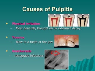
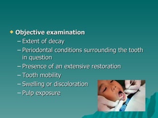
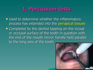
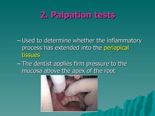
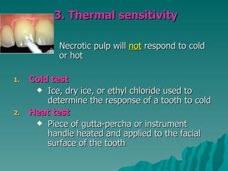
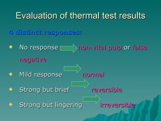
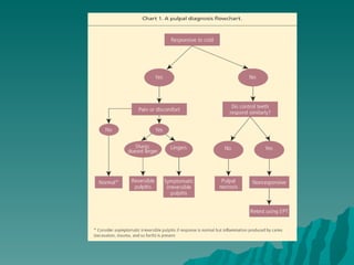
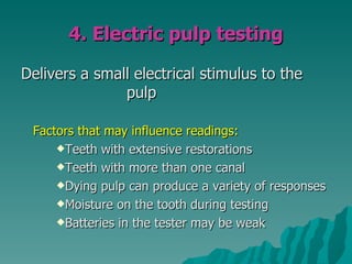
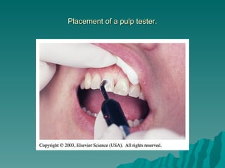
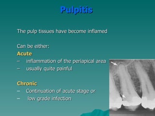
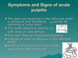
Diagnosis &
Treatment Planning in
    Endodontics
    Dr Saidah Tootla
Introduction
 Endodontics  is the specialty of dentistry
 that manages the prevention,
 diagnosis, and treatment of the dental
 pulp and the periradicular tissues that
 surround the root of the tooth
Causes of Pulpitis

    Physical irritation
    – Most generally brought on by extensive decay.

    Trauma
    – Blow to a tooth or the jaw

    Anachoresis
    - retrograde infections
Signs and Symptoms
 Pain when biting down
 Pain when chewing

 Sensitivity with hot or cold beverages

 Facial swelling

 Discolouration of the tooth
Endodontic Diagnosis
   Subjective examination
     – Chief complaint
     – Character and duration of pain
     – Painful stimuli
     – Sensitivity to biting and pressure
     – Discolouration of tooth
Important questions?
 What do you think the problem is?
 Does it hurt to hot or cold?
 Does it hurt when you’re chewing?
 When does it start hurting?
 How bad is the pain?
 What type of pain is it?
 How long does the pain last?
 Does anything relieve it?
 How long has it been hurting?
   Objective examination
    – Extent of decay
    – Periodontal conditions surrounding the tooth
      in question
    – Presence of an extensive restoration
    – Tooth mobility
    – Swelling or discoloration
    – Pulp exposure
Challenges in diagnosis of pulpitis
 Referred pain & the lack of
  proprioceptors in the pulp          localizing
  the problem to the correct tooth can often
  be a considerable diagnostic challenge
 Also of significance is the difficulty in
  relating the clinical status of a tooth to
  histopathology of the pulp in concern
 Unfortunately, no reliable symptoms or
  tests consistently correlate the two.
Diagnostic Tests
   Percussion
   Palpation
   Thermal
   Electrical
   Radiographs
1. Percussion tests
 Used to determine whether the inflammatory
  process has extended into the periapical tissues
 Completed by the dentist tapping on the incisal
  or occlusal surface of the tooth in question with
  the end of the mouth mirror handle held parallel
  to the long axis of the tooth
2. Palpation tests

– Used to determine whether the inflammatory
  process has extended into the periapical
  tissues
– The dentist applies firm pressure to the
  mucosa above the apex of the root
3. Thermal sensitivity

            Necrotic pulp will not respond to cold
            or hot

1.   Cold test
       Ice, dry ice, or ethyl chloride used to
         determine the response of a tooth to cold
2.   Heat test
       Piece of gutta-percha or instrument
         handle heated and applied to the facial
         surface of the tooth
Evaluation of thermal test results
4 distinct responses:

   No response        non-vital pulp or false
    negative

   Mild response       normal

   Strong but brief       reversible

   Strong but lingering      irreversible
Causes of false
         positives/negative
   Calcified canals
   Immature apex – usually seen in
    young patients
   Trauma
   Premedication of the patient – pulp
    sedated
4. Electric pulp testing

Delivers a small electrical stimulus to the
               pulp

 Factors that may influence readings:
     Teeth with extensive restorations

     Teeth with more than one canal

     Dying pulp can produce a variety of responses

     Moisture on the tooth during testing

     Batteries in the tester may be weak
Placement of a pulp tester.
5. Radiographs
1.    Pre-operative radiograph
     – Invaluable diagnostic tool
     – Periapical radiolucency
     – Widening of PDL
     – Deep caries
     – Resorption
     – Pulp stones
     – Large restorations
     – Root fractures
Requirements of Endodontic Films
 Show  4-5 mm beyond the apex of the
  tooth and the surrounding bone or
  pathologic condition.
 Present an accurate image of the tooth
  without elongation or fore-shortening.
 Exhibit good contrast so all pertinent
  structures are readily identifiable.
Quality radiograph in endodontics.
Diagnostic Conclusions


• Normal pulp

• Pulpitis
Normal pulp

There are no subjective symptoms or objective
signs. The pulp responds normally to sensory
stimuli, and a healthy layer of dentine
surrounds the pulp
Pulpitis

The pulp tissues have become inflamed

Can be either:
Acute
– inflammation of the periapical area
– usually quite painful

Chronic
– Continuation of acute stage or
–   low grade infection
Acute Pulpitis
 mainly  occurs in children teeth and
  adolescent
 pain is more pronounced than in
  chronic
Symptoms and Signs of acute
             pulpitis
 The pain not localized in the affected tooth
  is constant and throbbing      worse by
  reclining or lying down
 The tooth becomes painful
  with hold or cold stimuli
 The pain may be sharp and stabbing
 Change of color is obvious in the affected
  tooth
 swelling of the gum or face in the
  area of the affected tooth
Forms of acute pulpitis
1. Form of purulent acute where the
 pulp is totally inflamed
2. Form of gangrenous acute where
 the pulp begins to die in a less
 painful manner that can lead into the
 formation of an abscess
Chronic Pulpitis
1.   Reversible
2.   Irreversible
Reversible pulpitis

– The pulp is irritated, and the patient is
  experiencing pain to thermal stimuli
– Sharp shooting pain
– Duration of the pain episode lasts for
  seconds
– The tooth pulp can be saved
– Usually this condition is caused by
  average caries
Irreversible pulpitis

–   The tooth will display symptoms of lingering pain
–   pain occurs spontaneously or lingers minutes
    after the stimulus is removed
–   patient may have difficulty locating the tooth
    from which the pain originates
–   As infection develops and extends through the
    apical foramen, the tooth becomes exquisitely
    sensitive to pressure and percussion
–   A periapical abscess elevates the tooth from its
    socket and feels “high” when the patient bites down
Periradicular abscess

An inflammatory reaction to pulpal infection
that can be chronic or have rapid onset with
pain, tenderness of the tooth to palpation and
percussion, pus formation, and swelling of the
tissues.
Periodontal abscess
 An inflammatory reaction frequently
  caused by bacteria entrapped in the
  periodontal sulcus for a long time. A
  patient will experience rapid onset, pain,
  tenderness to palpation and percussion,
  pus formation, and swelling.
 Destruction of the

 periodontium occurs
Periradicular cyst

 A cyst that develops at or near the root of a
necrotic pulp. These types of cysts develop
as an inflammatory response to pulpal
infection and necrosis of the pulp
Pulp fibrosis

The decrease of living cells within the pulp
causing fibrous tissue to take over the pulpal
canal
Necrotic tooth

– Also referred to as non-vital. Used to
  describe a pulp that does not respond to
  sensory stimulus
– Tooth is usually discoloured
Plan of Treatment
 Depends   widely on the diagnosis
Simple plan of treatment
Visit 1:
 Medical history
 History of the tooth
 Access cavity
 Place rubberdam
 Extirpation + irrigation with sodium hypochlorite
 Placed intra-canal medication (calcium hydroxide)
 Place cotton pellet
 Placed temporary restoration (IRM/Kalzinol)
Visit 2:
 Working length determination

 Debridement using the hybrid technique

 Irrigation

 Placed intra-canal medication (calcium
  hydroxide)
 Place cotton pellet

 Placed temporary restoration
  (IRM/Kalzinol)
Visit 3:
 Obturation with GP using lateral
  condensation

 Placed   temporary/permanent
             restoration (IRM/Kalzinol)
Referral
 To   appropriate discipline
Remember
    Access cavity shapes:
2.   Anterior – inverted triangle
3.   Premolars – round
4.   Molars – rhomboid
    Always use rubberdam
    Never to use Cavit as a temporary
     restoration
    Always place an intra-canal
     medication….calcium hydroxide???
    Always use RC Prep or Glyde when filing
Contraindications for RCT
   Caries extending beyond bone level
   Rubberdam cannot be placed
   Crown of tooth cannot be restored in restorative
    dentistry nor prosthodontics
   Patient is physically/mentally handicapped and
    therefore cannot follow OH instructions
   Putrid OH
   Unmotivated patient
   Severe root resorption
   Vertical root fractures
   Cost factor
Inter & cross-departmental
               diagnosis
 Mobile teeth
 Teeth associated with severe periodontal
  problems
 Confusion between TMJ dysfunctional
  symptoms and RCT pain
 Many decayed teeth
 Sclerosed canal due to trauma
 Uncertainty of prognosis related to
  abscess, severe caries, facial swelling,
  cellulites, and medical condition of patient
Referral to post-grad clinics
 Extensive   internal or external root
  resorption
 Severely curved, narrow, tortuous
  canals
 Full-mouth rehabilitation required
 Multiple exposures due to
  attrition/abrasion
 Problems with occlusion causing the
  need for RCT
Questions????

Diagnosis tx-planning

  • 1.
    Diagnosis & Treatment Planningin Endodontics Dr Saidah Tootla
  • 2.
    Introduction  Endodontics is the specialty of dentistry that manages the prevention, diagnosis, and treatment of the dental pulp and the periradicular tissues that surround the root of the tooth
  • 3.
    Causes of Pulpitis  Physical irritation – Most generally brought on by extensive decay.  Trauma – Blow to a tooth or the jaw  Anachoresis - retrograde infections
  • 4.
    Signs and Symptoms Pain when biting down  Pain when chewing  Sensitivity with hot or cold beverages  Facial swelling  Discolouration of the tooth
  • 5.
    Endodontic Diagnosis  Subjective examination – Chief complaint – Character and duration of pain – Painful stimuli – Sensitivity to biting and pressure – Discolouration of tooth
  • 6.
    Important questions?  Whatdo you think the problem is?  Does it hurt to hot or cold?  Does it hurt when you’re chewing?  When does it start hurting?  How bad is the pain?  What type of pain is it?  How long does the pain last?  Does anything relieve it?  How long has it been hurting?
  • 7.
    Objective examination – Extent of decay – Periodontal conditions surrounding the tooth in question – Presence of an extensive restoration – Tooth mobility – Swelling or discoloration – Pulp exposure
  • 8.
    Challenges in diagnosisof pulpitis  Referred pain & the lack of proprioceptors in the pulp localizing the problem to the correct tooth can often be a considerable diagnostic challenge  Also of significance is the difficulty in relating the clinical status of a tooth to histopathology of the pulp in concern  Unfortunately, no reliable symptoms or tests consistently correlate the two.
  • 9.
    Diagnostic Tests  Percussion  Palpation  Thermal  Electrical  Radiographs
  • 10.
    1. Percussion tests Used to determine whether the inflammatory process has extended into the periapical tissues  Completed by the dentist tapping on the incisal or occlusal surface of the tooth in question with the end of the mouth mirror handle held parallel to the long axis of the tooth
  • 11.
    2. Palpation tests –Used to determine whether the inflammatory process has extended into the periapical tissues – The dentist applies firm pressure to the mucosa above the apex of the root
  • 12.
    3. Thermal sensitivity Necrotic pulp will not respond to cold or hot 1. Cold test  Ice, dry ice, or ethyl chloride used to determine the response of a tooth to cold 2. Heat test  Piece of gutta-percha or instrument handle heated and applied to the facial surface of the tooth
  • 13.
    Evaluation of thermaltest results 4 distinct responses:  No response non-vital pulp or false negative  Mild response normal  Strong but brief reversible  Strong but lingering irreversible
  • 15.
    Causes of false positives/negative  Calcified canals  Immature apex – usually seen in young patients  Trauma  Premedication of the patient – pulp sedated
  • 16.
    4. Electric pulptesting Delivers a small electrical stimulus to the pulp Factors that may influence readings: Teeth with extensive restorations Teeth with more than one canal Dying pulp can produce a variety of responses Moisture on the tooth during testing Batteries in the tester may be weak
  • 17.
    Placement of apulp tester.
  • 19.
    5. Radiographs 1. Pre-operative radiograph – Invaluable diagnostic tool – Periapical radiolucency – Widening of PDL – Deep caries – Resorption – Pulp stones – Large restorations – Root fractures
  • 20.
    Requirements of EndodonticFilms  Show 4-5 mm beyond the apex of the tooth and the surrounding bone or pathologic condition.  Present an accurate image of the tooth without elongation or fore-shortening.  Exhibit good contrast so all pertinent structures are readily identifiable.
  • 21.
  • 22.
  • 23.
    Normal pulp There areno subjective symptoms or objective signs. The pulp responds normally to sensory stimuli, and a healthy layer of dentine surrounds the pulp
  • 24.
    Pulpitis The pulp tissueshave become inflamed Can be either: Acute – inflammation of the periapical area – usually quite painful Chronic – Continuation of acute stage or – low grade infection
  • 25.
    Acute Pulpitis  mainly occurs in children teeth and adolescent  pain is more pronounced than in chronic
  • 26.
    Symptoms and Signsof acute pulpitis  The pain not localized in the affected tooth is constant and throbbing worse by reclining or lying down  The tooth becomes painful with hold or cold stimuli  The pain may be sharp and stabbing  Change of color is obvious in the affected tooth  swelling of the gum or face in the area of the affected tooth
  • 28.
    Forms of acutepulpitis 1. Form of purulent acute where the pulp is totally inflamed 2. Form of gangrenous acute where the pulp begins to die in a less painful manner that can lead into the formation of an abscess
  • 29.
    Chronic Pulpitis 1. Reversible 2. Irreversible
  • 30.
    Reversible pulpitis – Thepulp is irritated, and the patient is experiencing pain to thermal stimuli – Sharp shooting pain – Duration of the pain episode lasts for seconds – The tooth pulp can be saved – Usually this condition is caused by average caries
  • 31.
    Irreversible pulpitis – The tooth will display symptoms of lingering pain – pain occurs spontaneously or lingers minutes after the stimulus is removed – patient may have difficulty locating the tooth from which the pain originates – As infection develops and extends through the apical foramen, the tooth becomes exquisitely sensitive to pressure and percussion – A periapical abscess elevates the tooth from its socket and feels “high” when the patient bites down
  • 32.
    Periradicular abscess An inflammatoryreaction to pulpal infection that can be chronic or have rapid onset with pain, tenderness of the tooth to palpation and percussion, pus formation, and swelling of the tissues.
  • 34.
    Periodontal abscess  Aninflammatory reaction frequently caused by bacteria entrapped in the periodontal sulcus for a long time. A patient will experience rapid onset, pain, tenderness to palpation and percussion, pus formation, and swelling.  Destruction of the periodontium occurs
  • 36.
    Periradicular cyst Acyst that develops at or near the root of a necrotic pulp. These types of cysts develop as an inflammatory response to pulpal infection and necrosis of the pulp
  • 37.
    Pulp fibrosis The decreaseof living cells within the pulp causing fibrous tissue to take over the pulpal canal
  • 38.
    Necrotic tooth – Alsoreferred to as non-vital. Used to describe a pulp that does not respond to sensory stimulus – Tooth is usually discoloured
  • 39.
    Plan of Treatment Depends widely on the diagnosis
  • 40.
    Simple plan oftreatment Visit 1:  Medical history  History of the tooth  Access cavity  Place rubberdam  Extirpation + irrigation with sodium hypochlorite  Placed intra-canal medication (calcium hydroxide)  Place cotton pellet  Placed temporary restoration (IRM/Kalzinol)
  • 41.
    Visit 2:  Workinglength determination  Debridement using the hybrid technique  Irrigation  Placed intra-canal medication (calcium hydroxide)  Place cotton pellet  Placed temporary restoration (IRM/Kalzinol)
  • 42.
    Visit 3:  Obturationwith GP using lateral condensation  Placed temporary/permanent restoration (IRM/Kalzinol)
  • 43.
    Referral  To appropriate discipline
  • 44.
    Remember  Access cavity shapes: 2. Anterior – inverted triangle 3. Premolars – round 4. Molars – rhomboid  Always use rubberdam  Never to use Cavit as a temporary restoration  Always place an intra-canal medication….calcium hydroxide???  Always use RC Prep or Glyde when filing
  • 45.
    Contraindications for RCT  Caries extending beyond bone level  Rubberdam cannot be placed  Crown of tooth cannot be restored in restorative dentistry nor prosthodontics  Patient is physically/mentally handicapped and therefore cannot follow OH instructions  Putrid OH  Unmotivated patient  Severe root resorption  Vertical root fractures  Cost factor
  • 46.
    Inter & cross-departmental diagnosis  Mobile teeth  Teeth associated with severe periodontal problems  Confusion between TMJ dysfunctional symptoms and RCT pain  Many decayed teeth  Sclerosed canal due to trauma  Uncertainty of prognosis related to abscess, severe caries, facial swelling, cellulites, and medical condition of patient
  • 47.
    Referral to post-gradclinics  Extensive internal or external root resorption  Severely curved, narrow, tortuous canals  Full-mouth rehabilitation required  Multiple exposures due to attrition/abrasion  Problems with occlusion causing the need for RCT
  • 48.