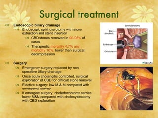
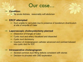
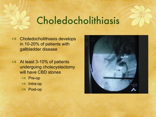
Cholangitis &
 Management of
Choledocholithiasis
         Ruby Wang MS 3
           Surg 300A
             8/20/07
Content
 Case

 Cholangitis
    Clinical manifestations
    Diagnosis
    Treatment

 Diagnosis and management of choledocholithiasis
    Pre-operative
    Intra-operative
    Post-operative
Case
 HPI:
    86 yo lady p/w 3-4 episodes of RUQ/mid-epigastric
      abdominal pain over the last year, lasting generally several
      hours, accompanied by occasional emesis, anorexia, and
      sensation of shaking chills.
    ROS: negative otherwise
 PE:
    VS: T 36.2, P98 , RR 18, BP 124/64
    Abdominal exam significant for RUQ TTP
 Labs
    AST 553, ALT 418. Alk Phos 466. Bilirubin 2.7
    WBC 30.3
 Imaging
    Abdominal US: multiple gallstones, no pericholecystic fluid,
      no extrahepatic/intrahepatic/CBD dilatation
Introduction
 Cholangitis is bacterial infection superimposed on biliary obstruction

 First described by Jean-Martin Charcot in 1850s as a serious and
  life-threatening illness

 Causes
      Choledocholithiasis
      Obstructive tumors
             Pancreatic cancer
             Cholangiocarcinoma
             Ampullary cancer
             Porta hepatis
      Others
             Strictures/stenosis
             ERCP
             Sclerosing cholangitis
             AIDS
             Ascaris lumbricoides
Epidemiology
   Nationality
      U.S: uncommon, and occurs in association with biliary obstruction and causes of
       bactibilia (s/p ERCP)
      Internationally:
          Oriental cholangiohepatitis endemic in SE Asia- recurrent pyogenic
             cholangitis with intrahepatic/extrahepatic stones in 70-80%
          Gallstones highest in N European descent, Hispanic populations, Native
             Americans
          Intestinal parasites common in Asia
   Sex
      Gallstones more common in
       women
      M: F ratio equal in
       cholangitis
   Age
      Median age between 50-60
      Elderly patients more likely
       to progress from
       asymptomatic gallstones to
       cholangitis without colic
Pathogenesis
   Normally, bile is sterile due to constant flush,
    bacteriostatic bile salts, secretory IgA, and biliary
    mucous; Sphincter of Oddi forms effective barrier to
    duodenal reflux and ascending infection

   ERCP or biliary stent insertion can disrupt the Sphincter
    of Oddi barrier mechanism, causing pathogeneic
    bacteria to enter the sterile biliary system.

   Obstruction from stone or tumor increases intrabiliary
    pressure

   High pressure diminishes host antibacterial defense-
    IgA production, bile flow- causing immune dysfunction,
    increasing small bowel bacterial colonization.
                                                                                     Adam.about.com

   Bacteria gain access to biliary tree by retrograde ascent

   Biliary obstruction (stone or stricture) causes bactibilia
      E Coli (25-50%)
      Klebsiella (15-20%),
      Enterobacter (5-10%)

   High pressure pushes infection into biliary canaliculi,
    hepatic vein, and perihepatic lymphatics, favoring
    migration into systemic circulation- bacteremia
    (20-40%).                                                    Gpnotebook.co.uk   Pathology.med.edu
Clinical Manifestations
 RUQ pain (65%)                          Charcot’s
 Fever (90%)                             Triad:
                                          Found in    Reynold’s
     May be absent in elderly patients   50-70%      Pentad:
 Jaundice (60%)                          of
                                          patients
 Hypotension (30%)
 Altered mental status (10%)

 Additional History
        Pruitus, acholic stools
        PMH for gallstones, CBD stones,
        Recent ERCP, cholangiogram
 Additional Physical
        Tachycardia
        Mild hepatomegaly
Diagnosis: lab values
 CBC
    79% of patients have WBC > 10,000, with mean of 13,600
    Septic patients may be neutropenic
 Metabolic panel
      Low calcium if pancreatitis
      88-100% have hyperbilirubinemia
      78% have increased alkaline phosphatase
      AST and ALT are mildly elevated
         Aminotransferase can reach 1000U/L- microabscess formation in the
          liver
    GGT most sensitive marker of choledocholithiasis
 Amylase/Lipase
    Involvement of lower CBD may cause 3-4x elevated amylase
 Blood cultures
    20-30% of blood cultures are positive
Diagnosis: first-line imaging
Ultrasonography
    Advantage:
        Sensitive for intrahepatic/extrahepatic/CBD dilatation
             CBD diameter > 6 mm on US associated with high
                prevalence of choledocholithaisis
             Of cholangitis patients, dilated CBD found in 64%,
        Rapid at bedside
        Can image aorta, pancreas, liver
        Identify complications: perforation, empyema, abscess
    Disadvantage
        Not useful for choledocholithiasis:
             Of cholangitis patients, CBD stones observed in 13%
        10-20% falsely negative - normal U/S does not r/o cholangitis
                                                                            Med.virgina.edu
             acute obstruction when there is no time to dilate
             Small stones in bile duct in 10-20% of cases
CT
    Advantages
        CT cholangiograhy enhances CBD stones and increases detection
          of biliary pathology
              Sensitivity for CBD stones is 95%
        Can image other pathologies: ampullary tumors, pericholecystic
          fluid, liver abscess
        Can visualize other pathologies- cholangitis: diverticuliits,
          pyelonephritis, mesenteric ischemia, ruptured appendix
    Disadvantages
        Sensitivity to contrast
        Poor imaging of gallstones                                       Soto et al. J. Roenterology. 2000
Diagnostic: MRCP and ERCP
Magnetic resonance cholangiopancreatography (MRCP)
 Advantage
       Detects choledocholithiasis, neoplasms, strictures, biliary dilations
       Sensitivity of 81-100%, specificity of 92-100% of
           choledocholithiasis
       Minimally invasive- avoid invasive procedure in 50% of patients
 Disadvantage:
       cannot sample bile, test cytology, remove stone
       Contraindications: pacemaker, implants, prosthetic valves
 Indications
       If cholangitis not severe, and risk of ERCP high, MRCP useful
       If Charcot’s triad present, therapeutic ERCP with drainage should
           not be delayed.
Endoscopic retrograde cholangiopancreatography (ERCP)
 Gold standard for diagnosis of CBD stones, pancreatitis, tumors,
    sphincter of Oddi dysfunction
 Advantage
       Therapeutic option when CBD stone identified
       Stone retrieval and sphincterotomy
 Disadvantage
       Complications: pancreatitis, cholangitis, perforation of duodenum
           or bile duct, bleeding
       Diagnostic ERCP complication rate 1.38% , mortality rate 0.21%
Medical Treatment
 Resucitate, Monitor, Stabilize if patient unstable
    Consider cholangitis in all patients with sepsis

 Antibiotics
    Empiric broad-spectrum Abx after blood cultures drawn
          Ampicillin (2g/4h IV) plus gentamicin (4-6mg/kg IV daily)
          Carbapenems: gram negative, enterococcus, anaerobes
          Levofloxacin (250-500mgIV qD) for impaired renal fxn.

   - 80% of patients can be managed conservatively 12-24 hrs Abx

   - If fail medical therapy, mortality rate 100% without surgical
   decompression: ERCP or open

   - Indication: persistent pain, hypotension, fever, mental confusion
Surgical treatment
   Endoscopic biliary drainage
      Endoscopic sphincterotomy with stone
        extraction and stent insertion
           CBD stones removed in 90-95% of
              cases
           Therapeutic mortality 4.7% and
              morbidity 10%, lower than surgical
              decompression

   Surgery
      Emergency surgery replaced by non-
        operative biliary drainage
      Once acute cholangitis controlled, surgical
        exploration of CBD for difficult stone removal
      Elective surgery: low M & M compared with
        emergency survey
      If emergent surgery, choledochotomy carries
        lower M&M compared with cholecystectomy
        with CBD exploration
Our case…
 Condition:
     No acute distress, reasonably soft abdomen

 ERCP attempted
     Duct unable to cannulate due to presence of duodenum diverticulum
      at site of ampulla of Vater

 Laparoscopic cholecystectomy planned
       Dissection of triangle of Calot
       Cystic duct and artery visualized and dissected
       Cystic duct ductotomy
       Insertion of cholangiogram catheter advanced and contrast bolused
        into cystic duct for IOC

 Intraoperative cholangiogram
     Several common duct filling defects consistent with stones
     Decision to proceed with CBD exploration
Choledocholithiasis
 Choledocholithiasis develops
  in 10-20% of patients with
  gallbladder disease

 At least 3-10% of patients
  undergoing cholecystectomy
  will have CBD stones
     Pre-op
     Intra-op
     Post-op
Pre-op diagnosis & management
 Diagnosis: Clinical history and exam, LFTs, Abdominal U/S, CT, MRCP
    High risk (>50%) of choledocholithiasis:
        clinical jaundice, cholangitis,
        CBD dilation or choledocholithiasis on ultrasound
        Tbili > 3 mg/dL correlates to 50-70% of CBD stone
    Moderate risk (10-50%):
        h/o pancreatitis, jaundice correlates to CBD stone in 15%
        elevated preop bili and AP,
        multiple small gallstones on U/S
    Low risk (<5%):
        large gallstones on U/S
        no h/o jaundice or pancreatitis,
        normal LFTs

 Treatment:
      ERCP
      Surgery
Intra-op diagnosis and management
   Diagnosis: intraoperative cholangiography (IOC)
      Cannulation of cystic duct, filling of L and R hepatic ducts, CBD and
        common hepatic duct diameter, presence or absence of filling defects.
      Detect CBD stones
      Potentially identify bile duct abnormalities, including iatrogenic injuries
      Sensitivity 98%, specificity 94%
      Morbidity and mortality low

   Treatment
      Open CBD exploration
            Most surgeons prefer less invasive techniques
      Laparoscopic CBD exploration
              via choledochotomy: CBD dilatation > 6mm
              via cystic duct (66-82.5%)
              CBD clearance rate 97%
              Morbidity rate 9.5%
              Stones impacted at Sphincter of Oddi most difficult to extract
      Intraoperative ERCP
Early years: Open CBD exploration &
    Introduction of endoscopic sphincterotomy
   1889, 1st CBD exploration by Ludwig
    Courvoisier, a Swiss surgeon
      Kocherization of duodenum and short
         longitudinal choledochotomy
      Stones removed with palpation, irrigation
         with flexible catheters, forceps,
      Completion with T-tube drainage
      For many years, this was the standard
         treatment for cholecystocholedocholithiasis

   1970s, endoscopic sphincterotomy (ES)
      Gained wide acceptance as good, less
        invasive, effective alternative
      In patients with CBD stones who have
        previously undergone cholecystectomy, ES
        is the method of choice
Open surgery vs Endoscopic sphincterotomy
   In patients with intact gallbladders, ES or open choledochotomy?
        Design: 237 patients with CBD stone and intact gallbladders, 66% managed with ES and rest
         with open choledochotomy
        Results: No significant difference in morbidity and mortality rates
            Lower incidence of retained stones after open choledochotomy
        Conclusion: open surgery superior to ES in those with intact gallbladders
                  Miller et al. Ann Surg 1988; 207: 135-41

   Is ES followed by open CCY superior to open CCY+ CBDE?
        Results: Initial stone clearance higher with open surgery (88% vs 65%, p< 0.05)
        Conclusion: routine preoperative ES not indicated
                  Stain et al. Ann Surg 1991; 213: 627-34

   Cochraine database of systematic reviews
        Design: 8 trials randomized 760 patients comparing ERCP with open surgical clearance
        Results: Open surgery more successful in CBD stone clearance, associated with lower
         mortality
        Conclusion: open bile duct surgery superior to ES
                    Cochrane database of systematic reviews 2007

   In patients with severe cholangitis, open or ES?
        Study design:          Randomized, prospsective trial of 82 patients with choledocholithiasis and
         severe toxic cholangitis managed endoscopically or with open choledochotomy
        Results: In group managed initially with endoscopic drainage, need for ventilatory support
         (29% vs 63%) and mortality (33% vs 66%) significantly less
        Conclusion: toxic cholangitis should managed with endoscopic sphincterotomy
                  Lai et al. J Engl J Med 1992; 326: 1582-6
Laparoscopic CBD Exploration
   In 1989, laparoscopic removal of gallbladder replaced open surgery
        In the past decade, laparoscopic CBD exploration (LCBDE) developed
   Techniques
        IOC define biliary anatomy: size and length of cystic duct, size of bile duct stones
        Choledochotomy
                 If cystic duct < CBD stone, If CBD > 6mm
                 If stone located proximal to cystic duct-common bile duct junction
                 If stone impacted in bile duct or papilla
        Transcystic approach
                 If CBD < 6mm in diameter
                 Cystic duct dissected close to junction with CBD, transverse incision made
                 Guidewire into CBd through cholangiogram catheter under fluoroscopy
                 Osotonic NaCl irrigate CBD to flush small stones through sphincter of Oddi
                 Unsuccessful in 10-20% of patients
                 Contraindications: pancreatitis, sphincter anomalies,
   Results
        High rate of lap CBD clearance: 73-100%
                 Similar success rates between transcystic and choledochotomy
        Conversion to open 5.2-19.6%
                 Causes : multiple/impacted stones, bleeding, unclear anatomy,equipment failure
        Length of hospital stay shorter in LCBDE than ES
        Mortality and Morbidity
                 No difference between LCBDE and ES
                                    Cochrane database of systematic reviews 2007
Post-op Diagnosis and
           Management
 T-tube cholangiography
    T-tube placed following CBDE to diagnosis and
     manage retained stones
    Retained CBD stones in 2-10% of patients after
     CBD exploration
    If not obstruction, tube is clamped and left for 6
     weeks.
    Cholangiogram repeat after 6 wks
 ERCP
    Treatment of retained stones undetected or left
     behind
In summary
   Non-surgical care first line
      Goal: extract stone, but if not possible, drain bile to improve condition until
         definitive surgical intervention
      ERCP: both diagnostic and therapeutic
            Stones> 1cm - Sphincterotomy needed before extraction
            Stones > 2cm: require lithotripsy or chemical dissolution
      PTC
   Surgical Care if endoscopy and IR drainage fail
      Issues
            Exploration of CBD
            Fate of gallbladder
      CBD exploration: laparoscopy first line
            Transcystic:
            Choledochotomy
      CBD exploration: open
            If laparoscopy has failed or contraindicated
            T-tube cholangiogram 10-14 days posto
            Open CBD is safe option, but limited to setting of concomitant open surgery
…our case
 Open procedure
     Due to previous failure of ERCP due to duodenum diverticulum
     Incision joining epigastric port with subcostal inciion
     Dis
 Cholecystectomy
     Gallbladder was dissected free from liver bed
     Cystic artery/duct identified, ligated.
 CBD exploration
     2 suture splaced in direction of common duct through anterior wall in the
      same longitudinal direction
     Choledochotomy- extended in both proximal and distal directions of
      CBD
     4 CBD stones evacuated
     Catheter advanced within CBD to perform sphincterotomy
     T-tube placed within common bile duct.

Surgery cholangitis[1]

  • 1.
    Cholangitis & Managementof Choledocholithiasis Ruby Wang MS 3 Surg 300A 8/20/07
  • 2.
    Content  Case  Cholangitis  Clinical manifestations  Diagnosis  Treatment  Diagnosis and management of choledocholithiasis  Pre-operative  Intra-operative  Post-operative
  • 3.
    Case  HPI:  86 yo lady p/w 3-4 episodes of RUQ/mid-epigastric abdominal pain over the last year, lasting generally several hours, accompanied by occasional emesis, anorexia, and sensation of shaking chills.  ROS: negative otherwise  PE:  VS: T 36.2, P98 , RR 18, BP 124/64  Abdominal exam significant for RUQ TTP  Labs  AST 553, ALT 418. Alk Phos 466. Bilirubin 2.7  WBC 30.3  Imaging  Abdominal US: multiple gallstones, no pericholecystic fluid, no extrahepatic/intrahepatic/CBD dilatation
  • 4.
    Introduction  Cholangitis isbacterial infection superimposed on biliary obstruction  First described by Jean-Martin Charcot in 1850s as a serious and life-threatening illness  Causes  Choledocholithiasis  Obstructive tumors  Pancreatic cancer  Cholangiocarcinoma  Ampullary cancer  Porta hepatis  Others  Strictures/stenosis  ERCP  Sclerosing cholangitis  AIDS  Ascaris lumbricoides
  • 5.
    Epidemiology  Nationality  U.S: uncommon, and occurs in association with biliary obstruction and causes of bactibilia (s/p ERCP)  Internationally:  Oriental cholangiohepatitis endemic in SE Asia- recurrent pyogenic cholangitis with intrahepatic/extrahepatic stones in 70-80%  Gallstones highest in N European descent, Hispanic populations, Native Americans  Intestinal parasites common in Asia  Sex  Gallstones more common in women  M: F ratio equal in cholangitis  Age  Median age between 50-60  Elderly patients more likely to progress from asymptomatic gallstones to cholangitis without colic
  • 6.
    Pathogenesis  Normally, bile is sterile due to constant flush, bacteriostatic bile salts, secretory IgA, and biliary mucous; Sphincter of Oddi forms effective barrier to duodenal reflux and ascending infection  ERCP or biliary stent insertion can disrupt the Sphincter of Oddi barrier mechanism, causing pathogeneic bacteria to enter the sterile biliary system.  Obstruction from stone or tumor increases intrabiliary pressure  High pressure diminishes host antibacterial defense- IgA production, bile flow- causing immune dysfunction, increasing small bowel bacterial colonization. Adam.about.com  Bacteria gain access to biliary tree by retrograde ascent  Biliary obstruction (stone or stricture) causes bactibilia  E Coli (25-50%)  Klebsiella (15-20%),  Enterobacter (5-10%)  High pressure pushes infection into biliary canaliculi, hepatic vein, and perihepatic lymphatics, favoring migration into systemic circulation- bacteremia (20-40%). Gpnotebook.co.uk Pathology.med.edu
  • 7.
    Clinical Manifestations  RUQpain (65%) Charcot’s  Fever (90%) Triad: Found in Reynold’s  May be absent in elderly patients 50-70% Pentad:  Jaundice (60%) of patients  Hypotension (30%)  Altered mental status (10%) Additional History Pruitus, acholic stools PMH for gallstones, CBD stones, Recent ERCP, cholangiogram Additional Physical Tachycardia Mild hepatomegaly
  • 8.
    Diagnosis: lab values CBC  79% of patients have WBC > 10,000, with mean of 13,600  Septic patients may be neutropenic  Metabolic panel  Low calcium if pancreatitis  88-100% have hyperbilirubinemia  78% have increased alkaline phosphatase  AST and ALT are mildly elevated  Aminotransferase can reach 1000U/L- microabscess formation in the liver  GGT most sensitive marker of choledocholithiasis  Amylase/Lipase  Involvement of lower CBD may cause 3-4x elevated amylase  Blood cultures  20-30% of blood cultures are positive
  • 9.
    Diagnosis: first-line imaging Ultrasonography  Advantage:  Sensitive for intrahepatic/extrahepatic/CBD dilatation  CBD diameter > 6 mm on US associated with high prevalence of choledocholithaisis  Of cholangitis patients, dilated CBD found in 64%,  Rapid at bedside  Can image aorta, pancreas, liver  Identify complications: perforation, empyema, abscess  Disadvantage  Not useful for choledocholithiasis:  Of cholangitis patients, CBD stones observed in 13%  10-20% falsely negative - normal U/S does not r/o cholangitis Med.virgina.edu  acute obstruction when there is no time to dilate  Small stones in bile duct in 10-20% of cases CT  Advantages  CT cholangiograhy enhances CBD stones and increases detection of biliary pathology  Sensitivity for CBD stones is 95%  Can image other pathologies: ampullary tumors, pericholecystic fluid, liver abscess  Can visualize other pathologies- cholangitis: diverticuliits, pyelonephritis, mesenteric ischemia, ruptured appendix  Disadvantages  Sensitivity to contrast  Poor imaging of gallstones Soto et al. J. Roenterology. 2000
  • 10.
    Diagnostic: MRCP andERCP Magnetic resonance cholangiopancreatography (MRCP)  Advantage  Detects choledocholithiasis, neoplasms, strictures, biliary dilations  Sensitivity of 81-100%, specificity of 92-100% of choledocholithiasis  Minimally invasive- avoid invasive procedure in 50% of patients  Disadvantage:  cannot sample bile, test cytology, remove stone  Contraindications: pacemaker, implants, prosthetic valves  Indications  If cholangitis not severe, and risk of ERCP high, MRCP useful  If Charcot’s triad present, therapeutic ERCP with drainage should not be delayed. Endoscopic retrograde cholangiopancreatography (ERCP)  Gold standard for diagnosis of CBD stones, pancreatitis, tumors, sphincter of Oddi dysfunction  Advantage  Therapeutic option when CBD stone identified  Stone retrieval and sphincterotomy  Disadvantage  Complications: pancreatitis, cholangitis, perforation of duodenum or bile duct, bleeding  Diagnostic ERCP complication rate 1.38% , mortality rate 0.21%
  • 11.
    Medical Treatment  Resucitate,Monitor, Stabilize if patient unstable  Consider cholangitis in all patients with sepsis  Antibiotics  Empiric broad-spectrum Abx after blood cultures drawn  Ampicillin (2g/4h IV) plus gentamicin (4-6mg/kg IV daily)  Carbapenems: gram negative, enterococcus, anaerobes  Levofloxacin (250-500mgIV qD) for impaired renal fxn. - 80% of patients can be managed conservatively 12-24 hrs Abx - If fail medical therapy, mortality rate 100% without surgical decompression: ERCP or open - Indication: persistent pain, hypotension, fever, mental confusion
  • 12.
    Surgical treatment  Endoscopic biliary drainage  Endoscopic sphincterotomy with stone extraction and stent insertion  CBD stones removed in 90-95% of cases  Therapeutic mortality 4.7% and morbidity 10%, lower than surgical decompression  Surgery  Emergency surgery replaced by non- operative biliary drainage  Once acute cholangitis controlled, surgical exploration of CBD for difficult stone removal  Elective surgery: low M & M compared with emergency survey  If emergent surgery, choledochotomy carries lower M&M compared with cholecystectomy with CBD exploration
  • 13.
    Our case…  Condition:  No acute distress, reasonably soft abdomen  ERCP attempted  Duct unable to cannulate due to presence of duodenum diverticulum at site of ampulla of Vater  Laparoscopic cholecystectomy planned  Dissection of triangle of Calot  Cystic duct and artery visualized and dissected  Cystic duct ductotomy  Insertion of cholangiogram catheter advanced and contrast bolused into cystic duct for IOC  Intraoperative cholangiogram  Several common duct filling defects consistent with stones  Decision to proceed with CBD exploration
  • 14.
    Choledocholithiasis  Choledocholithiasis develops in 10-20% of patients with gallbladder disease  At least 3-10% of patients undergoing cholecystectomy will have CBD stones  Pre-op  Intra-op  Post-op
  • 15.
    Pre-op diagnosis &management  Diagnosis: Clinical history and exam, LFTs, Abdominal U/S, CT, MRCP  High risk (>50%) of choledocholithiasis:  clinical jaundice, cholangitis,  CBD dilation or choledocholithiasis on ultrasound  Tbili > 3 mg/dL correlates to 50-70% of CBD stone  Moderate risk (10-50%):  h/o pancreatitis, jaundice correlates to CBD stone in 15%  elevated preop bili and AP,  multiple small gallstones on U/S  Low risk (<5%):  large gallstones on U/S  no h/o jaundice or pancreatitis,  normal LFTs  Treatment:  ERCP  Surgery
  • 16.
    Intra-op diagnosis andmanagement  Diagnosis: intraoperative cholangiography (IOC)  Cannulation of cystic duct, filling of L and R hepatic ducts, CBD and common hepatic duct diameter, presence or absence of filling defects.  Detect CBD stones  Potentially identify bile duct abnormalities, including iatrogenic injuries  Sensitivity 98%, specificity 94%  Morbidity and mortality low  Treatment  Open CBD exploration  Most surgeons prefer less invasive techniques  Laparoscopic CBD exploration  via choledochotomy: CBD dilatation > 6mm  via cystic duct (66-82.5%)  CBD clearance rate 97%  Morbidity rate 9.5%  Stones impacted at Sphincter of Oddi most difficult to extract  Intraoperative ERCP
  • 17.
    Early years: OpenCBD exploration & Introduction of endoscopic sphincterotomy  1889, 1st CBD exploration by Ludwig Courvoisier, a Swiss surgeon  Kocherization of duodenum and short longitudinal choledochotomy  Stones removed with palpation, irrigation with flexible catheters, forceps,  Completion with T-tube drainage  For many years, this was the standard treatment for cholecystocholedocholithiasis  1970s, endoscopic sphincterotomy (ES)  Gained wide acceptance as good, less invasive, effective alternative  In patients with CBD stones who have previously undergone cholecystectomy, ES is the method of choice
  • 18.
    Open surgery vsEndoscopic sphincterotomy  In patients with intact gallbladders, ES or open choledochotomy?  Design: 237 patients with CBD stone and intact gallbladders, 66% managed with ES and rest with open choledochotomy  Results: No significant difference in morbidity and mortality rates  Lower incidence of retained stones after open choledochotomy  Conclusion: open surgery superior to ES in those with intact gallbladders  Miller et al. Ann Surg 1988; 207: 135-41  Is ES followed by open CCY superior to open CCY+ CBDE?  Results: Initial stone clearance higher with open surgery (88% vs 65%, p< 0.05)  Conclusion: routine preoperative ES not indicated  Stain et al. Ann Surg 1991; 213: 627-34  Cochraine database of systematic reviews  Design: 8 trials randomized 760 patients comparing ERCP with open surgical clearance  Results: Open surgery more successful in CBD stone clearance, associated with lower mortality  Conclusion: open bile duct surgery superior to ES  Cochrane database of systematic reviews 2007  In patients with severe cholangitis, open or ES?  Study design: Randomized, prospsective trial of 82 patients with choledocholithiasis and severe toxic cholangitis managed endoscopically or with open choledochotomy  Results: In group managed initially with endoscopic drainage, need for ventilatory support (29% vs 63%) and mortality (33% vs 66%) significantly less  Conclusion: toxic cholangitis should managed with endoscopic sphincterotomy  Lai et al. J Engl J Med 1992; 326: 1582-6
  • 19.
    Laparoscopic CBD Exploration  In 1989, laparoscopic removal of gallbladder replaced open surgery  In the past decade, laparoscopic CBD exploration (LCBDE) developed  Techniques  IOC define biliary anatomy: size and length of cystic duct, size of bile duct stones  Choledochotomy  If cystic duct < CBD stone, If CBD > 6mm  If stone located proximal to cystic duct-common bile duct junction  If stone impacted in bile duct or papilla  Transcystic approach  If CBD < 6mm in diameter  Cystic duct dissected close to junction with CBD, transverse incision made  Guidewire into CBd through cholangiogram catheter under fluoroscopy  Osotonic NaCl irrigate CBD to flush small stones through sphincter of Oddi  Unsuccessful in 10-20% of patients  Contraindications: pancreatitis, sphincter anomalies,  Results  High rate of lap CBD clearance: 73-100%  Similar success rates between transcystic and choledochotomy  Conversion to open 5.2-19.6%  Causes : multiple/impacted stones, bleeding, unclear anatomy,equipment failure  Length of hospital stay shorter in LCBDE than ES  Mortality and Morbidity  No difference between LCBDE and ES Cochrane database of systematic reviews 2007
  • 20.
    Post-op Diagnosis and Management  T-tube cholangiography  T-tube placed following CBDE to diagnosis and manage retained stones  Retained CBD stones in 2-10% of patients after CBD exploration  If not obstruction, tube is clamped and left for 6 weeks.  Cholangiogram repeat after 6 wks  ERCP  Treatment of retained stones undetected or left behind
  • 21.
    In summary  Non-surgical care first line  Goal: extract stone, but if not possible, drain bile to improve condition until definitive surgical intervention  ERCP: both diagnostic and therapeutic  Stones> 1cm - Sphincterotomy needed before extraction  Stones > 2cm: require lithotripsy or chemical dissolution  PTC  Surgical Care if endoscopy and IR drainage fail  Issues  Exploration of CBD  Fate of gallbladder  CBD exploration: laparoscopy first line  Transcystic:  Choledochotomy  CBD exploration: open  If laparoscopy has failed or contraindicated  T-tube cholangiogram 10-14 days posto  Open CBD is safe option, but limited to setting of concomitant open surgery
  • 22.
    …our case  Openprocedure  Due to previous failure of ERCP due to duodenum diverticulum  Incision joining epigastric port with subcostal inciion  Dis  Cholecystectomy  Gallbladder was dissected free from liver bed  Cystic artery/duct identified, ligated.  CBD exploration  2 suture splaced in direction of common duct through anterior wall in the same longitudinal direction  Choledochotomy- extended in both proximal and distal directions of CBD  4 CBD stones evacuated  Catheter advanced within CBD to perform sphincterotomy  T-tube placed within common bile duct.

Editor's Notes

  • #15 We know that she has elevated LFTs.