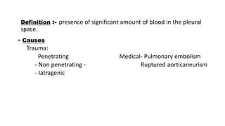
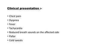
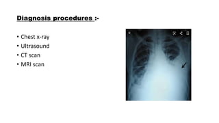
2. Definition :- presence of significant amount of blood in the pleural
space.
• Causes
Trauma:
Penetrating Medical- Pulmonary embolism
- Non penetrating - Ruptured aorticaneurism
- Iatragenic
3. Clinical presentation :-
• Chest pain
• Dyspnea
• Fever
• Tachycardia
• Reduced breath sounds on the affected side
• Pallor
• Cold sweats
4. source of blood
• - Chest wall
• - Lung
• - Mediastinum
• - Diaphragm
Blood entering the pleural space coagulate rapidly and defibrinated due
to physiologic movement of heart & lung.
5. Traumatic hemothorax
• High incidence in blunt chest trauma
• Hemothorax most common with displaced rib #
• Concomitant occurrence of hemothorax & pneumothorax is common
whether the trauma is blunt or penetrating.
Diagnosis: CXR ,CT scan
Treatment:
Immediate ICT insertion
VATS
6. VATS ( video–assisted thoracoscopic surgery )
Indications:
• no precise criteria available but
• - If Bleeding >200 ml/hr & no signs of slowing bleeding
• (bleeding is not from misplaced central line should be ensured)
• - Exsanguinating hemorrhage through the chest tube
VATS is very effective in –
- Hemodynamically stable patient with
persistent bleeding.
- Controlling bleeding from intercostal
vessels with lung lacerations
7. Complications of Haemothorax
• Clot Retention(3%)
• Pleural infection (3 to 4 %)
• Pleural effusion (13-34%)
• Fibrothorax(<1%)
8. Iatragenic Hemothorax
Causes-
• Central venous catheterization
• Thoracocentesis
• Pleural biopsy
• Rare cause
• percutaneous lung aspiration or biopsy
• trans bronchial biopsy
• In ICU- common following invasive procedure in patient with CRF
Treatment - ICT
11. Haemothorax complication
anticoagulant therapy :-
• Occurs mainly in treatment of pulmonary embolism
• Drugs responsible
• heparin
• warfarin
• enoxaparin
• Hemothorax is apperent 4-7 days of initiation of treatment mainly
Treatment
• - Discontinuation of anticoagulant
• - Immediate ICT
12. Physiotherapy treatment :-
• The patient clinical picture should lead the physiotherepist in
deciding what treatment is suitable.
• If the patient has a chest tube and intercostel drain in ,the treatment
might be different from when the patient had surgery .
• Help to improve ventilation , oxygenation and to re-inflate atelectatic
lung areas. This could be done through Deep breathing exercise
technique.
• Assisting with mobalisation or strengthening exercises to improve
patient’s exercise tolerance and mobility.
• Help to maintain airway clearance. This could be done by assisted
coughing techniques to help clear any secretions.