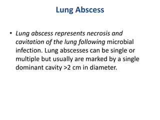
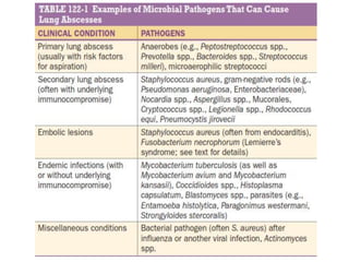
Lung Abscess
Lia Trapaidze
Lung Abscess
• Lung abscess represents necrosis and
cavitation of the lung following microbial
infection. Lung abscesses can be single or
multiple but usually are marked by a single
dominant cavity >2 cm in diameter.
Lung Abscess
• ETIOLOGY
• Lung abscesses are usually characterized as either
primary (~80% of cases) or secondary.
• Primary lung abscesses usually arise from aspiration,
are often caused principally by anaerobic bacteria, and
occur in the absence of an underlying pulmonary or
systemic condition.
• Secondary lung abscesses arise in the setting of an
underlying condition, such as a postobstructive process
(e.g., a bronchial foreign body or tumor) or a systemic
process (e.g., HIV infection or another
immunocompromising condition).
• Lung abscesses can also be characterized as acute (<4–
6 weeks in duration) or chronic (~40% of cases).
Lung Abscess
• PATHOGENESIS
• Primary Lung Abscesses
• The development of primary lung abscesses is
thought to originate when chiefly anaerobic
bacteria (as well as microaerophilic streptococci)
in the gingival crevices are aspirated into the lung
parenchyma in a susceptible host.
• Pneumonitis develops initially (exacerbated in
part by tissue damage caused by gastric acid);
then, over a period of 7–14 days, the anaerobic
bacteria produce parenchymal necrosis and
cavitation whose extent depends on host–
pathogen interaction.
Lung Abscess
• PATHOGENESIS
• Secondary Lung Abscesses
• The pathogenesis of secondary abscesses depends on the
predisposing factor. For example, in cases of bronchial obstruction
from malignancy or a foreign body, the obstructing lesion prevents
clearance of oropharyngeal secretions, leading to abscess
development.
• With underlying systemic conditions (e.g., immunosuppression
after bone marrow or solid organ transplantation), impaired host
defense mechanisms lead to increased susceptibility to the
development of lung abscesses caused by a broad range of
pathogens, including opportunistic organisms.
• Lung abscesses also arise from septic emboli, either in tricuspid
valve endocarditis (often involving Staphylococcus aureus) or in
Lemierre’s syndrome, in which an infection begins in the pharynx
(classically involving Fusobacterium necrophorum) and then spreads
to the neck and the carotid sheath (which contains the jugular vein)
to cause septic thrombophlebitis.
Lung Abscess
• PATHOLOGY AND MICROBIOLOGY
• Primary Lung Abscesses
• The dependent segments (posterior upper lobes
and superior lower lobes) are the most common
locations of primary lung abscesses, given the
predisposition of aspirated materials to be
deposited in these areas.
• Generally, the right lung is affected more
commonly than the left because the right
mainstem bronchus is less angulated.
• The microbiology of primary lung abscesses is
often polymicrobial, primarily including anaerobic
organisms as well as microaerophilic streptococci.
Lung Abscess
• PATHOLOGY AND MICROBIOLOGY
• Secondary Lung Abscesses
• The location of secondary abscesses may vary with the
underlying cause. The microbiology of secondary lung
abscesses can encompass quite a broad bacterial spectrum,
with infection by Pseudomonas aeruginosa and other
gram-negative rods most common.
• In addition, a broad array of pathogens can be identified in
patients from certain endemic areas and in specific clinical
scenarios (e.g., a significant incidence of fungal infections
among immunosuppressed patients following bone marrow
or solid organ transplantation).
• Because immunocompromised hosts and patients without
the classic presentation of a primary lung abscess can be
infected with a wide array of unusual organisms.
Lung Abscess
• CLINICAL MANIFESTATIONS
• Clinical manifestations may initially be similar
to those of pneumonia, with fevers, cough,
sputum production, and chest pain;
• A more chronic and indolent presentation that
includes night sweats, fatigue, and anemia is
often observed with anaerobic lung abscesses.
Lung Abscess
• CLINICAL MANIFESTATIONS
• A subset of patients with putrid lung abscesses may
report discolored phlegm and foul-tasting or foul-
smelling sputum.
• Patients with lung abscesses due to non-anaerobic
organisms, such as S. aureus, may present with a more
fulminant course characterized by high fevers and rapid
progression.
• Findings on physical examination may include fevers,
poor dentition, and/or gingival disease as well as
amphoric and/or cavernous breath sounds on lung
auscultation.
• Additional findings may include digital clubbing and
the absence of a gag reflex.
Lung Abscess
• DIFFERENTIAL DIAGNOSIS
• The differential diagnosis of lung abscesses is broad
and includes other noninfectious processes that result
in cavitary lung lesions, including lung infarction,
malignancy, sequestration, cryptogenic organizing
pneumonia, sarcoidosis, vasculitides and other
autoimmune diseases (e.g., granulomatosis with
polyangiitis), lung cysts or bullae containing fluid, and
septic emboli (e.g., from tricuspid valve endocarditis).
• Other less common entities can include pulmonary
manifestations of diseases that usually present at
locations other than the chest (e.g., inflammatory
bowel disease, pyoderma gangrenosum).
Lung Abscess
• DIAGNOSIS
• Lung abscesses are documented by chest imaging.
Although a chest radiograph usually detects a thick-walled
cavity with an air-fluid level, CT permits better definition
and may provide earlier evidence of cavitation. CT may also
yield additional information regarding a possible
underlying cause of lung abscess, such as malignancy, and
may help distinguish a peripheral lung abscess from a
pleural infection. This distinction has important
implications for treatment because a pleural space
infection, such as an empyema, may require urgent
drainage.
• While sputum can be collected noninvasively for Gram’s
stain and culture, which may yield a pathogen, the infection
is likely to be polymicrobial, and culture results may not
reflect the presence of anaerobic organisms.
Lung Abscess
• DIAGNOSIS
• When a secondary lung abscess is present or empirical
therapy fails to elicit a response, sputum and blood
cultures are advised in addition to serologic studies for
opportunistic pathogens (e.g., viruses and fungi causing
infections in immunocompromised hosts).
• Additional diagnostics, such as bronchoscopy with
bronchoalveolar lavage or protected brush specimen
collection and CT-guided percutaneous needle aspiration,
can be undertaken.
• Early diagnostics in secondary abscesses, especially in
immunocompromised hosts, are particularly important,
because the patients involved may be especially fragile, at
risk for infection with a broad array of pathogens, and
therefore less likely than other patients to respond to
empirical therapy.
Lung Abscess
• TREATMENT
• For primary lung abscesses, the recommended
regimens are:
• (1) clindamycin (600 mg IV three times daily; then, with
the disappearance of fever and clinical improvement,
300 mg PO four times daily) or
• (2) an IV-administered ß-lactam/ß-lactamase
combination, followed—once the patient’s condition is
stable—by orally administered amoxicillin-clavulanate.
• This therapy should be continued until imaging
demonstrates that the lung abscess has cleared or
regressed to a small scar.
• Treatment duration may range from 3–4 weeks to as
long as 14 weeks.
Lung Abscess
• TREATMENT
• In secondary lung abscesses, antibiotic
coverage should be directed at the identified
pathogen, and a prolonged course (until
resolution of the abscess is documented) is
often required.
• Treatment regimens and courses vary widely,
depending on the immune state of the host
and the identified pathogen.
Lung Abscess
• TREATMENT
• An abscess >6–8 cm in diameter is less likely to
respond to antibiotic therapy without additional
interventions.
• Options for patients who do not respond to
antibiotics and whose additional diagnostic
studies fail to identify an additional pathogen
that can be treated include surgical resection and
percutaneous drainage of the abscess, especially
when the patient is a poor surgical candidate.
• Possible complications of percutaneous drainage
include bacterial contamination of the pleural
space as well as pneumothorax and
hemothorax.

Lung Abscess.pptx for medical students..

  • 1.
  • 2.
    Lung Abscess • Lungabscess represents necrosis and cavitation of the lung following microbial infection. Lung abscesses can be single or multiple but usually are marked by a single dominant cavity >2 cm in diameter.
  • 3.
    Lung Abscess • ETIOLOGY •Lung abscesses are usually characterized as either primary (~80% of cases) or secondary. • Primary lung abscesses usually arise from aspiration, are often caused principally by anaerobic bacteria, and occur in the absence of an underlying pulmonary or systemic condition. • Secondary lung abscesses arise in the setting of an underlying condition, such as a postobstructive process (e.g., a bronchial foreign body or tumor) or a systemic process (e.g., HIV infection or another immunocompromising condition). • Lung abscesses can also be characterized as acute (<4– 6 weeks in duration) or chronic (~40% of cases).
  • 4.
    Lung Abscess • PATHOGENESIS •Primary Lung Abscesses • The development of primary lung abscesses is thought to originate when chiefly anaerobic bacteria (as well as microaerophilic streptococci) in the gingival crevices are aspirated into the lung parenchyma in a susceptible host. • Pneumonitis develops initially (exacerbated in part by tissue damage caused by gastric acid); then, over a period of 7–14 days, the anaerobic bacteria produce parenchymal necrosis and cavitation whose extent depends on host– pathogen interaction.
  • 6.
    Lung Abscess • PATHOGENESIS •Secondary Lung Abscesses • The pathogenesis of secondary abscesses depends on the predisposing factor. For example, in cases of bronchial obstruction from malignancy or a foreign body, the obstructing lesion prevents clearance of oropharyngeal secretions, leading to abscess development. • With underlying systemic conditions (e.g., immunosuppression after bone marrow or solid organ transplantation), impaired host defense mechanisms lead to increased susceptibility to the development of lung abscesses caused by a broad range of pathogens, including opportunistic organisms. • Lung abscesses also arise from septic emboli, either in tricuspid valve endocarditis (often involving Staphylococcus aureus) or in Lemierre’s syndrome, in which an infection begins in the pharynx (classically involving Fusobacterium necrophorum) and then spreads to the neck and the carotid sheath (which contains the jugular vein) to cause septic thrombophlebitis.
  • 8.
    Lung Abscess • PATHOLOGYAND MICROBIOLOGY • Primary Lung Abscesses • The dependent segments (posterior upper lobes and superior lower lobes) are the most common locations of primary lung abscesses, given the predisposition of aspirated materials to be deposited in these areas. • Generally, the right lung is affected more commonly than the left because the right mainstem bronchus is less angulated. • The microbiology of primary lung abscesses is often polymicrobial, primarily including anaerobic organisms as well as microaerophilic streptococci.
  • 9.
    Lung Abscess • PATHOLOGYAND MICROBIOLOGY • Secondary Lung Abscesses • The location of secondary abscesses may vary with the underlying cause. The microbiology of secondary lung abscesses can encompass quite a broad bacterial spectrum, with infection by Pseudomonas aeruginosa and other gram-negative rods most common. • In addition, a broad array of pathogens can be identified in patients from certain endemic areas and in specific clinical scenarios (e.g., a significant incidence of fungal infections among immunosuppressed patients following bone marrow or solid organ transplantation). • Because immunocompromised hosts and patients without the classic presentation of a primary lung abscess can be infected with a wide array of unusual organisms.
  • 10.
    Lung Abscess • CLINICALMANIFESTATIONS • Clinical manifestations may initially be similar to those of pneumonia, with fevers, cough, sputum production, and chest pain; • A more chronic and indolent presentation that includes night sweats, fatigue, and anemia is often observed with anaerobic lung abscesses.
  • 11.
    Lung Abscess • CLINICALMANIFESTATIONS • A subset of patients with putrid lung abscesses may report discolored phlegm and foul-tasting or foul- smelling sputum. • Patients with lung abscesses due to non-anaerobic organisms, such as S. aureus, may present with a more fulminant course characterized by high fevers and rapid progression. • Findings on physical examination may include fevers, poor dentition, and/or gingival disease as well as amphoric and/or cavernous breath sounds on lung auscultation. • Additional findings may include digital clubbing and the absence of a gag reflex.
  • 12.
    Lung Abscess • DIFFERENTIALDIAGNOSIS • The differential diagnosis of lung abscesses is broad and includes other noninfectious processes that result in cavitary lung lesions, including lung infarction, malignancy, sequestration, cryptogenic organizing pneumonia, sarcoidosis, vasculitides and other autoimmune diseases (e.g., granulomatosis with polyangiitis), lung cysts or bullae containing fluid, and septic emboli (e.g., from tricuspid valve endocarditis). • Other less common entities can include pulmonary manifestations of diseases that usually present at locations other than the chest (e.g., inflammatory bowel disease, pyoderma gangrenosum).
  • 13.
    Lung Abscess • DIAGNOSIS •Lung abscesses are documented by chest imaging. Although a chest radiograph usually detects a thick-walled cavity with an air-fluid level, CT permits better definition and may provide earlier evidence of cavitation. CT may also yield additional information regarding a possible underlying cause of lung abscess, such as malignancy, and may help distinguish a peripheral lung abscess from a pleural infection. This distinction has important implications for treatment because a pleural space infection, such as an empyema, may require urgent drainage. • While sputum can be collected noninvasively for Gram’s stain and culture, which may yield a pathogen, the infection is likely to be polymicrobial, and culture results may not reflect the presence of anaerobic organisms.
  • 14.
    Lung Abscess • DIAGNOSIS •When a secondary lung abscess is present or empirical therapy fails to elicit a response, sputum and blood cultures are advised in addition to serologic studies for opportunistic pathogens (e.g., viruses and fungi causing infections in immunocompromised hosts). • Additional diagnostics, such as bronchoscopy with bronchoalveolar lavage or protected brush specimen collection and CT-guided percutaneous needle aspiration, can be undertaken. • Early diagnostics in secondary abscesses, especially in immunocompromised hosts, are particularly important, because the patients involved may be especially fragile, at risk for infection with a broad array of pathogens, and therefore less likely than other patients to respond to empirical therapy.
  • 15.
    Lung Abscess • TREATMENT •For primary lung abscesses, the recommended regimens are: • (1) clindamycin (600 mg IV three times daily; then, with the disappearance of fever and clinical improvement, 300 mg PO four times daily) or • (2) an IV-administered ß-lactam/ß-lactamase combination, followed—once the patient’s condition is stable—by orally administered amoxicillin-clavulanate. • This therapy should be continued until imaging demonstrates that the lung abscess has cleared or regressed to a small scar. • Treatment duration may range from 3–4 weeks to as long as 14 weeks.
  • 16.
    Lung Abscess • TREATMENT •In secondary lung abscesses, antibiotic coverage should be directed at the identified pathogen, and a prolonged course (until resolution of the abscess is documented) is often required. • Treatment regimens and courses vary widely, depending on the immune state of the host and the identified pathogen.
  • 17.
    Lung Abscess • TREATMENT •An abscess >6–8 cm in diameter is less likely to respond to antibiotic therapy without additional interventions. • Options for patients who do not respond to antibiotics and whose additional diagnostic studies fail to identify an additional pathogen that can be treated include surgical resection and percutaneous drainage of the abscess, especially when the patient is a poor surgical candidate. • Possible complications of percutaneous drainage include bacterial contamination of the pleural space as well as pneumothorax and hemothorax.