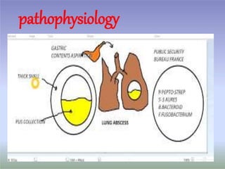
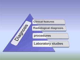
Lung abscess
Definition:-
 Microbial infection causes suppurative necrosis of the
lung parenchyma producing a cavity. Often communicate
with large airways resulting in cough, expectoration of
purulent sputum and AFL on imaging studies .
 May be more than one cavity, but usually one is large
and dominant .
 Majority of cases due to anaerobic mouth bacterial and
follow aspiration
 More common in pre-abx era with progression bacterial
pneumonia +/- empyema
Lung Abscess is;-
Classification
 Duration of symptoms prior to diagnosis;
 acute < 1month,
 chronic > 1 month
 Primary lung abscess or Secondary lung abscess;
Primary lung abscess:
used when abscess develops in
individuals prone to aspiration or in
general good health
80% of lung abscess is primary (50%
of these associated with putrid sputum)
Secondary lung abscess:
obstructive airway neoplasm as a
complication of intrathoracic surgery or
systemic condition/treatment that
compromises host immune defense
mechanisms
Putrid lung abscess (foul odor of expectorated sputum)
Microbiology
 Bacteria
o Usually: mouthflora anaerobes,Most frequently isolated anaerobes:
• Peptostreptococcus,
• Fusobacterium nucleatum,
• Prevotella melaningogenica
o Less common:
• Staphylococcus aureus, . Streptococcus pyogenes,
• Pseudomonas aeruginosa, Klebsiella pneumoniae ,
• Streptococcus pneumoniae
• gram-negative bacilli, such as E. coli , Haemophilus influenzae type B
• Legionella,, Nocardia asteroides
NoninfectiousCauses
 Neoplasms
Primary lung cancer, metastatic carcinoma, lymphoma
 Pulmonary infarction
Due to bland embolus (may be secondarily infected in <5%)
 Septic embolism
Tricuspid endocarditis due to S. aureus and others
(typically with positive blood cultures), jugular venous septic
phlebitis due to Fusobacterium necrophorum (Lemierre
syndrome)
Noninfectious Causes
Vasculitis
Wegener's granulomatosis, rheumatoid lung nodule
Airway disease
Bullae, blebs, or cystic bronchiectasis (usually thin-walled)
Developmental
Pulmonary sequestration
Other
Sarcoidosis, transdiaphragmatic bowel herniation giving appearance
of cavity with airfluid level
pathophysiology
RiskFactorsforlungabscess:
 Predisposition to aspiration
 stroke, drugs, general anesthesia, dysphasia, respiratory muscle
dysfunction, tooth extraction, mechanical interference with anatomic,
physiologic barriers to aspiration ,seizers .
 45% of health adults aspirate during sleep; 70% of patients with
AMS aspirate
 1ml of saliva with > 109 live bacteria.
 Poor dentition with gingivitis
 resulting in an unusually high density of oral anaerobic organism
particular in gingival crevices .
 Airway Obstruction
 Neoplasm: 50% of lung abscesses in patients >50 are associated
with lung carcinoma (post-obstruction vs. necrotic tumor)
 Foreign bodies, extrinsic compression (enlarged lymph node
 Patients with cell-mediated immunity (AIDS,
transplant):
OI pathogens such as mycobacteria, Nocardia,Aspergillus,
Rhodococccus
 Neutropenic patients:
 P. aeruginosa, S. aureus, fungi (Aspergillus, Zygomycetes)
 The onset may be abrupt or gradual.
Clinical features
 Symptoms include fever, sweating, cough and chest pain
simulating pneumonia.
 The cough is often non productive at first or may produce mucoid
or mucopurulent expectorate from bronchial inflammation close to
the abscess area and sometimes there is blood streaking.
Symptoms
 There is an expectoration of foul-smelling brown or gray sputum
(in anaerobic organisms) or green or yellow sputum
 If this happens suddenly in large quantities, this denotes a
rupture of the abscess cavity into the bronchus and blood
streaking is common.
 Pleuritic chest pain, especially with coughing is common
because the abscess is near to the pleura.
 Weight loss, anaemia, and clubbing or pulmonary osteo
arthropathy appear when the abscess becomes chronic (8-12
weeks after onset).
 May be minimal. Consolidation due to pneumonia surrounding
the abscess is the most frequent finding.
 Inspiratory rales and pleural rub may be heard.
 Rarely the amphoric or cavernous breath sounds are heard
 Rupture into the pleural space produces signs of effusion or
hydro pneumothorax.
Signs
 Digital clubbing may develop rapidly.
Amebic lung abscess
Patients who develop an amebic lung abscess often have symptoms
associated with a liver abscess.
These may include right upper quadrant pain and fever.
After perforation of the liver abscess into the lung, the patient may
develop a cough and expectorate a chocolate or anchovy paste–like
sputum that has no odor.
B) Radiological diagnosis
 Later this opacity develops a cavity with fluid level. may have
smooth and regular walls.
 Chest films may also reveal associated primary lesions,
e.g., a bronchogenic carcinoma.
malignant abcess shows eccenteric cavitation with thick
rough irregular walls.
 Low-attenuation central region and rim enhancement on
CT scan
 A dense shadow is the initial finding before rupture.
 Lung abscesses as a result of aspiration most frequently
occur in the posterior segments of the upper lobes or the
superior segments of the lower lobes.
PA and lateral view show Rt upper lobe consolidation
Contrast-enhanced (CT)
scan demonstrates large
focal area of decreased
attenuation with rim
enhancement (arrow)
characteristic of lung
abscess.
PAand lateral chest
radiographs 3 weeks later show
decreased size of lung abscess
and development of cavitation
with fluid level . The patient was
a 43-year-old woman with lung
abscess secondary to
Haemophilus infection
A cavity with an air fluid
level at apical segment of
upper lobe of right lung
due to lung abscess.
Pneumococcal pneumonia
complicated by lung necrosis
and abscess formation.
A lateral chest radiographshows
air-fluid level characteristic of
lung abscess.
A 54-year-old patient developed cough with foul-smellingsputum
production. A chest radiograph shows lung abscess in the left
lower lobe, superior segment.
Ultrasonography
Peripheral lung abscesses with pleural contact or included inside
a lung consolidation aredetectable using lung ultrasonography
 Lung abscess appears as a rounded hypoechoic lesion with
an outer margin.
 If a cavity is present, additional nondependent hyperechoic signs
are generated by the gas-tissue interface.
Diagnostic material uncontaminated by bacteria colonizing the upper
airway may be obtained for anaerobic culture from the following:
Blood cultures are infrequently positive in patients with lung abscess.
Transtracheal aspirateBlood culture
Pleural fluid (if empyema present) Transthoracic pulmonary aspirate
Surgical specimens Fiberoptic bronchoscopy with
protected brush
Bronchoalveolar lavage with
quantitative cultures
Expectorated sputum and other methods of sampling the upper airway do not
yield useful results for anaerobic culture because the oral cavity is extensively
colonized with anaerobes.
Procedures
Flexible fiberoptic bronchoscopy is performed to exclude bronchogenic
carcinoma whenever bronchial obstruction is suspected.
Laboratory Studies
complete white blood cell count with differential may reveal leukocytosis.
Gram stain, culture, and sensitivity.
If tuberculosis is suspected, ZeilNelsen and mycobacterial culture is requested.
Routine investigations:
e.g blood glucose , liver functions tests , renal functions test, electrolytes
Complications of lung abscess
 Rupture into pleural space causing empyema
 Pleural fibrosis
 Trapped lung
 Respiratory failure
 Bronchopleural fistula
 Pleural cutaneous fistula
In a patient with coexisting empyema and lung abscess,
draining the empyema while continuing prolonged antibiotic therapy is
often necessary.
 Cavitating lung cancer
 Localized empyema
 Infected bulla containing a fluid level
 Infected congenital pulmonary lesion,
such as bronchogenic cyst or sequestration
 Pulmonary hematoma
 Cavitating pneumoconiosis
 Hiatus hernia
 Lung parasites (eg, hydatid cyst, Paragonimus infection)
 Actinomycosis
 Wegener granulomatosis and other vasculitides
 Cavitating lung infarcts
 Cavitating sarcoidosis
Differential diagnosis
Treatment
Treatment of lung abscess is guided by the available microbiology and knowledge
of the underlying or associated conditions.
No treatment recommendations have been issued by major societies specifically
for lung abscess;
Can combine Penicillin + Flagyl
Flagyl as monotherapy is inferior to clindamycin as it is not active against micro
aerophilic streptococci and anaerobic cocci that are common constituents of
anaerobic lung infection (which are sensitive to Penicillin).
Several anaerobes may produce beta-lactamase , develop resistance to penicillin.
Antibiotic therapy
Anaerobic lung infection
Clindamycin is the treatment of choice ..
Other options: carbopenems, quionlones with good anaerobic activity (Moxiflox,
Gatiflox)
Bronchoscopy
No advantage of bronchscopic/postural drainage compared to antibiotics
Reserve for patients who do not respond
Indications for early bronchoscopy
underlying malignancy
lack of aspiration risk
edentulous patients
age >50 with long smoking history
lack of systemic symptoms
Endoscopic drainage is considered if an airway connection to the cavity can
be demonstrated.
o
o
o
Surgical intervention
 Surgery is rarely required for patients with uncomplicated lung abscess
The usual indications for surgery are
-- failure to respond to medical management,
-- suspected neoplasm, or
-- congenital lung malformation.
 When conventional therapy fails, either percutaneous catheter drainage
or surgical resection is usually considered.
 The surgical procedure performed is either lobectomy or pneumonectomy
 Consider lobectomy / pneumonectomry with large cavities (>8cm),
resistant organisms lik Pseudomonas, obstructing neoplasm,
massive hemorrhage
.

lung abscess

  • 1.
  • 3.
    Definition:-  Microbial infectioncauses suppurative necrosis of the lung parenchyma producing a cavity. Often communicate with large airways resulting in cough, expectoration of purulent sputum and AFL on imaging studies .  May be more than one cavity, but usually one is large and dominant .  Majority of cases due to anaerobic mouth bacterial and follow aspiration  More common in pre-abx era with progression bacterial pneumonia +/- empyema Lung Abscess is;-
  • 4.
    Classification  Duration ofsymptoms prior to diagnosis;  acute < 1month,  chronic > 1 month  Primary lung abscess or Secondary lung abscess; Primary lung abscess: used when abscess develops in individuals prone to aspiration or in general good health 80% of lung abscess is primary (50% of these associated with putrid sputum) Secondary lung abscess: obstructive airway neoplasm as a complication of intrathoracic surgery or systemic condition/treatment that compromises host immune defense mechanisms Putrid lung abscess (foul odor of expectorated sputum)
  • 5.
    Microbiology  Bacteria o Usually:mouthflora anaerobes,Most frequently isolated anaerobes: • Peptostreptococcus, • Fusobacterium nucleatum, • Prevotella melaningogenica o Less common: • Staphylococcus aureus, . Streptococcus pyogenes, • Pseudomonas aeruginosa, Klebsiella pneumoniae , • Streptococcus pneumoniae • gram-negative bacilli, such as E. coli , Haemophilus influenzae type B • Legionella,, Nocardia asteroides
  • 6.
    NoninfectiousCauses  Neoplasms Primary lungcancer, metastatic carcinoma, lymphoma  Pulmonary infarction Due to bland embolus (may be secondarily infected in <5%)  Septic embolism Tricuspid endocarditis due to S. aureus and others (typically with positive blood cultures), jugular venous septic phlebitis due to Fusobacterium necrophorum (Lemierre syndrome)
  • 7.
    Noninfectious Causes Vasculitis Wegener's granulomatosis,rheumatoid lung nodule Airway disease Bullae, blebs, or cystic bronchiectasis (usually thin-walled) Developmental Pulmonary sequestration Other Sarcoidosis, transdiaphragmatic bowel herniation giving appearance of cavity with airfluid level
  • 8.
  • 9.
    RiskFactorsforlungabscess:  Predisposition toaspiration  stroke, drugs, general anesthesia, dysphasia, respiratory muscle dysfunction, tooth extraction, mechanical interference with anatomic, physiologic barriers to aspiration ,seizers .  45% of health adults aspirate during sleep; 70% of patients with AMS aspirate  1ml of saliva with > 109 live bacteria.  Poor dentition with gingivitis  resulting in an unusually high density of oral anaerobic organism particular in gingival crevices .
  • 10.
     Airway Obstruction Neoplasm: 50% of lung abscesses in patients >50 are associated with lung carcinoma (post-obstruction vs. necrotic tumor)  Foreign bodies, extrinsic compression (enlarged lymph node  Patients with cell-mediated immunity (AIDS, transplant): OI pathogens such as mycobacteria, Nocardia,Aspergillus, Rhodococccus  Neutropenic patients:  P. aeruginosa, S. aureus, fungi (Aspergillus, Zygomycetes)
  • 12.
     The onsetmay be abrupt or gradual. Clinical features  Symptoms include fever, sweating, cough and chest pain simulating pneumonia.  The cough is often non productive at first or may produce mucoid or mucopurulent expectorate from bronchial inflammation close to the abscess area and sometimes there is blood streaking. Symptoms
  • 13.
     There isan expectoration of foul-smelling brown or gray sputum (in anaerobic organisms) or green or yellow sputum  If this happens suddenly in large quantities, this denotes a rupture of the abscess cavity into the bronchus and blood streaking is common.  Pleuritic chest pain, especially with coughing is common because the abscess is near to the pleura.  Weight loss, anaemia, and clubbing or pulmonary osteo arthropathy appear when the abscess becomes chronic (8-12 weeks after onset).
  • 14.
     May beminimal. Consolidation due to pneumonia surrounding the abscess is the most frequent finding.  Inspiratory rales and pleural rub may be heard.  Rarely the amphoric or cavernous breath sounds are heard  Rupture into the pleural space produces signs of effusion or hydro pneumothorax. Signs  Digital clubbing may develop rapidly.
  • 15.
    Amebic lung abscess Patientswho develop an amebic lung abscess often have symptoms associated with a liver abscess. These may include right upper quadrant pain and fever. After perforation of the liver abscess into the lung, the patient may develop a cough and expectorate a chocolate or anchovy paste–like sputum that has no odor.
  • 16.
    B) Radiological diagnosis Later this opacity develops a cavity with fluid level. may have smooth and regular walls.  Chest films may also reveal associated primary lesions, e.g., a bronchogenic carcinoma. malignant abcess shows eccenteric cavitation with thick rough irregular walls.  Low-attenuation central region and rim enhancement on CT scan  A dense shadow is the initial finding before rupture.  Lung abscesses as a result of aspiration most frequently occur in the posterior segments of the upper lobes or the superior segments of the lower lobes.
  • 17.
    PA and lateralview show Rt upper lobe consolidation
  • 18.
    Contrast-enhanced (CT) scan demonstrateslarge focal area of decreased attenuation with rim enhancement (arrow) characteristic of lung abscess.
  • 19.
    PAand lateral chest radiographs3 weeks later show decreased size of lung abscess and development of cavitation with fluid level . The patient was a 43-year-old woman with lung abscess secondary to Haemophilus infection
  • 20.
    A cavity withan air fluid level at apical segment of upper lobe of right lung due to lung abscess.
  • 21.
    Pneumococcal pneumonia complicated bylung necrosis and abscess formation. A lateral chest radiographshows air-fluid level characteristic of lung abscess.
  • 22.
    A 54-year-old patientdeveloped cough with foul-smellingsputum production. A chest radiograph shows lung abscess in the left lower lobe, superior segment.
  • 23.
    Ultrasonography Peripheral lung abscesseswith pleural contact or included inside a lung consolidation aredetectable using lung ultrasonography  Lung abscess appears as a rounded hypoechoic lesion with an outer margin.  If a cavity is present, additional nondependent hyperechoic signs are generated by the gas-tissue interface.
  • 24.
    Diagnostic material uncontaminatedby bacteria colonizing the upper airway may be obtained for anaerobic culture from the following: Blood cultures are infrequently positive in patients with lung abscess. Transtracheal aspirateBlood culture Pleural fluid (if empyema present) Transthoracic pulmonary aspirate Surgical specimens Fiberoptic bronchoscopy with protected brush Bronchoalveolar lavage with quantitative cultures Expectorated sputum and other methods of sampling the upper airway do not yield useful results for anaerobic culture because the oral cavity is extensively colonized with anaerobes. Procedures Flexible fiberoptic bronchoscopy is performed to exclude bronchogenic carcinoma whenever bronchial obstruction is suspected.
  • 25.
    Laboratory Studies complete whiteblood cell count with differential may reveal leukocytosis. Gram stain, culture, and sensitivity. If tuberculosis is suspected, ZeilNelsen and mycobacterial culture is requested. Routine investigations: e.g blood glucose , liver functions tests , renal functions test, electrolytes
  • 26.
    Complications of lungabscess  Rupture into pleural space causing empyema  Pleural fibrosis  Trapped lung  Respiratory failure  Bronchopleural fistula  Pleural cutaneous fistula In a patient with coexisting empyema and lung abscess, draining the empyema while continuing prolonged antibiotic therapy is often necessary.
  • 27.
     Cavitating lungcancer  Localized empyema  Infected bulla containing a fluid level  Infected congenital pulmonary lesion, such as bronchogenic cyst or sequestration  Pulmonary hematoma  Cavitating pneumoconiosis  Hiatus hernia  Lung parasites (eg, hydatid cyst, Paragonimus infection)  Actinomycosis  Wegener granulomatosis and other vasculitides  Cavitating lung infarcts  Cavitating sarcoidosis Differential diagnosis
  • 28.
    Treatment Treatment of lungabscess is guided by the available microbiology and knowledge of the underlying or associated conditions. No treatment recommendations have been issued by major societies specifically for lung abscess; Can combine Penicillin + Flagyl Flagyl as monotherapy is inferior to clindamycin as it is not active against micro aerophilic streptococci and anaerobic cocci that are common constituents of anaerobic lung infection (which are sensitive to Penicillin). Several anaerobes may produce beta-lactamase , develop resistance to penicillin. Antibiotic therapy Anaerobic lung infection Clindamycin is the treatment of choice .. Other options: carbopenems, quionlones with good anaerobic activity (Moxiflox, Gatiflox)
  • 29.
    Bronchoscopy No advantage ofbronchscopic/postural drainage compared to antibiotics Reserve for patients who do not respond Indications for early bronchoscopy underlying malignancy lack of aspiration risk edentulous patients age >50 with long smoking history lack of systemic symptoms Endoscopic drainage is considered if an airway connection to the cavity can be demonstrated.
  • 30.
    o o o Surgical intervention  Surgeryis rarely required for patients with uncomplicated lung abscess The usual indications for surgery are -- failure to respond to medical management, -- suspected neoplasm, or -- congenital lung malformation.  When conventional therapy fails, either percutaneous catheter drainage or surgical resection is usually considered.  The surgical procedure performed is either lobectomy or pneumonectomy  Consider lobectomy / pneumonectomry with large cavities (>8cm), resistant organisms lik Pseudomonas, obstructing neoplasm, massive hemorrhage .