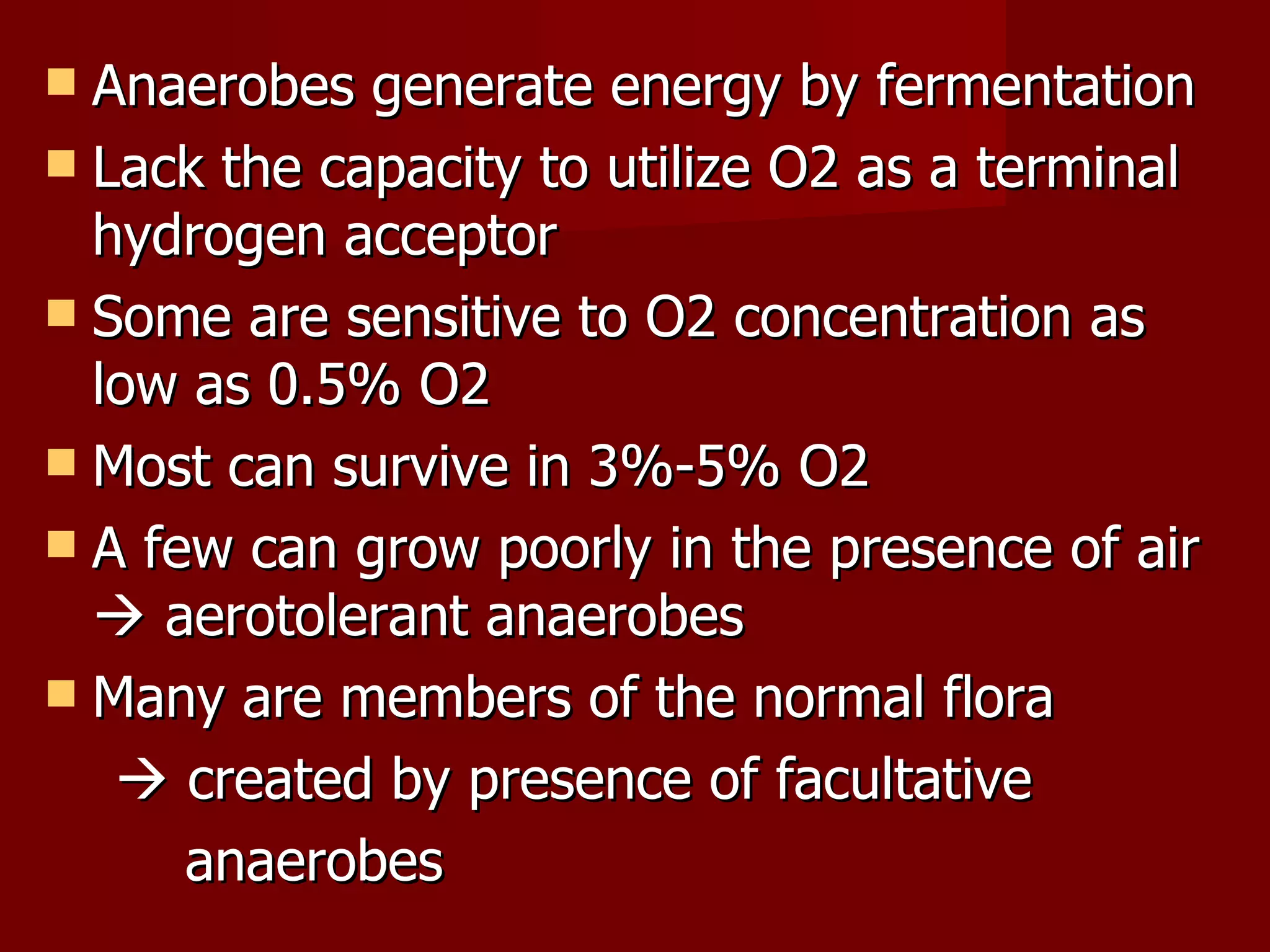
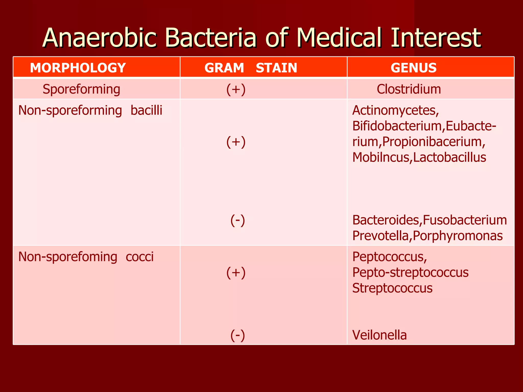
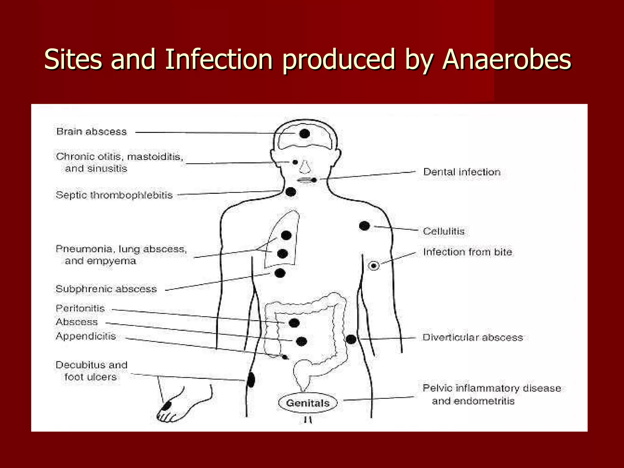
ANAEROBIC BACTERIOLOGY     Dr. Ma. Ellery M. Mendez   FPAMS , FPAAM, DPSMAP
Anaerobes generate energy by fermentation Lack the capacity to utilize O2 as a terminal  hydrogen acceptor Some are sensitive to O2 concentration as low as 0.5% O2 Most can survive in 3%-5% O2 A few can grow poorly in the presence of air    aerotolerant anaerobes Many are members of the normal flora    created by presence of facultative  anaerobes
FACTORS  THAT  INHIBIT  THE GROWTH  OF ANAEROBES  BY OXYGEN 1. Toxic compounds are produced e.g. H2O2 , Superoxides 2. Absence of catalase & Superoxide  dismutase 3. Oxidation of essential sulfhydyl groups in enzymes without sufficient reducing power to regenerate them
FACTORS  RESPONSIBLE  FOR  THEIR VIRULENCE 1. Lipopolysaccharide - promotes abscess formation, enhanced coagulation 2. Polysaccharide capsule - correlated with abscess production 3. Enzymes a. Collagenase b. Heparinase * develop thrombophlebitis & septic emboli   4. Short chained fatty acids a. Butyrate-  seen in dental plaque  b. succinic acid –  reduces phagocytic killing
Multiplication of the opportunistic pathogens is facilitated by: 1.  inhibition of phagocytosis & intracellular killing  by PMN in the presence of Bacteroides by: a. competition of opsonins b. inhibition by capsular materials 2.  protection of antibiotic susceptibility strains in  mixtures thru destruction by the ß-lactamases 3. utilization of O2 by facultative species that aids in  producing a suitable environment for growth of  anaerobe
Anaerobic Bacteria of Medical Interest MORPHOLOGY GRAM  STAIN GENUS Sporeforming (+) Clostridium Non-sporeforming  bacilli (+) (-) Actinomycetes, Bifidobacterium,Eubacte-rium,Propionibacerium, Mobilncus,Lactobacillus Bacteroides,Fusobacterium Prevotella,Porphyromonas Non-sporefoming  cocci  (+) (-) Peptococcus, Pepto-streptococcus Streptococcus Veilonella
CLINICAL  MANIFESTATION Clinical  hints  1. odor 2. tissue  3. location 4. necrotic tissue 5. endocarditis with (-) blood culture 6. infection associated with malignancy 7. black discoloration 8. blood containing exudates 9. associated with sulfur granules  10. Bacteremic feature with jaundice 11.  human bites
Sites and Infection produced by Anaerobes
 
LABORATORY  DIAGNOSIS A. COLLECTION Anaerobes are endogenous in nature I.  Appropriate specimens for anaerobic culture : 1. pus  2. pleural fluid 3. urine 4. pulmonary  secretions 5. uterine secretions or sinus tract material
II. Collection by needle aspiration is  preferrable than swab culture because of a. better survival of pathogen b. greater quantity of specimen c. less contamination with extraneous  organism are often achieved
B.  HANDLING If a swab must be used, a 2 tube system must be used    1 st   tube contains swab in O2 free  CO2    2 nd  tube contains PRAS (pre-reduced  anaerobically steilized culture media) Specimen should be placed in anaerobic  transport device with gas mixture
C. Isolation Gram stain should be done in the  laboratory : a. choice of appropriate media & methods for culture b. quality control for the types of  bacteria that laboratory culture  reveal
A solid or liquid medium maybe used & must provide an   anaerobic environment    Anaerobic Culture System ANAEROBIC  JAR 1. Candle Jar - reduces  O2 environment - only  ↑  CO2  tension 2. Gas Pak Jar a. Palladium aluminum coated pellets - catalyst - chemically reduces O2 - reacts with residual O2 in the presence of H2 to form  H2O
b. Gas Pak envelope  - generates CO2 & H2 gases c. Methylene blue strip  - indicator blue  ->  (+) O2 white  ->   (-) O2 II. Anaerobic Glove Chamber - close system - used for premature babies -  e.g. incubator  III. Roll Tube -  has a pedal    gas ( CO2 & H2 ) would come out  -  place test tube directly to the outlet
D. IDENTIFICATION Plates are checked at  > 18-24 hours for faster growing species like Cl. Perfringens & B.fragilis  & daily thereafter up to   >  5-7 days for slowly growing species like Actinomyces, Eubacterium & propionibacterium  Genus   is determined by - gram stain, cellular morphology, Gas-liquid  chromotography Species  determination is based on fermentation of sugars & other biochemical determination
TREATMENT -  Susceptibility testing should be done - surgical drainage & resection of necrotic tissue - most are resistant to aminoglycosides - for Bacteroides gr oup, if resistant to Penicillin &  Cephalosporin, they may use: a. Clindamycin  b. Metronidazole  c. Chloramphenicol
THE  END

Anaerobic Bacteriology Lecture

  • 1.
    ANAEROBICBACTERIOLOGY Dr. Ma. Ellery M. Mendez FPAMS , FPAAM, DPSMAP
  • 2.
    Anaerobes generate energyby fermentation Lack the capacity to utilize O2 as a terminal hydrogen acceptor Some are sensitive to O2 concentration as low as 0.5% O2 Most can survive in 3%-5% O2 A few can grow poorly in the presence of air  aerotolerant anaerobes Many are members of the normal flora  created by presence of facultative anaerobes
  • 3.
    FACTORS THAT INHIBIT THE GROWTH OF ANAEROBES BY OXYGEN 1. Toxic compounds are produced e.g. H2O2 , Superoxides 2. Absence of catalase & Superoxide dismutase 3. Oxidation of essential sulfhydyl groups in enzymes without sufficient reducing power to regenerate them
  • 4.
    FACTORS RESPONSIBLE FOR THEIR VIRULENCE 1. Lipopolysaccharide - promotes abscess formation, enhanced coagulation 2. Polysaccharide capsule - correlated with abscess production 3. Enzymes a. Collagenase b. Heparinase * develop thrombophlebitis & septic emboli 4. Short chained fatty acids a. Butyrate- seen in dental plaque b. succinic acid – reduces phagocytic killing
  • 5.
    Multiplication of theopportunistic pathogens is facilitated by: 1. inhibition of phagocytosis & intracellular killing by PMN in the presence of Bacteroides by: a. competition of opsonins b. inhibition by capsular materials 2. protection of antibiotic susceptibility strains in mixtures thru destruction by the ß-lactamases 3. utilization of O2 by facultative species that aids in producing a suitable environment for growth of anaerobe
  • 6.
    Anaerobic Bacteria ofMedical Interest MORPHOLOGY GRAM STAIN GENUS Sporeforming (+) Clostridium Non-sporeforming bacilli (+) (-) Actinomycetes, Bifidobacterium,Eubacte-rium,Propionibacerium, Mobilncus,Lactobacillus Bacteroides,Fusobacterium Prevotella,Porphyromonas Non-sporefoming cocci (+) (-) Peptococcus, Pepto-streptococcus Streptococcus Veilonella
  • 7.
    CLINICAL MANIFESTATIONClinical hints 1. odor 2. tissue 3. location 4. necrotic tissue 5. endocarditis with (-) blood culture 6. infection associated with malignancy 7. black discoloration 8. blood containing exudates 9. associated with sulfur granules 10. Bacteremic feature with jaundice 11. human bites
  • 8.
    Sites and Infectionproduced by Anaerobes
  • 9.
  • 10.
    LABORATORY DIAGNOSISA. COLLECTION Anaerobes are endogenous in nature I. Appropriate specimens for anaerobic culture : 1. pus 2. pleural fluid 3. urine 4. pulmonary secretions 5. uterine secretions or sinus tract material
  • 11.
    II. Collection byneedle aspiration is preferrable than swab culture because of a. better survival of pathogen b. greater quantity of specimen c. less contamination with extraneous organism are often achieved
  • 12.
    B. HANDLINGIf a swab must be used, a 2 tube system must be used  1 st tube contains swab in O2 free CO2  2 nd tube contains PRAS (pre-reduced anaerobically steilized culture media) Specimen should be placed in anaerobic transport device with gas mixture
  • 13.
    C. Isolation Gramstain should be done in the laboratory : a. choice of appropriate media & methods for culture b. quality control for the types of bacteria that laboratory culture reveal
  • 14.
    A solid orliquid medium maybe used & must provide an anaerobic environment  Anaerobic Culture System ANAEROBIC JAR 1. Candle Jar - reduces O2 environment - only ↑ CO2 tension 2. Gas Pak Jar a. Palladium aluminum coated pellets - catalyst - chemically reduces O2 - reacts with residual O2 in the presence of H2 to form H2O
  • 15.
    b. Gas Pakenvelope - generates CO2 & H2 gases c. Methylene blue strip - indicator blue -> (+) O2 white -> (-) O2 II. Anaerobic Glove Chamber - close system - used for premature babies - e.g. incubator III. Roll Tube - has a pedal  gas ( CO2 & H2 ) would come out - place test tube directly to the outlet
  • 16.
    D. IDENTIFICATION Platesare checked at > 18-24 hours for faster growing species like Cl. Perfringens & B.fragilis & daily thereafter up to > 5-7 days for slowly growing species like Actinomyces, Eubacterium & propionibacterium Genus is determined by - gram stain, cellular morphology, Gas-liquid chromotography Species determination is based on fermentation of sugars & other biochemical determination
  • 17.
    TREATMENT - Susceptibility testing should be done - surgical drainage & resection of necrotic tissue - most are resistant to aminoglycosides - for Bacteroides gr oup, if resistant to Penicillin & Cephalosporin, they may use: a. Clindamycin b. Metronidazole c. Chloramphenicol
  • 18.