Causes, Symptoms and Treatment of Lung Abscess
•
1 like•1,327 views

OM VERMA ASSISTANT PROFESSOR GRACIOUS COLLEGE OF NURSING ABHANPUR (C.G.)

Recommended
More Related Content
What's hot
What's hot (20)
Similar to Causes, Symptoms and Treatment of Lung Abscess
Similar to Causes, Symptoms and Treatment of Lung Abscess (20)
More from OM VERMA
More from OM VERMA (20)
Recently uploaded
Recently uploaded (20)
Causes, Symptoms and Treatment of Lung Abscess
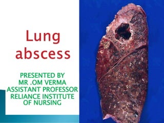
- 1. PRESENTED BY MR .OM VERMA ASSISTANT PROFESSOR RELIANCE INSTITUTE OF NURSING
- 3. 1.Localized suppurative (Suppuration is the process of pus forming.) Inflammation of the lung. According to Luckmann and Sorensen's 2. Necrosis of the pulmonary tissue (lungs) & formation of cavities containing necrotic debris or fluid caused by microbial infection. According to Brunner & Suddarth's 3. A localized area of destruction of lung parenchyma in which infection by pyogenic organisms results in tissue necrosis & suppuration . According toBrenda G. Bare
- 4. 4.Lung Abscess is a localized necrotic lesion of the lung parenchyma containing purulent material that collapses and forms a cavity. It is generally caused by aspiration of anaerobic bacteria. According to Lippincott 5. Lung abscess is a type of liquefactive necrosis of the lung tissue and formation of cavities (more than 2 cm) containing necrotic debris or fluid caused by microbial infection. This pus-filled cavity is often caused by aspiration, which may occur during anesthesia, sedation, or unconsciousness from injury. According to Lewis's
- 6. Location: Dependent areas (posterior segments of upper & lower lobes). Stages: Pneumonic stage: patch of consolidation Is a region of normally compressible lung tissue that has filled with liquid instead of air. The condition is marked by in duration (swelling or hardening of normally soft tissue) of a normally lung. Rupture: central necrosis, communication with a bronchus & expectoration (mucus ) of the liquid centre. Acute cavitations: irregular thick wall, shaggy ( shaggy means branches ) inner margin & surrounding consolidation (collection). Chronic cavitation: thin wall, smooth inner margin & subsidence of surrounding inflammatory reaction.
- 7. Complication of pneumonia: Staph pneumonia. Klebsiella pneumonia. T.B. Necrosis of pulmonary tissue and formation of cavities containing necrotic derbies or fluid causes microbial infection. the formation of multiple small abscesses is occasionally referred to as necrotizing pneumonia or lung gangrene.
- 8. Lung abscess with bulging fissure ( a long narrow opening or line of breakage ) sign.
- 9. 3.EMBOLIC ABSCESSES an abscess arising distal to the point of arrest of a septic embolus. A septic embolism is a type of embolism that is infected with bacteria, resulting in the formation of pus. a clot or other plug, usually part or all of a THROMBUS
- 10. Embolic abscesses primary Due to impaction(pressure) of septic embolus(a b/d clot air bubble ) in pulmonary artery secondary Due to infection of infracted lung
- 11. Infected catheters. Infected pacemakers. Tricuspid endocarditic ( IV drug abusers).
- 12. ETOLOGY
- 13. HEMATOGENOUS metastases of tumors or in infections; blood- borne. spread from a distal site) originating in the blood. 2. producing blood or components of blood. 3. distributed or spread by way of the bloodstream, • SEPTIC THROMBOPHLEBITIS • venous thrombosis, inflammation, and bacteremia.
- 14. Metabolism of alcohol reduces glutathione anti-oxidant levels in the lungs. Oxidation damage to the cells impairs the ability of the lungs to remove fluid. Oxidative damage to cells reduces immune response. IMMUNE SYSTEM ISN’T WORKING WELL: This can let in germs found in your mouth or throat, like fungi or the bacteria that cause tuberculosis, strep throat, lead to lungs abscess.
- 15. Blood-borne causes: It’s rare, but bacteria or infected blood clots from an infected part of your body can travel through your bloodstream and into your lung, where they cause an abscess. POOR ORAL HEALTH: People with gum disease are more likely to get an abscess
- 16. infective endocarditis (IE) caused by Staphylococcus aureus has been ... Our patient presented with metastatic abscesses in the lungs that led to
- 17. Lung Abscess pyogenic lung infection/pneumonia, necrotizing pneumonia. ... The most frequent cause is aspiration of anaerobic organisms from the mouth in those predisposed to ... Penetrating pulmonary trauma - eg, a stab wound.
- 18. • BRONCHIAL OBSTRUCTION : tumour, foreign body, Blocked airway: Mucus can form behind a tumor or foreign object in your windpipe and lead to an abscess. If bacteria get into the mucus, the blockage stops you from coughing it out. IMMUNODEFICIENCY immunodeficiency disease characterized by eczema, recurrent staphylococcal skin abscesses, recurrent lung infections, eosinophilia (a high number of eosinophils in the blood) and high serum levels of IgE.
- 19. lung disease: Lung conditions such as bronchiectasis, cystic fibrosis, lung contusions (bruises), and infected infarcts may lead to a lung abscess
- 20. Tuberculosis & non tuberculous mycobacterial infection – fluid filled cavities – upper lobes / apical segments of lower lobes Fungal infection – Histoplasma capsulatum Blastomyces dermatitidis Coccidiodes immitis Aspergillus Cryptococcus neoformans Candida
- 21. Aspiration of oropharyngeal or gastric secretion. 2) Septic emboli. Necrotizing pneumonia Necrotizing tumors Gram negative organisms. (klebsiella) Anaerobic bacilli (Bacterorides Carcinoma of the lung Parasitic and fungal diseases of the lung. TB
- 22. Lung abscess starts as an area of pneumonia Small zones of necrosis Coalesce together to from one or more large cavities of 1-2 c.m Progressive and enlargement to from the abscess cavity The abscess cavity well erode( increase) a bronchus
- 23. Expec-toration of purulent sputum with air fluid formation in the cavity Fate 1. infection of the other lung 2. Open into pleura –pyopneumothorax 3. Hematogenous spread
- 24. The presenting features of lung abscess vary considerably . 1. Symptoms progress over weeks to months 2. Fever, cough, and sputum production 3. Night sweats, weight loss & anemia 4. Hemoptysis, is the coughing up of blood or blood-stained mucus from the bronchi, larynx, trachea, or lungs.
- 25. Digital clubbing – develop within a few weeks if treatment is inadequate. Dullness to percussion Diminished breath sounds if abscess is too large and situated near the surface of lung. Amphoric / cavernous breath sounds Cough with foul smelling purulent sputum. Fever with shivering Night sweats Chest pain Shortness of breath Lethargy ) Finger clubbing
- 26. Leukocytosis refers to an increase in the total number of WBCs Anorexia Weight loss Weakness Dyspnea
- 27. Lung abscess Acute < 6 weeks. Chronic > 6 weeks
- 28. HISTORY TAKING PHYSICAL EXAMINATION During a physical exam, doctor will listen for abnormal sounds in your lungs and heart using a stethoscope.And detect any lungs fluid accumulation . BRONCHOSCOPY TO Identification of proximal airway obstruction by a tumour or foreign body CHEST X-RAYS = nearly Identifying the lung abscess as a cavity filled with fluid and air.
- 29. Sputum Gram Stain: May occasionally be helpful if there is a large number of white blood cells and bacteria consistent with oropharyngeal flora. -
- 30. Arterial Blood Gas Test The arterial blood gas test is a test used to check the level of oxygen and carbon dioxide in your blood. A doctor or nurse will take blood from the artery in your wrist. Then, they will send the blood to a lab for testing. The results of this test indicate the amount of oxygen and carbon dioxide in your blood.
- 31. Pulse Oximetry Test The doctor will measure your oxygen level using a small sensor that’s placed on the tip of your finger to see if you are getting enough oxygen. This is called the pulse oximetry test . CT SCAN = thick-walled, usually round cavity with irregular margins forming an acute angle with chest wall, no signs of compression of surrounding lung .
- 33. Drainage: You may need this if abscess is 6 centimeters or more in diameter. doctor will use a CT scan to guide him as he inserts the drain through your chest wall into the abscess.
- 35. COMPLICATION
- 36. Caused by spread of infection into the pleural space or by contamination of the pleural cavity after percutaneous drainage. 2.Amyloidosis is a group of diseases in which abnormal protein, known as amyloid fibrils, builds up in tissue. 3. Hemoptysis is the coughing up of blood or blood-stained mucus from the bronchi, larynx, trachea, or lungs.