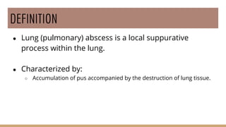
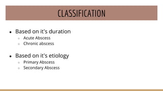
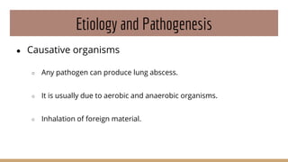
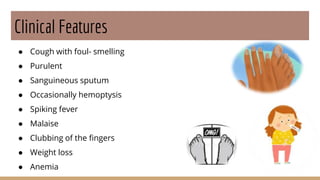
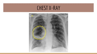
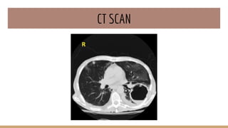
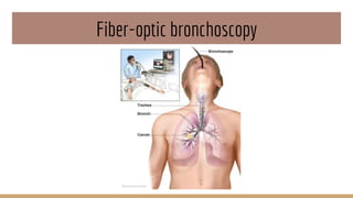
Presented by:
Ankita Roy
BSc Nursing 2nd Year
Royal School of Nursing
DEFINITION
● Lung (pulmonary) abscess is a local suppurative
process within the lung.
● Characterized by:
○ Accumulation of pus accompanied by the destruction of lung tissue.
CLASSIFICATION
● Based on it’s duration
○ Acute Abscess
○ Chronic abscess
● Based on it’s etiology
○ Primary Abscess
○ Secondary Abscess
Etiology and Pathogenesis
● Causative organisms
○ Any pathogen can produce lung abscess.
○ It is usually due to aerobic and anaerobic organisms.
○ Inhalation of foreign material.
● Causative agents
○ Anaerobic bacteria, with or without mixed aerobic infection
○ Staphylococcus aureus
○ Klebsiella pneumoniae
○ Streptococcus pyogenes
○ Type 3 Pneumococcus
Etiopathogenesis
● Aspiration of infective material
○ Common in acute alcoholism, coma, anesthesia etc.
● Antecedent primary lung infection
○ Microbes usually associated are:
■ S. aureus
■ K. pneumoniae
■ Type 3 Pneumococcus
● Bronchial Obstruction
● Septic embolism
● Miscellaneous
○ Direct penetrating trauma to the lungs.
○ Spread of infections from neighboring organs.
Morphology
● Gross
○ Size: Vary from few millimeters to large cavities of
5-6cm
○ Content:
■ Abscess cavity may contain suppurative
debris.
■ Communication with an air passage 
abscess partially drained to produce an air-
containing cavity
○ Continued infection may result  gangrene of
the lung
● Microscopy
○ Cardinal feature: It is characterized by
suppurative destruction of the lung
parenchyma within the central area of
cavitations.
○ Abscess shows numerous
polymorphonuclear leukocytes and
variable numbers of macrophages.
○ The abscess is surrounded by fibrous
wall infiltrated by inflammatory cells.
Clinical Features
● Cough with foul- smelling
● Purulent
● Sanguineous sputum
● Occasionally hemoptysis
● Spiking fever
● Malaise
● Clubbing of the fingers
● Weight loss
● Anemia
Diagnostic findings
● History taking
● Physical examination
● Chest X-Ray
● CT Scan
● CBC
● Sputum culture and sensitivity tests
● Fiberoptic bronchoscopy
CHEST X-RAY
CT SCAN
CBC TEST
Sputum Culture Test
Fiber-optic bronchoscopy
Prevention
● Appropriate antibiotic therapy before any dental procedures in
patients who must have teeth extracted while their teeth and
gums are infected.
● Adequate dental and oral hygiene, because anaerobic bacteria
play a role in the pathogenesis of lung abscess.
● Appropriate antimicrobial therapy for patients with pneumonia.
Management
● Pharmacological Management
○ Treatment focus on cause of cough
○ Symptomatic treatment elimination of irritant and pain relief analgesic
and antipyretic
○ Antibiotic treatment may be indicated depending on the symptoms,
sputum purulence, and results of the sputum culture
○ Antihistamine are usually not prescribed
● Surgical Management
○ LOBECTOMY
Complication
● Pneumothorax or empyema
● Hemorrhage into the abscess cavity
● Meningitis or brain abscess
● Secondary amyloidosis
THANK YOU

Lung abscess

  • 1.
    Presented by: Ankita Roy BScNursing 2nd Year Royal School of Nursing
  • 2.
    DEFINITION ● Lung (pulmonary)abscess is a local suppurative process within the lung. ● Characterized by: ○ Accumulation of pus accompanied by the destruction of lung tissue.
  • 3.
    CLASSIFICATION ● Based onit’s duration ○ Acute Abscess ○ Chronic abscess ● Based on it’s etiology ○ Primary Abscess ○ Secondary Abscess
  • 4.
    Etiology and Pathogenesis ●Causative organisms ○ Any pathogen can produce lung abscess. ○ It is usually due to aerobic and anaerobic organisms. ○ Inhalation of foreign material.
  • 5.
    ● Causative agents ○Anaerobic bacteria, with or without mixed aerobic infection ○ Staphylococcus aureus ○ Klebsiella pneumoniae ○ Streptococcus pyogenes ○ Type 3 Pneumococcus
  • 6.
    Etiopathogenesis ● Aspiration ofinfective material ○ Common in acute alcoholism, coma, anesthesia etc. ● Antecedent primary lung infection ○ Microbes usually associated are: ■ S. aureus ■ K. pneumoniae ■ Type 3 Pneumococcus ● Bronchial Obstruction
  • 7.
    ● Septic embolism ●Miscellaneous ○ Direct penetrating trauma to the lungs. ○ Spread of infections from neighboring organs.
  • 8.
    Morphology ● Gross ○ Size:Vary from few millimeters to large cavities of 5-6cm ○ Content: ■ Abscess cavity may contain suppurative debris. ■ Communication with an air passage  abscess partially drained to produce an air- containing cavity ○ Continued infection may result  gangrene of the lung
  • 9.
    ● Microscopy ○ Cardinalfeature: It is characterized by suppurative destruction of the lung parenchyma within the central area of cavitations. ○ Abscess shows numerous polymorphonuclear leukocytes and variable numbers of macrophages. ○ The abscess is surrounded by fibrous wall infiltrated by inflammatory cells.
  • 10.
    Clinical Features ● Coughwith foul- smelling ● Purulent ● Sanguineous sputum ● Occasionally hemoptysis ● Spiking fever ● Malaise ● Clubbing of the fingers ● Weight loss ● Anemia
  • 11.
    Diagnostic findings ● Historytaking ● Physical examination ● Chest X-Ray ● CT Scan ● CBC ● Sputum culture and sensitivity tests ● Fiberoptic bronchoscopy
  • 12.
  • 13.
  • 14.
  • 15.
  • 16.
  • 17.
    Prevention ● Appropriate antibiotictherapy before any dental procedures in patients who must have teeth extracted while their teeth and gums are infected. ● Adequate dental and oral hygiene, because anaerobic bacteria play a role in the pathogenesis of lung abscess. ● Appropriate antimicrobial therapy for patients with pneumonia.
  • 18.
    Management ● Pharmacological Management ○Treatment focus on cause of cough ○ Symptomatic treatment elimination of irritant and pain relief analgesic and antipyretic ○ Antibiotic treatment may be indicated depending on the symptoms, sputum purulence, and results of the sputum culture ○ Antihistamine are usually not prescribed ● Surgical Management ○ LOBECTOMY
  • 19.
    Complication ● Pneumothorax orempyema ● Hemorrhage into the abscess cavity ● Meningitis or brain abscess ● Secondary amyloidosis
  • 20.

Editor's Notes