
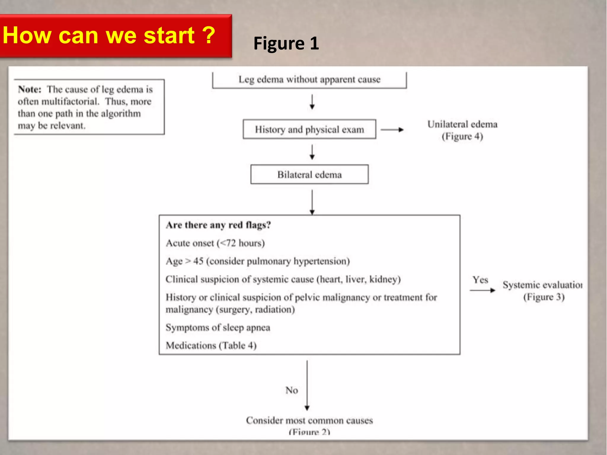

![History
Key elements of the history include
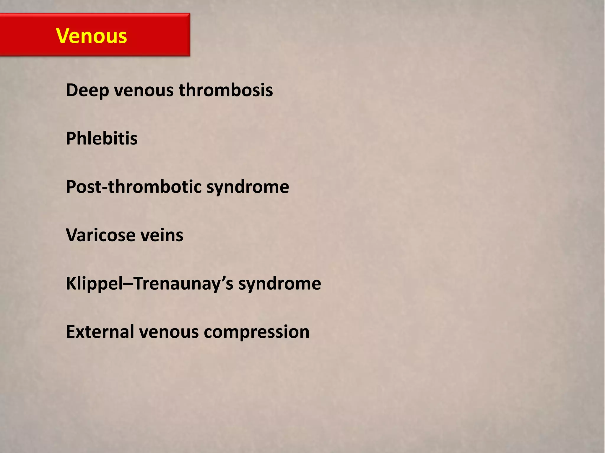
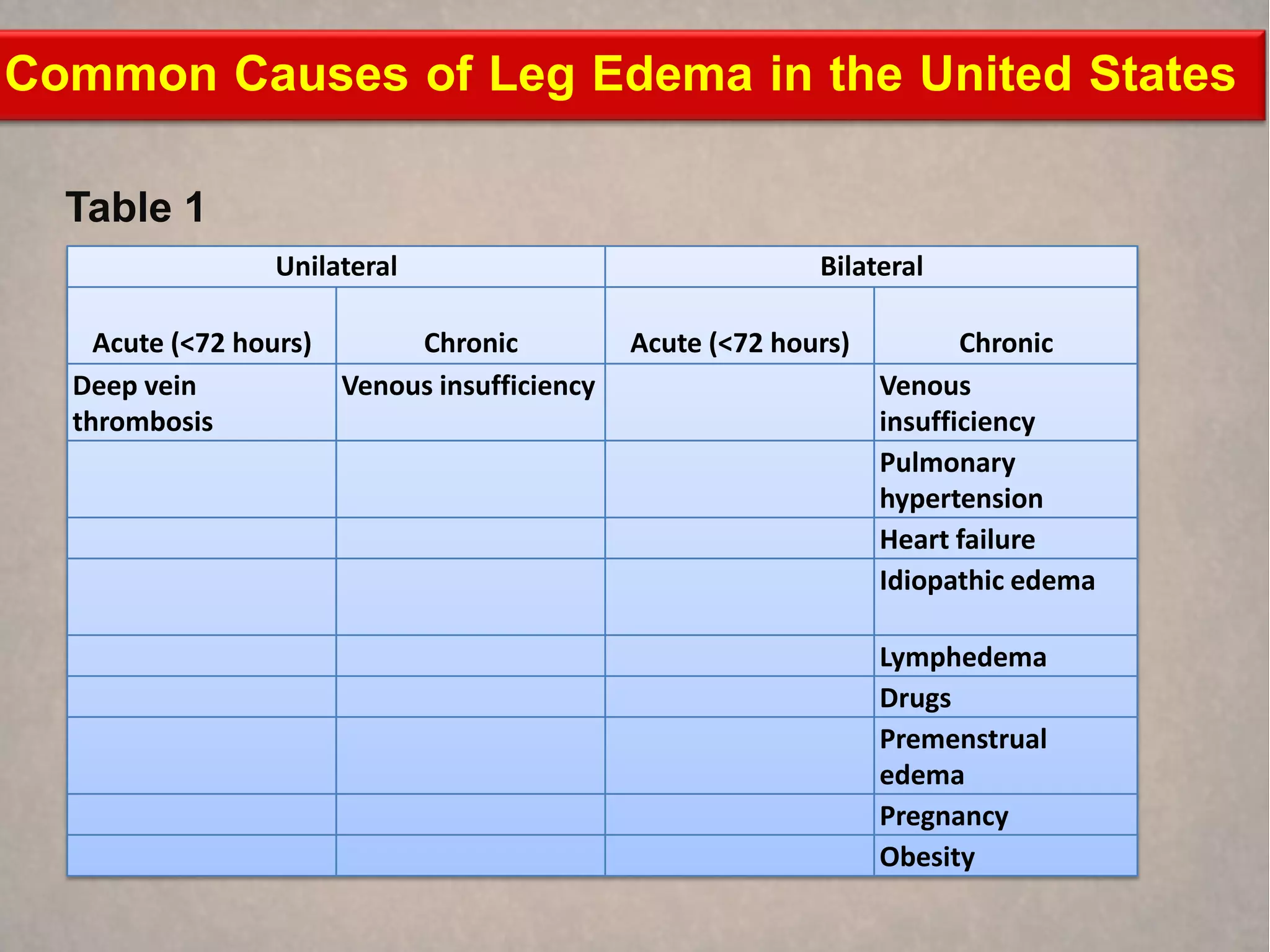
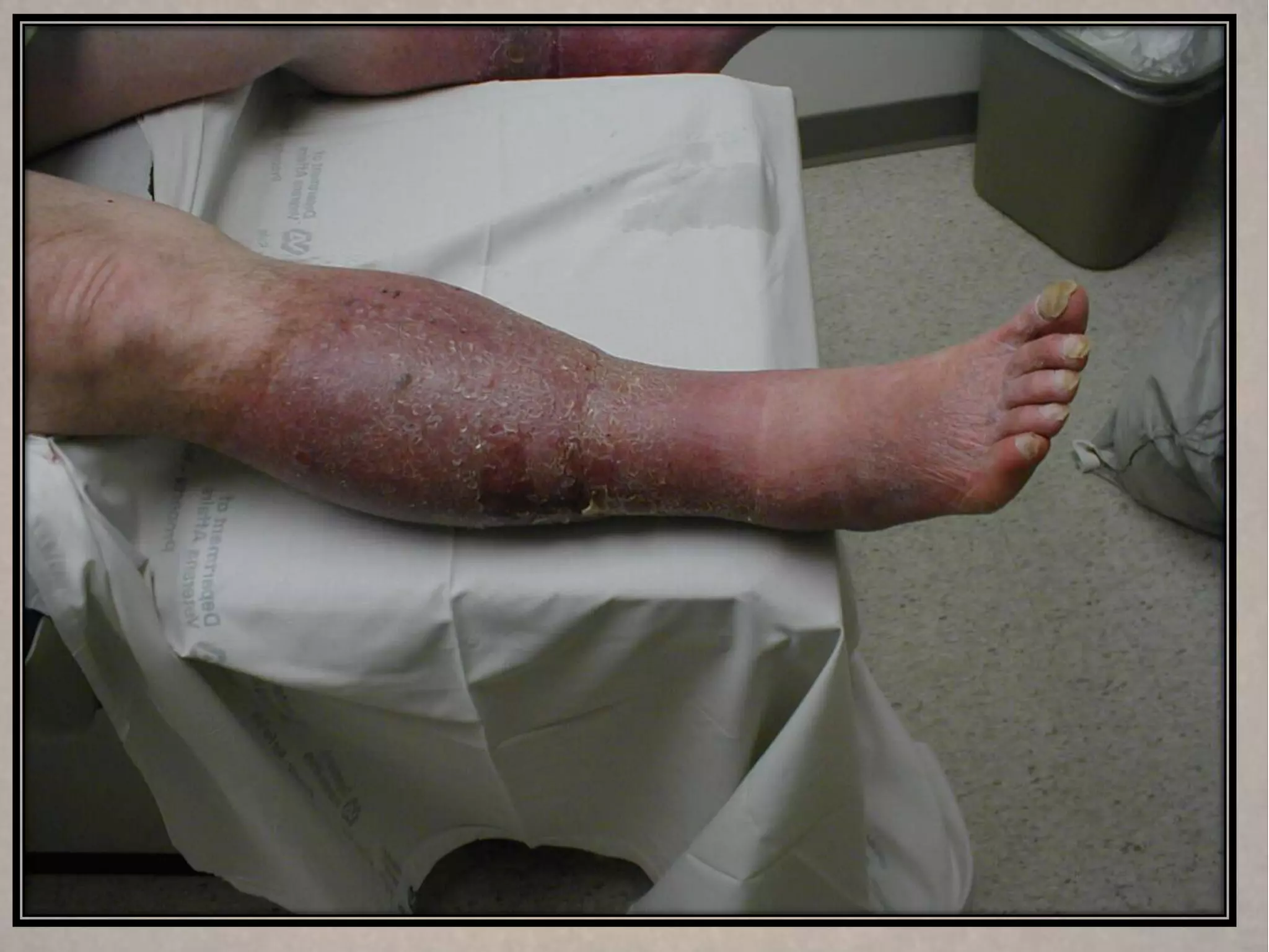
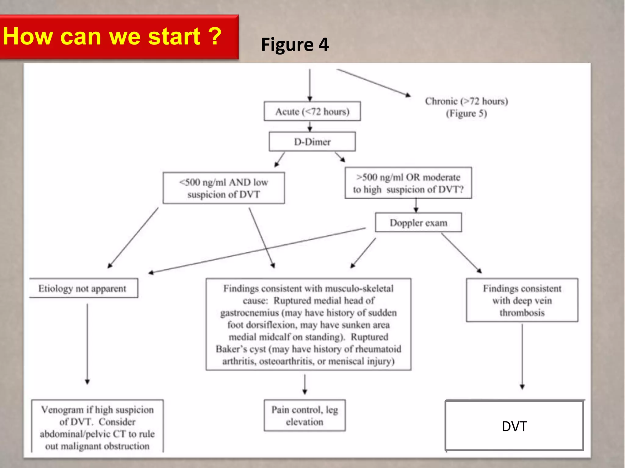
What is the duration of the edema (acute [<72 hours] vs. chronic)? If the onset is acute,
deep vein thrombosis should be strongly considered.
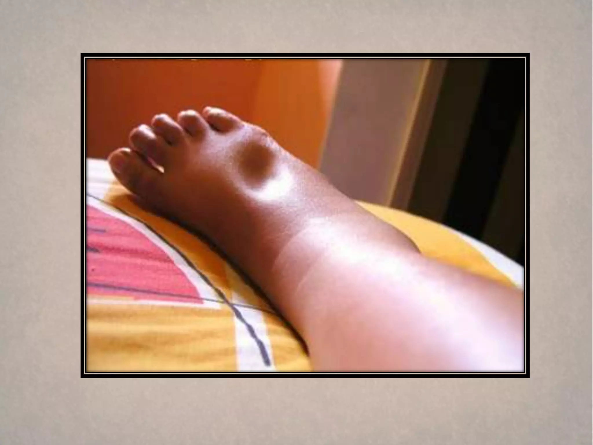
Is the edema painful ? Deep vein thrombosis and reflex sympathetic dystrophy are usually
painful. Chronic venous insufficiency can cause low-grade aching. Lymphedema is usually
painless.
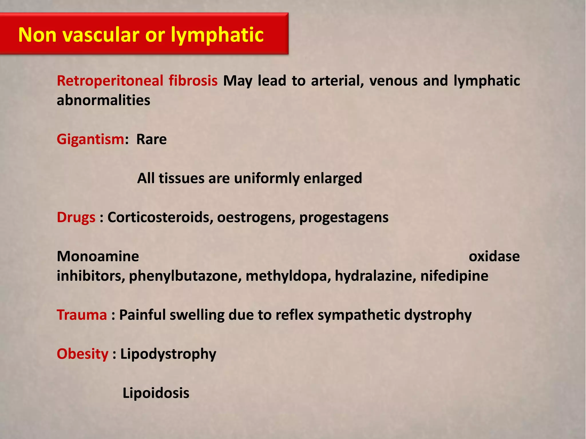
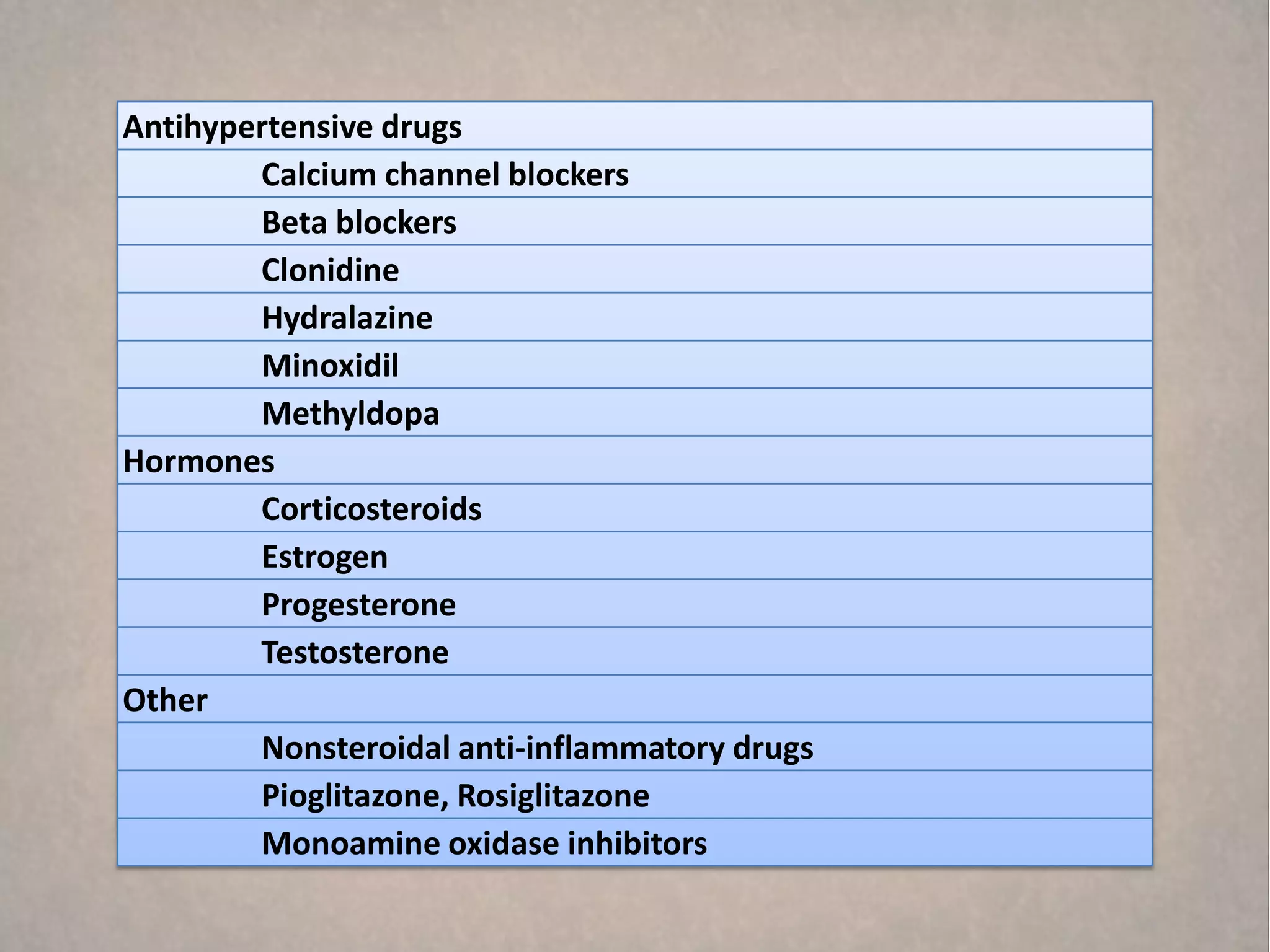
What drugs are being taken? Calcium channel blockers, prednisone, and anti-inflammatory
drugs are common causes of leg edema
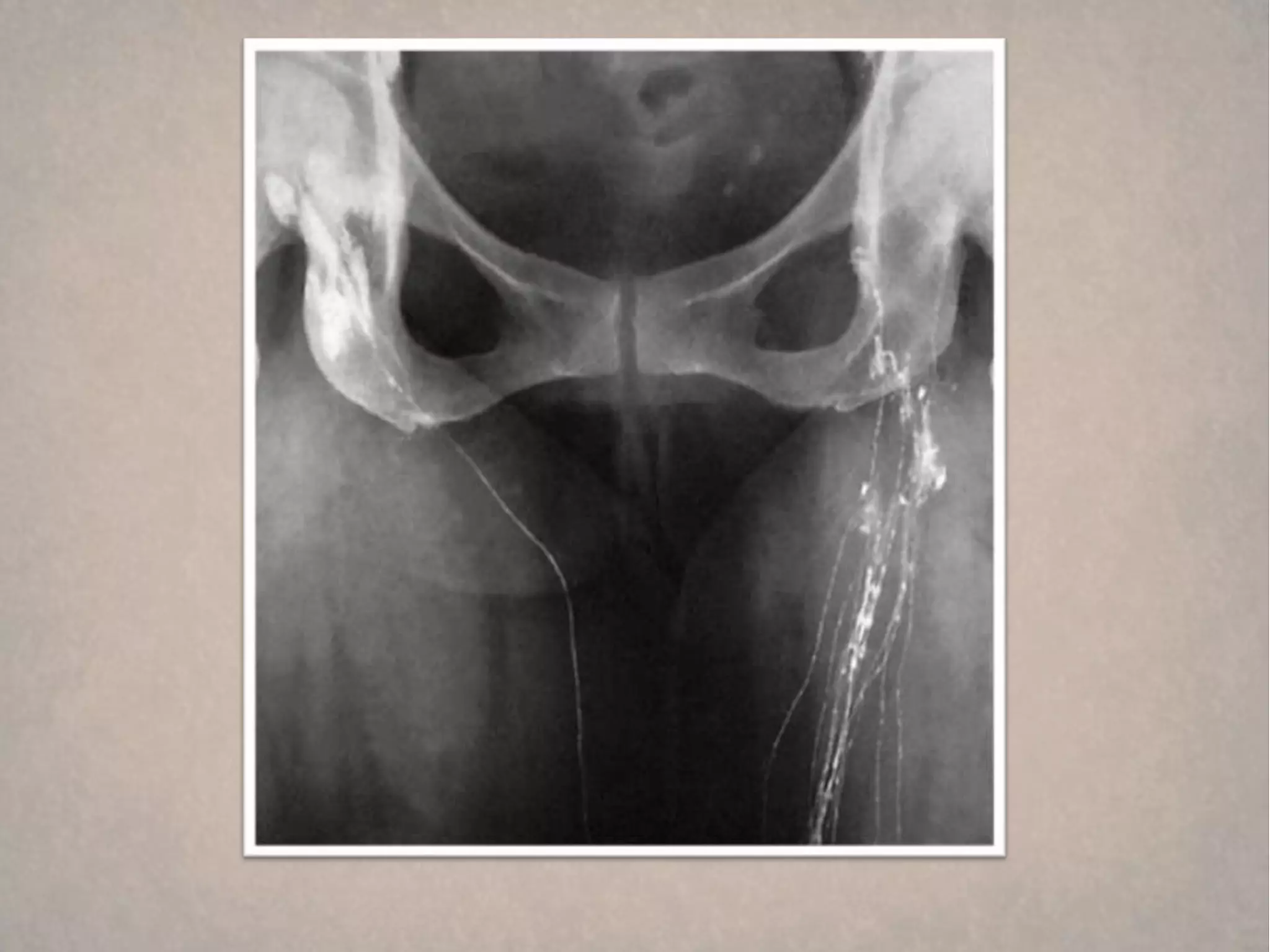
Is there a history of systemic disease (heart, liver, or kidney disease)?](https://image.slidesharecdn.com/lowerlimbswilling-130218163251-phpapp02/75/Lower-limb-swilling-25-2048.jpg)


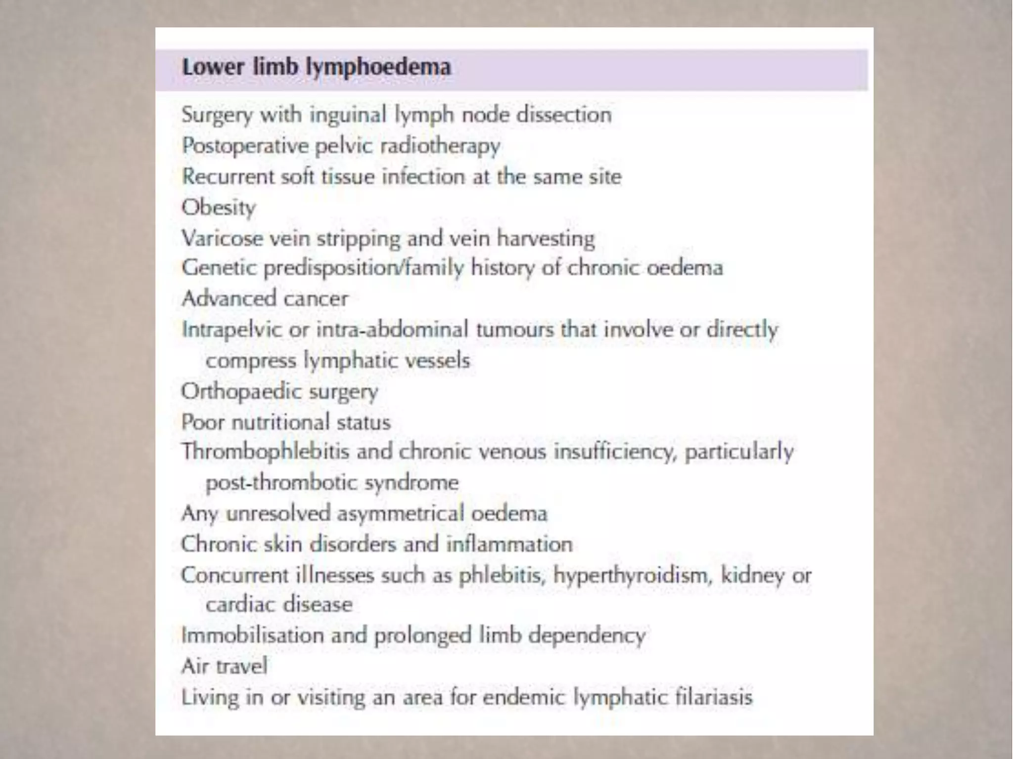
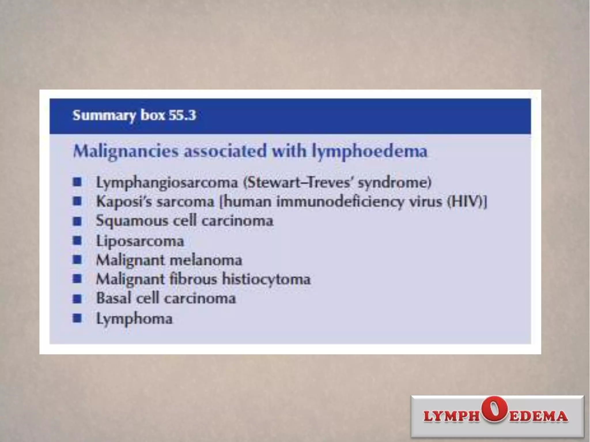
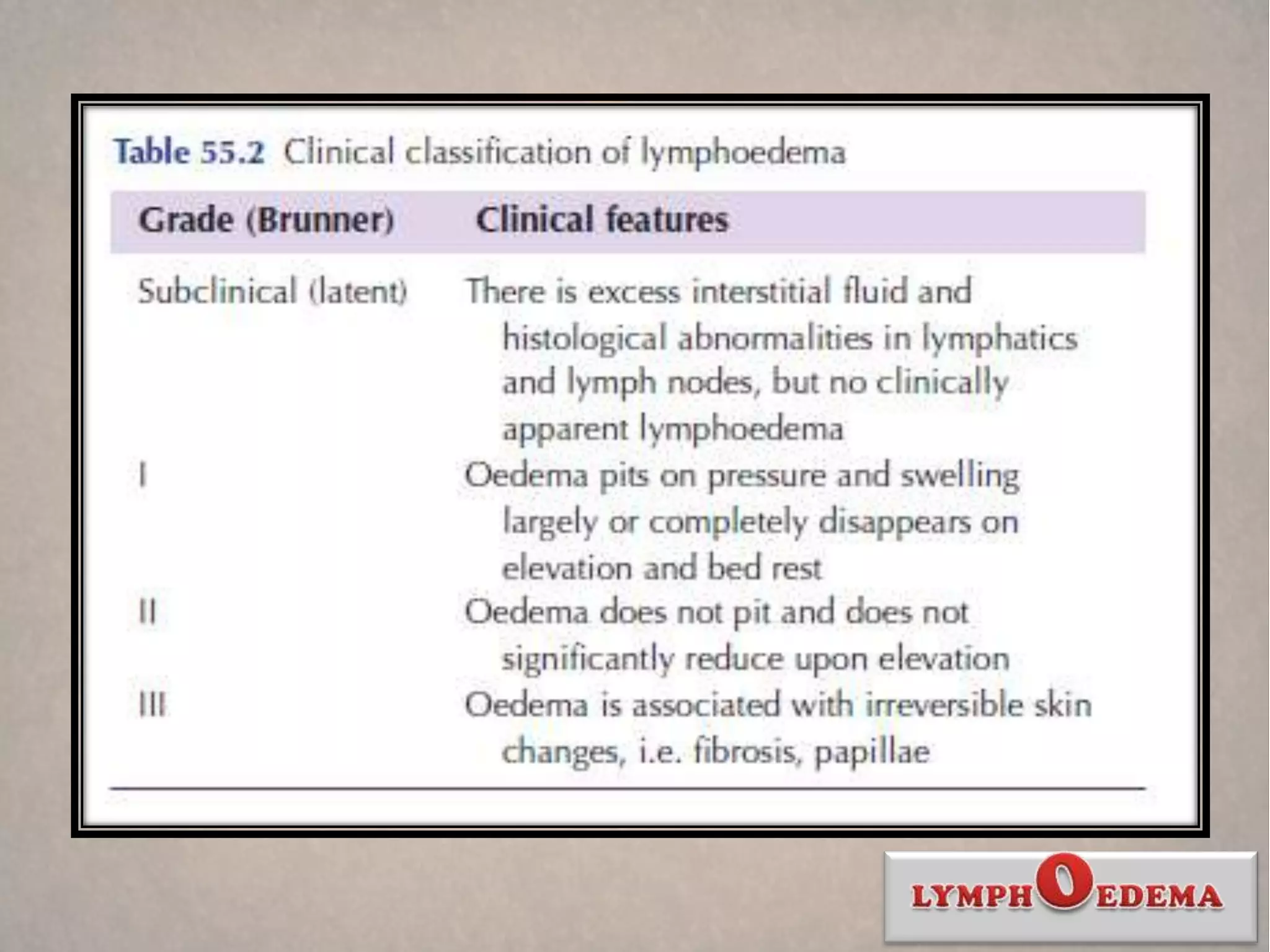

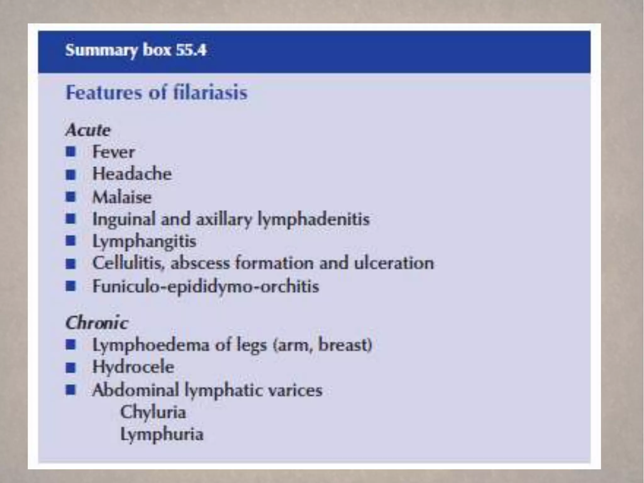


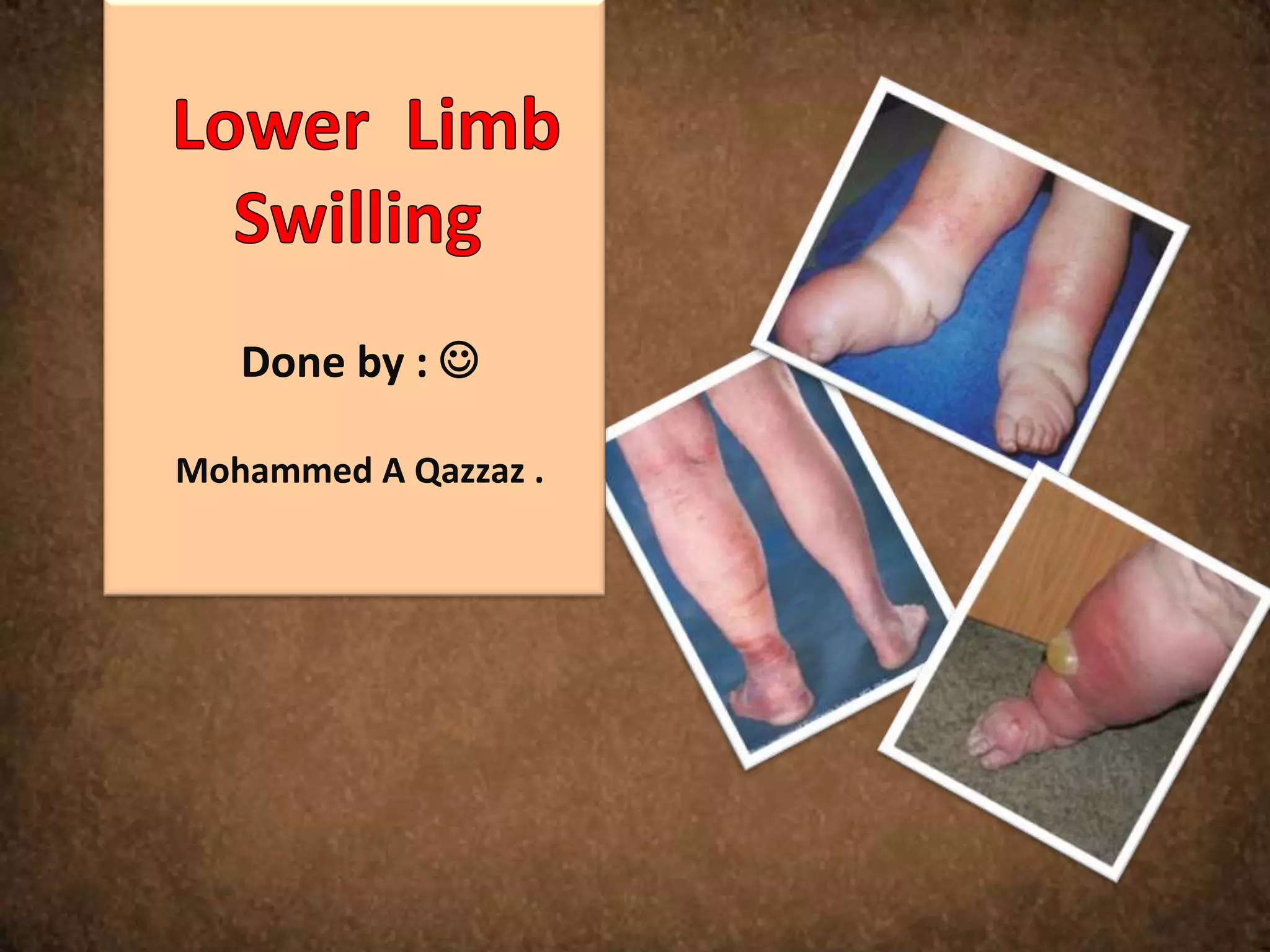
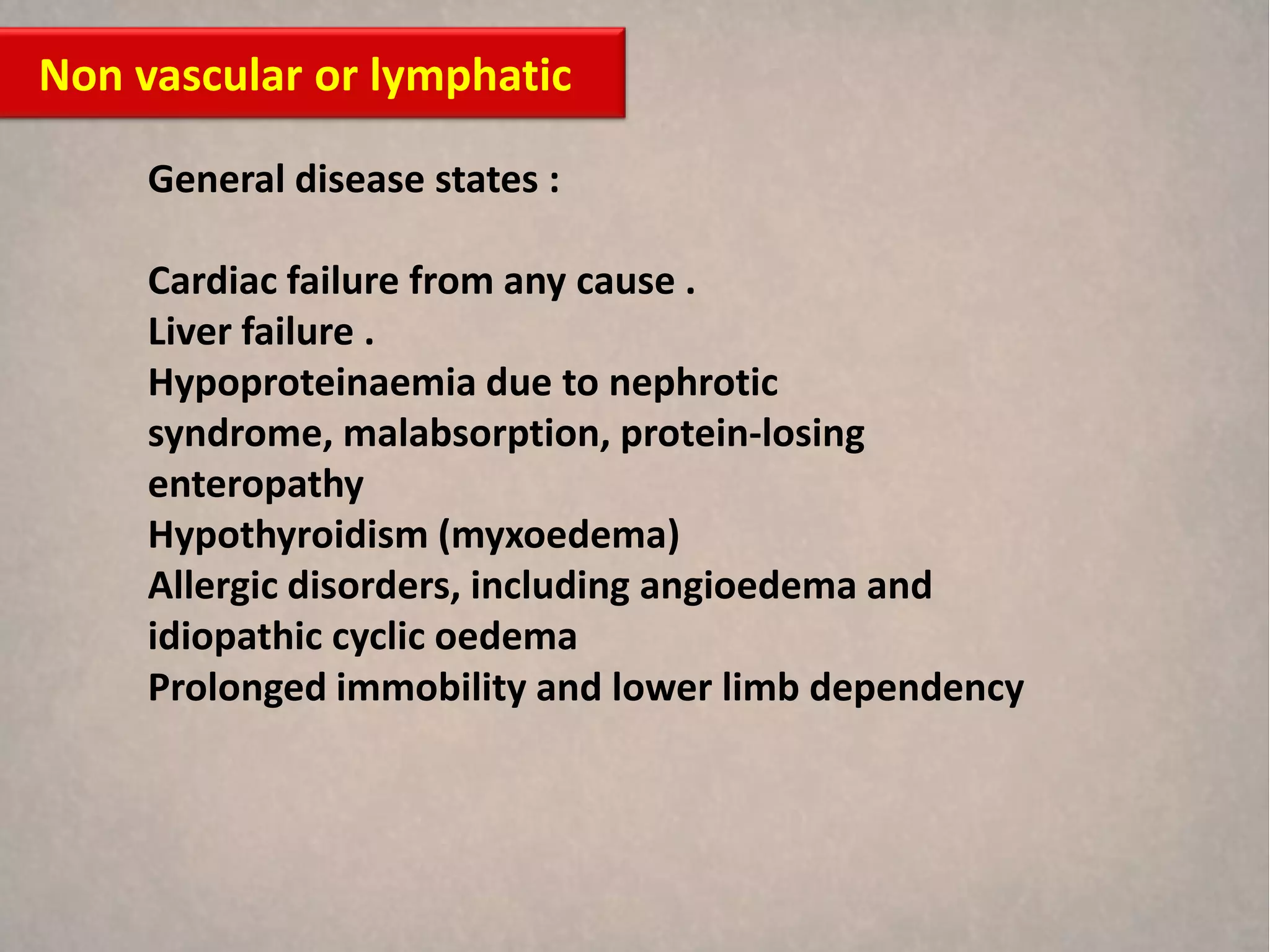
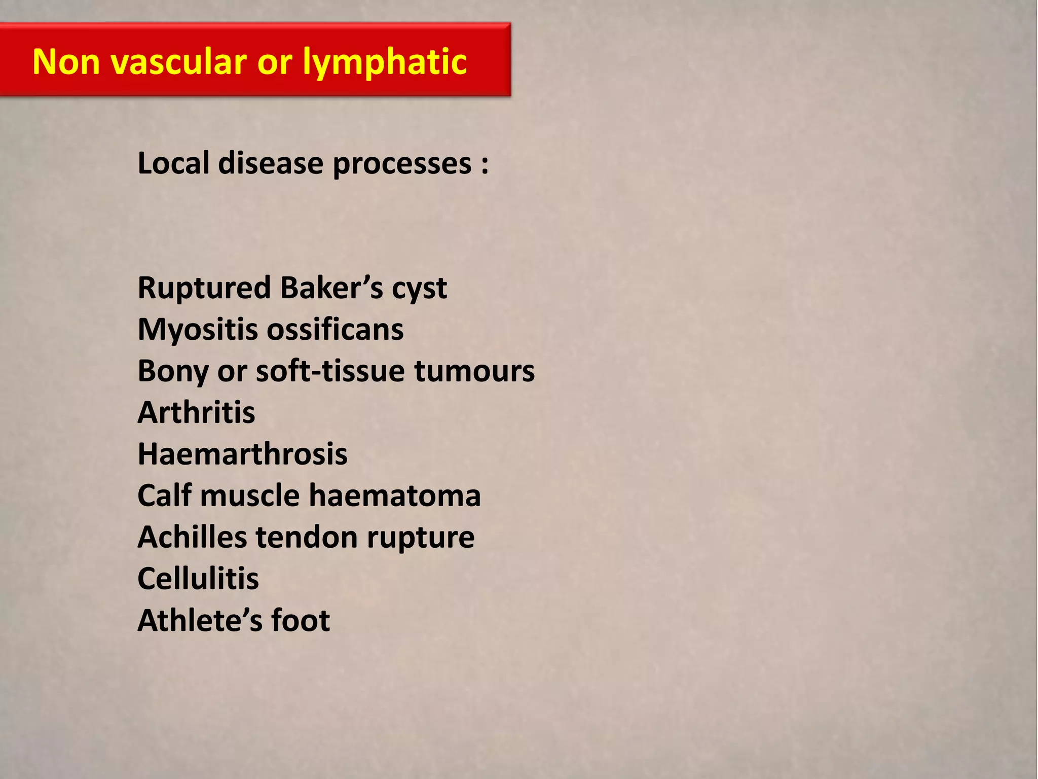
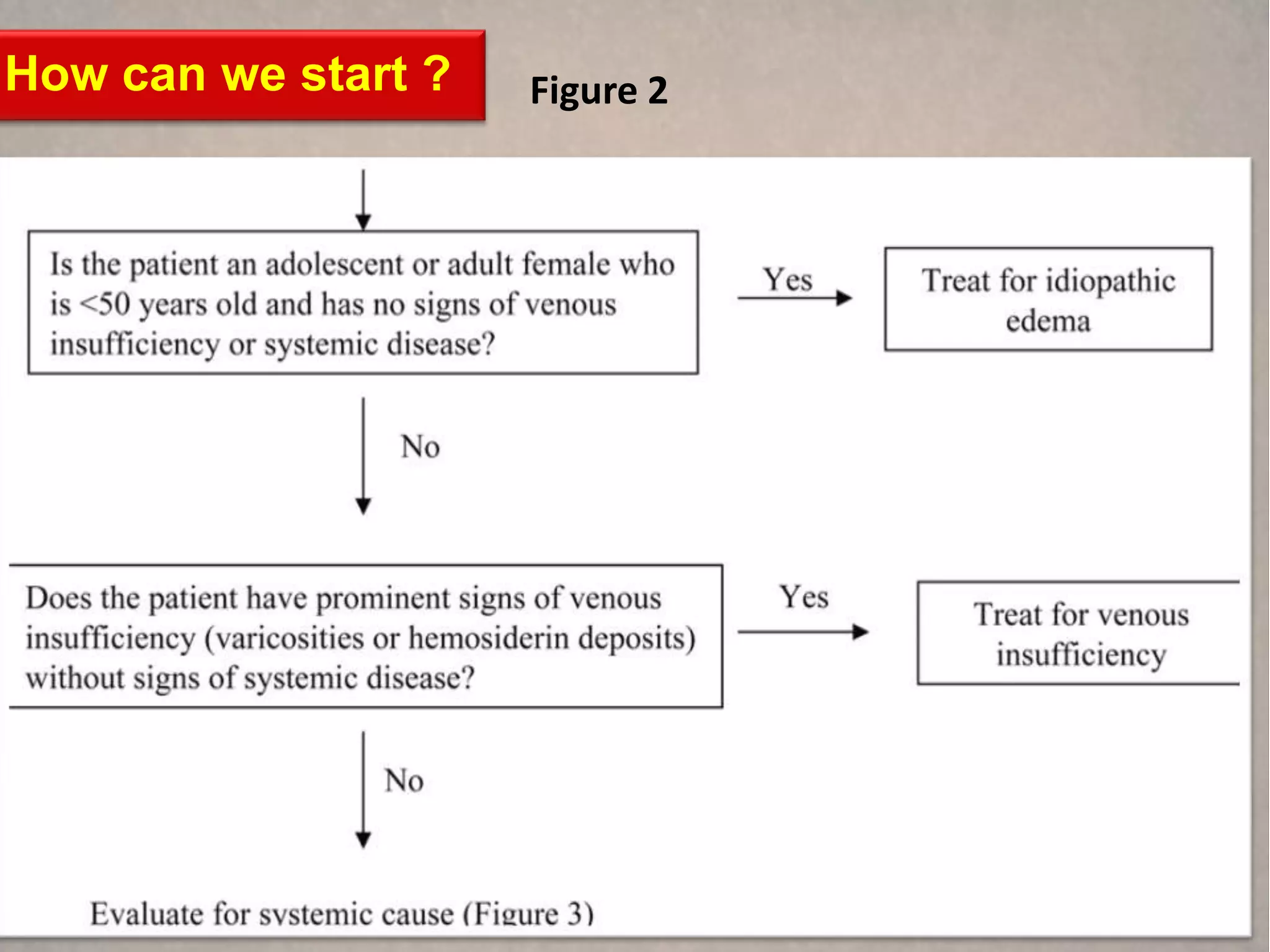
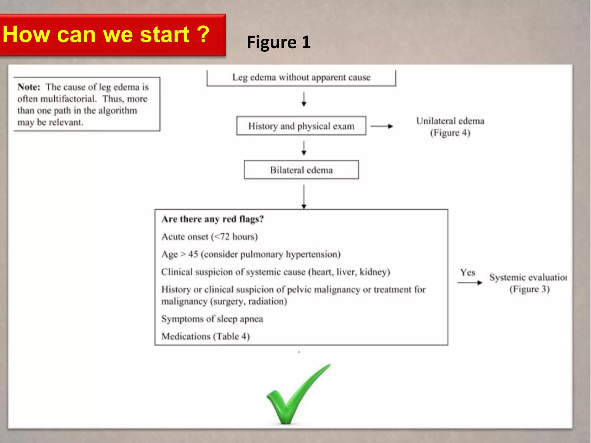
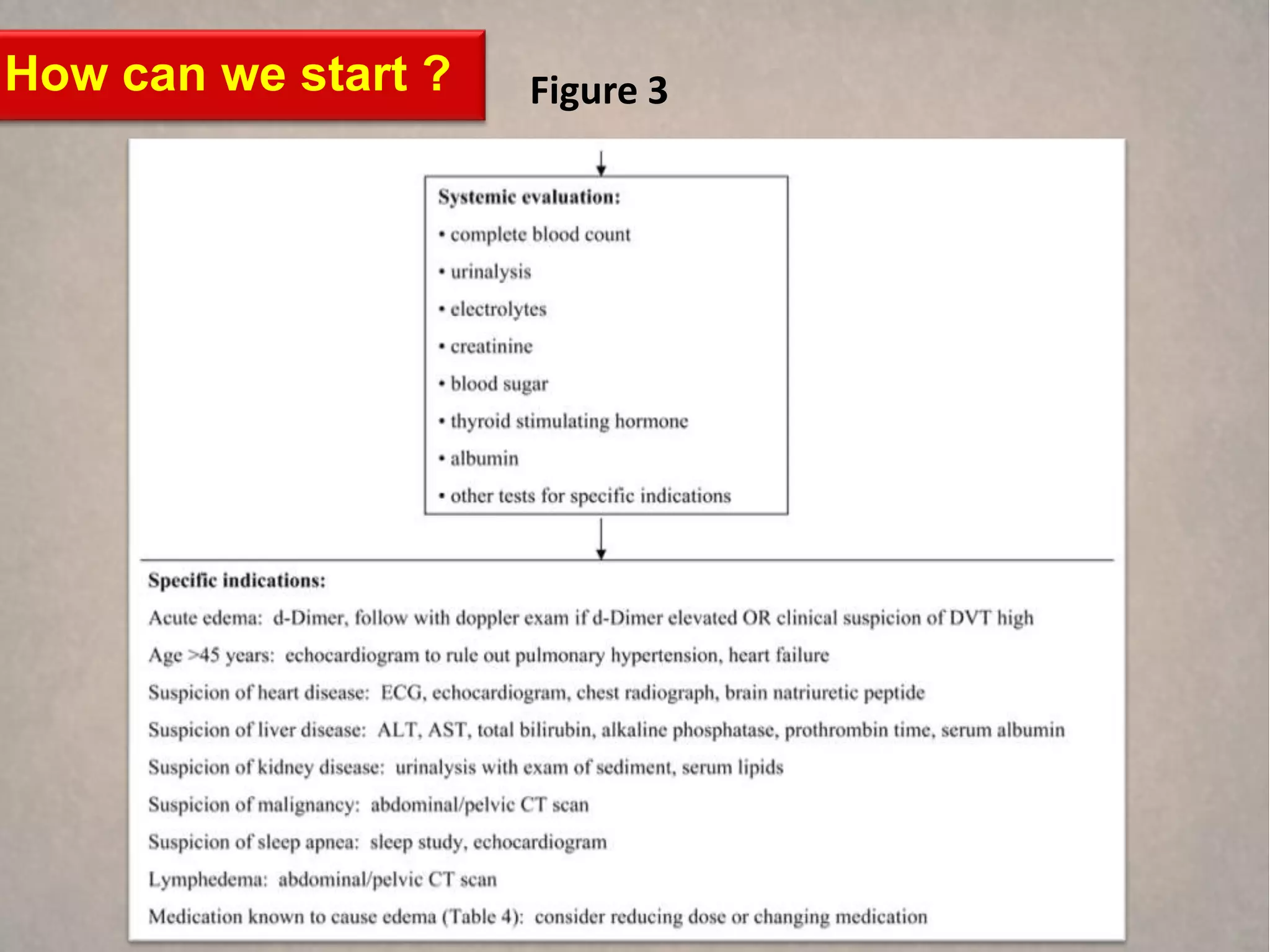
The document discusses the anatomy and functions of the lower limbs, primarily focusing on conditions that lead to leg swelling or edema. It categorizes causes of edema into vascular and non-vascular categories, detailing various underlying conditions, including heart failure, infections, and lymphatic dysfunctions. Additionally, it addresses idiopathic edema and chronic venous insufficiency while providing essential management strategies for such conditions.


