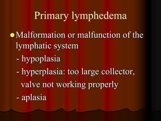
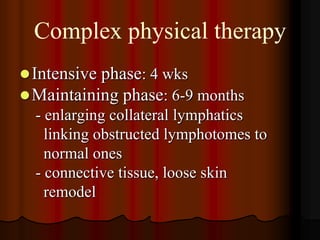
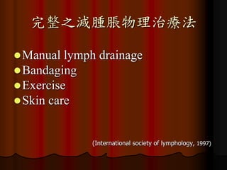
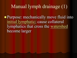
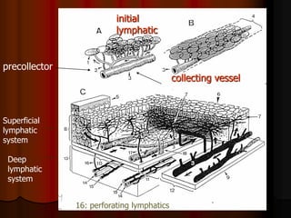
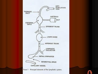
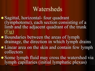
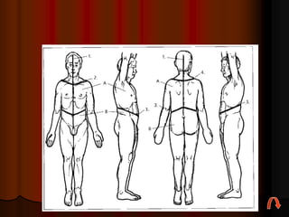
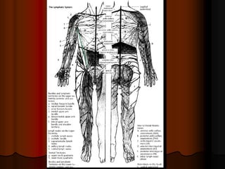
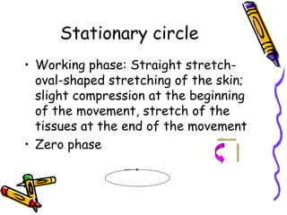
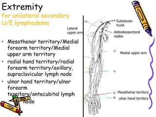
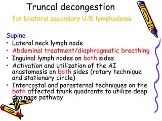
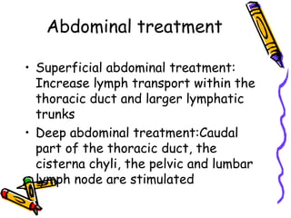
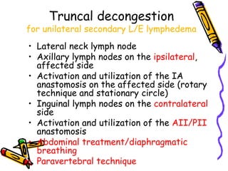
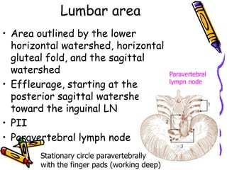
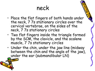
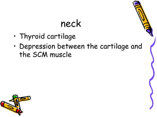
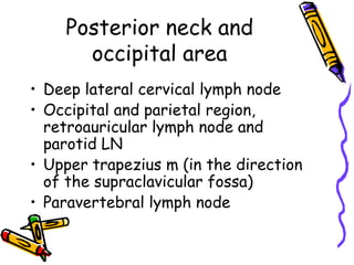
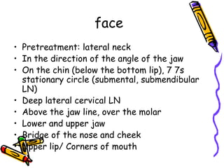
This document provides information on manual lymph drainage for treating lymphedema. It describes the lymphatic system and causes of lymphedema, stages of lymphedema, and complete decongestive physiotherapy. It details techniques for manual lymph drainage including direction of strokes, basic movements like pumps and circles, sequence of treatment, and truncal and extremity drainage patterns. The goal is to encourage lymph flow and reduce swelling through light skin stretching and movement toward lymph nodes.
1. The document discusses lymphedema, which is abnormal accumulation of tissue proteins and fluid caused by malformation or damage to the lymphatic system.
2. It describes primary lymphedema caused by genetic defects and secondary lymphedema caused by surgery, radiation, trauma, or other insults that damage the lymphatic system.
3. Treatment involves complete decongestive physiotherapy including manual lymph drainage, compression bandaging, exercise, and skin care to drain excess fluid and promote collateral lymphatic vessel growth.
venous & lymphatic drainage of upper limbIallu Reddy
1) The median cubital vein is a large communicating vein that shunts blood from the cephalic vein to the basilic vein near the elbow.
2) During an attempted blood draw from the median cubital vein, the technician noticed bright red blood indicating a puncture of the brachial artery, which lies deep to the median cubital vein and is separated by the bicipital aponeurosis fascia.
3) On a second attempt slightly medial, the patient felt sharp pain radiating to the lateral three digits, suggesting injury to the median nerve, which also lies deep in this region.
The document describes cardiovascular anatomy including:
- The dermatomal distribution of the T1 ventral ramus and location of the nipple at T4.
- Blood supply and innervation of the intercostal spaces from intercostal and posterior intercostal arteries.
- Location and components of the intercostal neurovascular bundle between internal and innermost intercostal muscles.
- Purpose and location of intercostal nerve blocks in the posterior angle of the rib.
This document discusses the veins of the lower limb. It begins by describing the anatomy and classifications of veins in general. It then focuses on the specific veins of the lower limb, including the superficial greater and small saphenous veins and deep femoral, profunda femoris, popliteal, peroneal, anterior tibial, and posterior tibial veins. The document concludes by discussing deep vein thrombosis (DVT), its causes, symptoms, diagnosis, and treatments including anticoagulation and compression stockings.
The chest cavity contains the lungs, heart, major blood vessels, and other structures. It is bounded by the ribs, sternum, vertebral column, and diaphragm. The chest cavity is further divided into the pleural cavities and mediastinum. The mediastinum is the median partition between the lungs and contains the esophagus, trachea, thymus, and major blood vessels. Each lung is surrounded by a pleural membrane made of visceral and parietal layers that create a pleural cavity containing fluid.
The document describes the anatomy and locations of lymph nodes in the mediastinum, dividing it into the anterior, middle and posterior compartments. It provides details on the contents and boundaries of each compartment, as well as labeled diagrams showing lymph nodes and other structures in different axial, coronal and sagittal views of the thorax. Locations and boundaries are given for specific lymph node stations, including the supraclavicular, lower paratracheal, and hilar nodes.
Radiological anatomy of chest including lungs,mediastinum and thoracic cagePankaj Kaira
The document describes the anatomy of the thoracic cage and its components. It discusses the sternum, ribs, costal cartilage, and their joints. It also describes the lungs and their lobes, as well as the structures of the mediastinum such as the trachea, bronchi, blood vessels, and nerves. Key details are provided on the segments of the lungs and the fissures that divide the lobes.
The lymphatic system drains tissue fluid, plasma proteins, and cellular debris from the lower limb into lymph vessels. The lymph is then filtered by lymph nodes and directed into the venous system. There are superficial and deep lymphatic vessels in the lower limb. The superficial vessels follow the great and small saphenous veins, while the deep vessels accompany the anterior tibial, posterior tibial, and peroneal arteries and drain into the popliteal lymph nodes. Lymphadenopathy is abnormal swelling of lymph nodes, usually due to infection, malignancy, or autoimmune conditions. Enlarged inguinal lymph nodes could indicate issues with the lower limb or genital region.
The document discusses the mediastinum, which is the central compartment of the thoracic cavity located between the lungs. It is divided into superior, anterior, middle and posterior mediastinum. The superior mediastinum contains structures such as the thymus gland, great vessels like the superior vena cava and aorta, and nerves like the vagus nerve. The anterior mediastinum contains the thymus gland in children and structures related to the heart. The middle mediastinum contains the heart enclosed in the pericardium. The posterior mediastinum contains the esophagus and descending aorta along with nerves and lymph nodes. Mediastinitis is an infection of the mediastinum which can
Prof. ghabisha saif posterior abdominal wall anatomy Aziz825933
The posterior abdominal wall is made up of bony, muscular, and fascial structures. The bony portion includes the lumbar vertebrae. Three muscles form most of the muscular portion on each side - the psoas major, iliacus, and quadratus lumborum. The great vessels of the abdomen, the abdominal aorta and inferior vena cava, pass along the posterior wall. Lymphatics and nerves, including the lumbar plexus and sympathetic trunks, are also located within the posterior abdominal wall. The coeliac and superior hypogastric plexuses are two autonomic nerve plexuses associated with the structures in the posterior abdominal wall.
The mediastinum is the space within the thorax bounded by the sternum anteriorly, the thoracic vertebral column posteriorly, and the mediastinal pleura on either side. It is divided into superior and inferior portions by a line passing through the sternal angle and lower border of T4 vertebra. The inferior mediastinum is further divided into the anterior, middle, and posterior mediastinum. The superior mediastinum contains structures such as the trachea, esophagus, thymus, thoracic duct, and arch of the aorta. The middle mediastinum includes the heart within the pericardium as well as the bifurcation of the trachea and principal bronchi.
This document provides an overview of the anatomy of the thorax, abdomen, pelvis, and related structures. It describes the bones and boundaries of the thoracic cage and inlet/outlet. It identifies structures in the mediastinum including the trachea, lungs, pleura, heart and its blood supply. For the abdomen, it describes the organs and their arrangement, abdominal blood supply, and normal abdominal x-ray findings. It also describes the bones, inlet/outlet of the pelvis, differences between the male and female pelvis, pelvic viscera and walls.
This document summarizes the anatomy of the thoracic wall. It describes the layers which include skin, superficial fascia, deep fascia and muscles. It outlines the major intrinsic and extrinsic muscles of the thorax. It also details the 11 intercostal spaces, noting they contain intercostal muscles, nerves and vessels. The document explains the anatomy of the intercostal muscles and nerves, as well as the thoracic vasculature including arteries and veins. It concludes with sections on the pleura, pleural recesses and pleural reflections.
The document provides detailed information on the anatomy and structures of the renal (kidney) system. It discusses the location and descriptions of the kidneys, ureters, urinary bladder, and urethra. It describes the internal structures of the kidneys including the cortex, medulla, renal pyramids, and nephrons. It discusses the microstructure of the nephrons and how they filter blood to form urine. It also discusses the blood supply, lymphatic drainage, and innervation of the kidneys.
The thoracic duct is the largest lymphatic vessel that drains lymph from most of the body into the venous system in the neck. It begins in the abdomen and ascends through the thorax and neck before terminating at the junction of the left internal jugular and subclavian veins. It receives tributaries along its course that drain lymph from various regions. Damage to the thoracic duct can cause leakage of chyle into body cavities.
The mediastinum is the central compartment of the thoracic cavity that contains the heart, lungs, esophagus, and other structures. It is divided into superior, anterior, middle, and posterior compartments. The superior mediastinum contains the thymus gland, major blood vessels like the aorta and superior vena cava, nerves like the vagus and phrenic nerves, and the esophagus. The middle mediastinum contains the heart surrounded by the pericardium. The anterior and posterior mediastinum contain the esophagus and major blood vessels.
The thoracic duct is the largest lymphatic vessel in the body. It begins in the abdomen at the lower border of the T12 vertebrae and extends upwards through the posterior and superior mediastinum to the neck. At the C7 vertebral level, it arches laterally and drains into the junction of the left internal jugular and subclavian veins. It drains lymph from the entire body below the diaphragm and left side of the body above the diaphragm, receiving tributaries from lymph nodes and vessels along its course. Injuries or obstructions to the thin-walled thoracic duct can lead to fluid accumulation in the thorax or abdomen.
The aorta is the largest artery that carries oxygenated blood from the left ventricle throughout the body. It can be divided into four sections - the ascending aorta, aortic arch, thoracic aorta, and abdominal aorta. The aortic arch arises from the ascending aorta and curves superiorly, posteriorly and to the left before descending as the thoracic aorta. It gives off three branches - the brachiocephalic trunk, left common carotid artery, and left subclavian artery. The thoracic aorta continues down through the thoracic cavity and passes through the aortic hiatus in the diaphragm to become the abdominal aorta.
The document describes the major veins of the lower limb, including the superficial veins like the great saphenous vein and small saphenous vein, as well as the deep veins such as the anterior tibial vein, posterior tibial vein, popliteal vein, and femoral vein. It provides details on the origin, drainage pathways, and anatomical relationships of each of these key lower limb veins. The document serves as an overview of venous drainage from the lower limbs back to the heart.
The lungs are a pair of elastic organs located in the chest cavity that are responsible for respiration. Each lung has a conical shape and is divided into lobes separated by fissures. The right lung has three lobes while the left has two. The lungs receive deoxygenated blood from the heart via the pulmonary arteries and return oxygenated blood to the heart via pulmonary veins. They also have a bronchial blood supply from the bronchial arteries. The major functions of the lungs are gas exchange and respiration.
Varicose veins are dilated, elongated and tortuous veins typically found in the legs. They are caused by increased pressure in the veins due to valve incompetence or obstruction. The great saphenous vein and small saphenous vein are the most commonly affected veins. Treatment options include conservative measures, compression stockings, sclerotherapy, and surgery depending on the severity and symptoms.
The document summarizes the anatomy of the pleura and lungs. It describes the pleura as a membrane that surrounds the lungs and lines the chest cavity. It has a parietal layer on the chest wall and visceral layer covering the lungs. The two layers form a pleural cavity containing pleural fluid. Each lung is cone-shaped and divided into lobes separated by fissures. The lungs receive deoxygenated blood from the pulmonary arteries and return oxygenated blood to the heart via pulmonary veins. Lymph drains from the lungs through plexuses and nodes in the hilum. The lungs are innervated by the pulmonary plexus and have segments supplied by segmental bronchi, arteries and veins.
This document provides an overview of the anatomy of the thorax seen from different views including axial, sagittal, and cross-sectional views. It lists over 40 structures visible in each cross-sectional view including muscles, bones, blood vessels, lymph nodes, and organs. The sagittal section lists 31 structures such as the internal jugular vein, sternocleidomastoid muscle, lungs, heart, liver, diaphragm, and muscles of the back and neck. The document serves as a reference for the anatomical structures visible in different cross-sections through the thorax.
Thoracic anatomy on various imaging modalitiesDev Lakhera
1. The document describes the normal thoracic anatomy as seen on various imaging modalities like chest radiography and computed tomography. It details the anatomy of structures like the trachea, bronchi, lungs, blood vessels, mediastinum and heart.
2. Key anatomical structures are defined, such as the lobes and fissures of the lungs, segments and zones. Common variations are also mentioned.
3. Imaging features of mediastinal lymph nodes, bones and soft tissues are provided. Different "windows" used for optimal visualization of soft tissue and lung structures on CT are explained.
4. Axial, sagittal and coronal reformats of CT images demonstrate the 3D anatomy of medi
This document provides an overview of the anatomy of the esophagus. It describes the course and relations of the esophagus in the neck, thorax, and abdomen. Key points include that the esophagus extends from the level of C6 to T12 and passes through the mediastinum in the thorax. It details the esophagus' anterior, posterior, and lateral relations at each level. The document also discusses the blood supply, nerve supply, and lymphatic drainage of the esophagus.
Lymphoscintigraphy As an Imaging Modality in Lymphatic SystemApollo Hospitals
Lymphedema is a chronic debilitating disease that results from chronic lymphatic insufficiency. Lymphoscintigraphy forms an authentic yet simple diagnostic and screening procedure in patients with preclinical and clinical lymphedema of different etiologies. Our study population consisted of 540 patients with diagnosed lymphedema of different etiologies and grading. Here we highlight our experience of lymphoscintigraphy in different clinical situations and staging of lymphedema. Lymphoscintigraphy is a simple, noninvasive procedure, which documents clinical diagnosis and guides the management of Lymphedema
This document summarizes lymphedema and its management in palliative care patients with advanced cancer. It defines lymphedema as excessive accumulation of lymph fluid in tissues due to lymphatic failure or damage. There are two main types: primary from congenital defects and secondary from causes like cancer, surgery, radiation. Palliative care aims to improve quality of life through modified lymphedema treatment including bandaging, exercises and skin care. Case studies demonstrate individualized management can reduce swelling and improve comfort at end of life. The conclusion states lymphedema in palliative patients can be effectively managed to improve quality of living.
This document provides an overview of peripheral vascular disease, including common causes of lower limb pain such as peripheral vascular disease, chronic venous disease, and DVT. It discusses the differential diagnosis and risk factors for arterial and venous disease. The clinical presentation, diagnostic tests like ABI and ultrasound, and treatment options including lifestyle changes, medications, endovascular and surgical procedures are described for PAD and chronic venous disease. Guidelines are provided for referring patients with intermittent claudication or varicose veins/chronic venous insufficiency based on symptoms, investigations, initial management, and indications for specialist referral.
DVT refers to deep vein thrombosis, which is the formation of a blood clot in the deep veins, usually of the legs. Risk factors include age, immobilization, pregnancy, surgery, cancer and genetic factors. Symptoms include leg swelling, pain, redness and tenderness. Diagnosis involves a clinical probability assessment, D-dimer testing and duplex ultrasonography. Treatment includes anticoagulation medications and compression stockings to prevent complications like pulmonary embolism and post-thrombotic syndrome.
This document discusses peripheral vascular disease and its management. It covers topics such as thrombosis, varicose veins, deep vein thrombosis, peripheral artery disease, aneurysms, Raynaud's phenomenon, Buerger's disease, venous disease, blood clots, lymphedema, risk factors, symptoms, diagnostic tests including Doppler ultrasound, ankle-brachial index, angiography and treatments including medications, surgery, and lifestyle changes like exercise and smoking cessation.
Leg ulcers are a common chronic condition affecting around 1% of the population. Treatment costs the UK around £600 million annually and prevalence is increasing with obesity and other comorbidities. Leg ulcers are classified based on their underlying cause, such as venous insufficiency, arterial disease, or neuropathy, and treatment depends on classification. Diagnosis involves patient history, clinical examination, and potential investigations. General management includes controlling risk factors, dressings, antibiotics, and correcting underlying issues. Specific treatments target the cause, such as compression therapy for venous ulcers. Management can be a long process due to the relapsing nature of leg ulcers.
DVT is the formation of a blood clot in the deep veins, usually in the legs. Risk factors include age, immobilization, pregnancy, cancer, family history. Symptoms are leg swelling, pain, shortness of breath. Diagnosis involves a physical exam, Wells score, D-dimer test, ultrasound or CT scan. Treatment is blood thinners like heparin or warfarin to prevent clots from getting worse or causing pulmonary embolisms.
The document outlines the case of a 47-year-old woman presenting with right calf pain and swelling who is diagnosed with deep vein thrombosis (DVT) based on symptoms, risk factors including smoking and previous DVT, and tests showing a clot in her right leg vein. It then provides details on the pathophysiology, risk factors, diagnosis, treatment including pharmacological options for both acute and chronic management, and prevention of DVT.
Deep vein thrombosis (DVT) is a blood clot that forms in the deep veins, usually of the legs. It can be asymptomatic or cause leg pain, swelling, warmth, and redness. Risk factors include prolonged bed rest, surgery, cancer, and inherited or acquired hypercoagulable states. Diagnosis involves the Wells criteria for pre-test probability followed by D-dimer testing and duplex ultrasound imaging of the legs. Treatment aims to prevent pulmonary embolism and includes bed rest, leg elevation, compression stockings, and anticoagulation medications like heparin or warfarin. Differential diagnoses include cellulitis, arthritis, and peripheral edema from other causes.
Deep Vein thrombosis and Pulmonary embolismIvan Luyimbazi
Deep vein thrombosis (DVT) is the formation of blood clots in the deep veins, most commonly in the legs. Risk factors include older age, obesity, surgery, trauma, pregnancy, oral contraceptives, and inherited coagulation disorders. Symptoms can include pain, swelling, warmth, and redness of the affected limb. Diagnosis is made through Doppler ultrasound, venography, or D-dimer testing. Treatment involves blood thinners like low molecular weight heparin or warfarin to prevent further clotting and potential pulmonary embolism. Complications can include pulmonary embolism, venous gangrene, recurrent DVT, and post-phlebitic syndrome. Prevention focuses on avoiding risk
Measuring for Lower Extremity Compression GarmentsOSUCCC - James
The document discusses how to measure for lower extremity compression garments. It outlines the objectives which are to understand how to determine the appropriate compression stocking for a patient, know the differences between circular and flat knit garments, learn the bony landmarks for custom garment measurements, measure asymmetrical limbs, and practice measuring. It then explains how and why compression stockings work, contraindications, and differences between circular and flat knit stockings. The key measurement points are defined and techniques for accurate measuring are provided.
Major trends in the digital world : “Fusion” “Share” and “Data"拓弥 宮田
- Presentation material delivered in the event “ What Will Drive U.S. and Japanese Economic Growth in 2015? “ by Nikkei America and Japan Society.
http://japansociety.org/event/what-will-drive-us-and-japanese-economic-growth-in-2015
- 3 important keywords that will impact many industries.
- “Fusion” : Hardware and software fusion. A software platform allows a physical product to extend beyond its physical and pre-programmed limitations.
- “Share” : “Sharing” or “Collaborative Consumption” trend. A shift away from ownership.
- “Data” : Data driven development. The company which owns a large and deep proprietary dataset will be the winner in many industry.
Join an expert panel put together by the Design World editorial team to examine the latest developments and challenges in the ever-changing field of robotics. We’ll learn about Clearpath Robotics’ unmanned vehicles, used for research and development, and what design challenges they faced in developing their products. Panelists will discuss what some of the best practices are for engineers involved in the design of robotics. We’ll also talk about safety issues in robotics and why ease of use of industrial robots is becoming more important. And we’ll examine what’s driving robotics technology today, as well as where the field is going in the coming years.
Responding the continuously increasing interest about robotics and autonomous vehicle applications in Oceania from the academics, research and the industry, we decided to deliver more in depth session about our robotics solutions at National Instruments Technical Symposium tour in Australia and New Zealand. In addition to our solutions, we also wanted to back-up our technologies by different user solutions, guest presentations from different areas of robotics and we were also looking to leverage achievements and experiences of our regional partners. After being introduced to the "Big Eye" solution for robotics at the recent A1 Meeting in Austin, we invited our colleagues at NI Korea to be our guest presenters at NITS. Chu Kim also extended the invitation to the Pohang Institute of Intelligent Robotics (PIRO) to present their LabVIEW powered solution for robot aided education at our two biggest NITS locations: Sydney and Melbourne.
The presentation, which featured in our ‘Robotics Showcase’, was delivered by Dr Tae Hun Kang, Research Team Manager of PIRO, and Gio Hwang, Marketing Manager from NI Korea. They presented a robot, known as ‘Big Eye’, a joint initiative between PIRO and NI Korea to provide very intuitive educational tools to tech students on the fundamentals of robotics, along with a guide to develop robotics application in LabVIEW. In addition, potential distributors for the Big Eye solution were invited to attend the session and later meet with PIRO and NI for private discussions at the events. Based on outstanding response from our audience and very good initial discussions with potential distributor in Australia, we believe the ‘Big Eye’ might salute to our students at local Universities in a very near future.
As well as these event and potential distribution successes, having our colleagues from Korea with us in Australia provided an excellent opportunity to exchange ideas, share successes and experiences and trigger ideas for future collaboration. We are looking forward to future partnerships such as this with our colleagues throughout the region.
Thanks to Chu Kim, Gio Hwang and Dr Kang for their enthusiasm, professionalism and support on this initiative.
Cyberknife is a robotic radiosurgery system that can treat tumors anywhere in the body with sub-millimeter accuracy. It contains a linear accelerator mounted on a robotic arm that moves in six degrees of freedom to accurately deliver radiation from numerous angles. Treatment is tracked in real-time using x-ray images and the robotic arm moves to correct for any tumor motion during treatment. Cyberknife allows for both single and multiple fraction stereotactic radiosurgery treatments without the need for invasive head frames.
The document describes a project to create an autonomous flying surveillance robot (FSR) that can operate indoors and outdoors under any weather conditions. A group of 4 students from Lokmanya Tilak College of Engineering are developing the FSR. It will use a camera for aerial surveillance and inspection. Sensors and a PID control system will stabilize the flying robot and make it easier to control. The FSR has applications in hazardous environments, defense, security, inspections, and more.
Wireless Indoor Localization with Dempster-Shafer Simple Support FunctionsVladimir Kulyukin
A mobile robot is localized in an indoor environment
using IEEE 802.11b wireless signals. Simple support
functions of the Dempster-Shafer theory are used to combine evidence
from multiple localization algorithms. Emperical results
are presented and discussed. Conclusions are drawn regarding
when the proposed sensor fusion methods may improve performance
and when they may not.
Open Source Event Processing for Sensor Fusion Applicationsguestc4ce526
This deck is from my Robodev 2008 presentation on sensor fusion using open source technologies. It includes a detailed description of my homebrew sensor fusion Roomba, which I demonstrated at the conference.
Lymphatic system of upper and lower limbs in human bodyDr Usha (Physio)
This document discusses lymphatic drainage in the upper and lower limbs. It begins by describing how lymph is formed from tissue fluid and travels through lymphatic vessels to lymph nodes before reentering circulation. For the upper limb, the main lymph nodes are the axillary nodes, which are divided into anterior, posterior, lateral, central and apical groups. Other lymph nodes of the upper limb include the infraclavicular, deltopectoral and superficial cubital nodes. For the lower limb, most lymph drains to the inguinal nodes, either directly or through the popliteal and anterior tibial nodes. The superficial and deep lymphatics of both limbs are also described.
In human anatomy, the thigh is the area between the hip (pelvis) and the knee. Anatomically, it is part of the lower limb. The single bone in the thigh is called the femur.
This document describes the anatomy of the anterior abdominal wall. It outlines the boundaries, planes, and quadrants of the abdominal wall. It discusses the skin, superficial fascia, vessels, and muscles of the anterior abdominal wall. Specifically, it provides details on the umbilicus, layers of superficial fascia, arteries, veins, nerves and lymphatics. It also describes the origin, course, insertion, innervation and actions of the three flat muscles of the anterior abdominal wall - external oblique, internal oblique, and transversus abdominis muscles.
Anatomy of Pelvic structures and It's correlationSairindri Sahoo
This document provides an overview of the surgical anatomy of the female pelvis. It describes the layers of the abdominal wall and pelvic floor muscles. It details the vascular and nerve supply, as well as structures in the vulva including the labia majora, labia minora, clitoris and vestibule. The perineal pouches and spaces are also outlined.
1. The anterior abdominal wall has borders defined by the costal cartilages superiorly, pubic bone and iliac crest inferiorly, and umbilicus at the level of the L3-L4 disc.
2. It is divided into quadrants and 9 regions by intersecting lines and planes and contains organs like the liver, stomach, intestines.
3. It has 3 muscle layers - external and internal oblique, and transversus abdominis - innervated by intercostal and lumbar nerves and separated by fascial layers.
4. Structures include the linea alba, linea semilunaris, arteries and veins, lymphatic drainage, and the
1. The document describes the anatomy of the pectoral region including bones like the clavicle and scapula, and muscles that connect the upper limb to the thoracic wall and vertebral column.
2. It also describes the anatomy of the breast including its composition of ducts and lobes, blood supply from intercostal and thoracodorsal arteries, and lymphatic drainage to axillary lymph nodes.
3. Key axillary lymph node groups are described including the anterior, posterior, lateral, central, apical, and infraclavicular nodes. Clinical correlations regarding breast cancer spread and surgical incisions are provided.
The document summarizes the lymphatic drainage of major organs. It describes how the lymphatic vessels from the gastrointestinal tract drain into lymph nodes along blood vessels and ultimately into the cisterna chyli and thoracic duct. It also outlines the lymphatic drainage patterns of the liver, spleen, male and female genital tracts, and mediastinum.
The document discusses the lymphatic system of the head and neck. It describes the anatomy and physiology of lymphatics, including the mechanisms of lymph flow. It details the lymph nodes of the head and neck region, organized by groups. These include the superficial and deep cervical lymph nodes. The document discusses clinical examination of the lymphatic system and conditions that cause lymph node enlargement in the head and neck region.
Lymphatic Drainage of whole Body by Faisal Azmi Faisal Azmi
The lymphatic system helps drain fluid from tissues, defend the body against pathogens, and absorb dietary fats. It consists of lymph vessels, lymph nodes, and lymph which circulates through the body. The main lymphatic ducts are the thoracic duct which drains the left side of the body and passes lymph to the left subclavian vein, and the smaller right lymphatic duct. There are defined regions that lymph nodes drain called lymphatic watersheds. Examining lymph nodes by region helps identify the source of disease spread or primary tumors.
The clinical Anatomy of the Thorax. eng.pdfSonyChowdary4
This document provides an overview of the clinical anatomy of the thorax. It discusses the structures and boundaries of the thorax, including landmarks like the clavicles, sternum, ribs, and costal margins. Methods for examining the thorax like percussion, auscultation, imaging tests, and endoscopy are outlined. Key contents of the thorax are described, such as the lungs, heart, blood vessels, nerves and fascial layers. Common thoracic anomalies and diseases involving the lungs and chest wall are also reviewed.
1. The document describes the muscles of the upper limb including their origins, insertions, and actions. It discusses the muscles of the pectoral girdle, back, thorax, shoulder, arm, forearm, hand, and their roles in moving the respective joints.
2. The arteries and veins of the upper limb are outlined, including the branches of the axillary, brachial, radial, and ulnar arteries. The lymphatic drainage and lymph nodes of the axilla and arm are also described.
3. The formation and branches of the brachial plexus are explained. Key nerves like the median, ulnar, radial, musculocutaneous and axillary nerves
This is a clinically oriented maternal anatomy, prepared by Dr Gebresilassie Andualem
You can get more books from our Telegram channel:
https://t.me/OBGYN_Note_Book
The anterior abdominal wall anatomy is summarized in 3 sentences:
The anterior abdominal wall is made up of skin, subcutaneous tissue, and layers of muscle. It confines the abdominal organs and provides surgical access. The muscles are innervated by intercostal, subcostal, and ilioinguinal nerves, while the epigastric vessels supply blood.
The document describes the anatomy of the upper limb muscles, including:
1) Muscles that move the pectoral girdle and scapula, stabilize the scapula, and direct scapular movement.
2) Muscles of the back, thorax, shoulder, arm, forearm, hand, and their actions.
3) Major nerves of the brachial plexus including the musculocutaneous, median, ulnar, radial, and axillary nerves and their distributions.
The lower limb can be divided into four main regions: the gluteal region, thigh, leg, and foot. The thigh specifically contains the femur bone and is divided into three fascial compartments by intermuscular septa. The major veins of the lower limb include the superficial great and small saphenous veins and deep femoral and popliteal veins. Lymph from the lower limb drains to either superficial or deep inguinal lymph nodes located in the femoral region.
The lymphatic system drains fluid from tissues into the bloodstream and is part of the body's immune system. The head and neck region is drained by a network of superficial and deep lymph nodes that drain into the right lymphatic duct or the thoracic duct. Metastasis of head and neck cancers commonly spreads to these lymph nodes. Accurate staging of head and neck cancers requires examination of the lymph node levels in the neck.
1. The document summarizes the anatomy and blood supply of the stomach, small intestine, large intestine, and rectum.
2. The stomach is J-shaped and located in the epigastrium. It receives blood supply from branches of the celiac trunk and superior mesenteric artery.
3. The small intestine extends from the pylorus to the ileocecal junction and consists of the duodenum, jejunum, and ileum. The duodenum receives its blood supply from the gastroduodenal artery while the jejunum and ileum receive branches from the superior mesenteric artery.
4. The large intestine consists of the cecum
The document contains 24 numbered sections, each containing only a date of 2013/10/13 and a section label from I-1 to I-24. It appears to be a log or record of items from October 13, 2013 but provides no other context or descriptive information for the numbered sections.
The document discusses palliative care, which aims to relieve suffering and improve quality of life for patients with serious illnesses. It provides an overview of palliative care goals, processes, interventions, symptoms addressed, and tools used to assess patients' physical functioning and set functional goals. The document also outlines examples of physical therapy interventions for common issues like pain, dyspnea, and fatigue in cancer patients.
This document summarizes treatment for breast cancer, including surgery, radiation therapy, chemotherapy, hormone therapy, and targeted therapy. It discusses different types of breast surgery including mastectomy, breast-conserving surgery, lymph node surgery, and breast reconstruction. It also covers common side effects of chemotherapy and hormone therapy. Physical therapy exercises for breast cancer recovery are proposed in four phases focusing on range of motion, strength, flexibility, and endurance.
The document discusses physical therapy interventions for cancer patients after surgery, focusing on early mobilization, lung hygiene exercises, and specific rehabilitation programs for common cancer types like breast cancer, lung cancer, and gastrointestinal cancers. Post-surgery goals include preventing complications, improving range of motion, and managing symptoms like pain, breathlessness, and bone metastasis. Precautions and considerations for different surgeries are also outlined.
The document provides information about lymphedema including its causes, stages, treatments, and skin complications. It discusses conservative therapies like decongestive lymphatic therapy (DLT) which involves manual lymphatic drainage, compression therapy, skin care, and self drainage exercises. DLT is an effective treatment for lymphedema of various degrees by reducing lymphatic load and improving transport capacity. The document also covers bandaging techniques and compression garments.
The document discusses exercise for cancer patients across the cancer care trajectory. It covers goals of cancer rehabilitation including prevention, restoration, support, and palliation. It then discusses exercise for prevention, detection, coping with treatment, rehabilitation after treatment, survival, health promotion, and palliation. Specific benefits of exercise discussed include reducing cancer risk and recurrence, managing treatment side effects, and improving physical and psychological well-being.
This document discusses physical therapy approaches for cancer patients experiencing common symptoms like fatigue, pain, and breathing difficulties. It covers:
1. Definitions and screening tools for cancer-related fatigue and pain. Fatigue is graded on a scale and can interfere with daily activities. Pain is also assessed for intensity.
2. Non-pharmacological treatments for fatigue and pain including energy conservation, exercise, massage, heat/ice therapy, and electrotherapy. Exercise is recommended for patients during and after cancer treatment.
3. Cancer pain has multiple causes and classifications. The WHO pain ladder provides guidance on pain management strategies from non-opioid to opioid approaches. Physical therapy can help address pain through techniques like
This document discusses bone and soft-tissue tumors. It provides information on different types of bone tumors like osteosarcoma, chondrosarcoma, and Ewing's sarcoma. It also discusses approaches to musculoskeletal tumors including history, imaging, biopsy, and differential diagnosis. Surgical treatment options for bone cancers include resection, reconstruction, and prosthesis replacement. Limb salvage is preferred over amputation when possible. Follow-up care after treatment is also covered.
Radiation therapy can cause complications and side effects. Common early side effects include mucositis, dermatitis, and hair loss affecting areas receiving radiation. Late effects include xerostomia, osteoradionecrosis, brain necrosis, myelitis, fibrosis, and endocrine changes. Trismus, a limited opening of the mouth, is a complication for head and neck cancers. Physiotherapists can help with trismus management and pulmonary rehabilitation. Precautions and symptom management strategies are needed depending on the radiation site and timeline.
2. Lymphedema
Abnormal accumulation of tissue
proteins, edema, and chronic
inflammation within an extremity
3. Primary lymphedema
Malformation or malfunction of the
lymphatic system
- hypoplasia
- hyperplasia: too large collector,
valve not working properly
- aplasia
8. 完整之減腫脹物理治療法
Manual lymph drainage
Bandaging
Exercise
Skin care
(International society of lymphology, 1997)
9. Manual lymph drainage (1)
Purpose: mechanically move fluid into
initial lymphatic; cause collateral
lymphatics that cross the watershed
become larger
10. initial
lymphatic
precollector
collecting vessel
Superficial
lymphatic
system
Deep
lymphatic
system
16: perforating lymphatics
11.
12. Watersheds
Sagittal,horizontal- four quadrant
(lymphotome), each section consisting of a
limb and the adjacent quadrant of the trunk
(Fig)
Boundaries between the areas of lymph
drainage, the direction in which lymph drains
Linear area on the skin and contain few lymph
collectors
Some lymph fluid may cross the watershed via
lymph capillaries (initial lymphatic plexus)
13.
14. Horizontal Watersheds
Upper horizontal watershed: a line from the
jugular notch (manubrium) to the aromion,
and continues posterior to the vertebral
levels between C7 and T2;separates the
neck and shoulder territory from the
territories of the arm and thorax
Lower horizontal watershed: start at the
umbilicus and follows the caudal limitation
of the rib cage to the vertebral column
17. Manual lymph drainage (2)
Skin movement:outer 0.3mm of the skin
Rich bed of lymph capillaries in the
superficial tissues
Stretches the microfilaments just below the
skin which control opening to the initial
lymphatic, thus allowing interstitial fluid to
enter the lymphatic system while also
stimulating lymph vessels to contract
18. Lymph collectors
Lymph angion: 6-20 mm, up to 10cm
Lymph transportation
19. Manual lymph drainage (3)
Pressure: very light, gentle; the softer
the tissue, the lighter the pressure, as
trying to move one Kleenex over the
surface of another Kleenex
30~40mmHg
1.5~8 ounces/square inch (pressure
found in the collecting lymphatics)
20. Manual lymph drainage (4)
40-90 mins on consecutive days
Direction: toward the lymph node
Speed: the greater the amount of fluid, the
slower the movement
Rhythm: maintain connection with the
same area for at least a minute, repeating
the stroke with the same pressure,
direction, and speed
21. Manual lymph drainage (5)
Direction: stretching the lymphatics
longitudinally, horizontally, and
diagonally; toward the lymph node
(neck, axilla, and groin)
Does not include long strokes, heavy
pressure, rapid movements (ex:
percussion)
22. Direction
Upper body: between the
waist and clavicle;
between the waist and
spine of the scapula on the
dorsum of the trunk
R’t axillary nodes: right
arm and right side of the
trunk
L’t axillary nodes: left arm
and left side of the trunk
Neck: medial side of each
breast, along the sternum
23. Direction
Lower body:
Inguinal nodes in front:
superficial lymphatics in the
buttocks drain laterally
around the body to the
inguinal nodes in front
Lateral area of the posterior
thigh draining laterally
around the leg
Medial area of the posterior
thigh draining medially
around the leg
24. Lymph node
Function: produce lymphocyte/filter
lymph
Do not regenerate
Sensitive to radiotherapy
Lymph circulation slows down at the
lymph nodes, prone to congestion
25. Factors influence lymph move
Do not have a central pump like the heart of the
blood circulatory system
Lymph angion
-Random spontaneous contraction of the smooth
muscle wall of the lymph vessel
-Stretch reflex of the angions, start and stop
depending on whether the pressure inside the
lymphatics exceeds or falls
Pumping of the arterial system
Pumping of the skeletal muscles during
activity(Fig)
Pressure changes in the thorax during breathing
26. Factors influence lymph move
Low amplitude body movement, ex:
walking 40 paces/min, tend to empty
lymphatics in the chest and abdomen
External mechanical factor: manual
lymphatic massage
27. Basic movements
• Stationary circle
• Thumb walk, thumb circle
• Pump, pump-chase
• Scoop
• “J” strokes, “Jay walk”
• Flat hand push: only stretches the
lymphatics in one direction
• Figure 8
28. Stationary circle
• Working phase: Straight stretch-
oval-shaped stretching of the skin;
slight compression at the beginning
of the movement, stretch of the
tissues at the end of the movement
• Zero phase
29. Thumb walk, thumb circle
• Applied with the palmer surface of
the thumb
• Primarily on the hand and foot, face,
bony protuberance
30. Pump
• on the extremities, gently compress
the tissues and scoop or stretch the
skin toward the appropriate lymph
node
• Working phase: the hand is placed on
the skin with ulnar deviation and
wrist flexion, finger extended, thumb
in opposition to the fingers, transit
to radial deviation and wrist
extension
31. Scoop
• Applied on distal extremities, spiral
shaped movement
• Working phase: hand in ulnar
deviation and pronation
(perpendicular to the pathway of
lymph collectors), web space between
the index finger and thumb is in
contact with the skin, gliding over the
skin in a spiral like movement
32. Rotary, “J” strokes, “Jay walk”
• on the back of the torso, thigh
• Hand in an elevated position and
parallel to the pathway of lymph
collectors, wrist in flexion, all finger
tips in contact with the skin
• Palm placed on the skin in an elliptical
movement (over the ulnar side)
33. Fibrosis technique
• Kneading: the fibrotic tissue is lifted
softly from the underlying tissue in
an S-shape
• Fibrotic tissue fold is lifted and the
other hand pressing down on it
• Contraindicated in the area of
radiation fibrosis
34. Sequence
• Begin on the well side first: massage
lymph node and trunk quadrant
opposite to the edematous side
• Clearing across the watersheds
• Clearance of deep truncal areas
• Massage the lymph nodes and trunk
quadrant closest to the edematous
limb
• Massage the proximal area of the
edematous limb
• Massage the distal area of the
edematous limb
• Lymph node
35. Truncal decongestion
for unilateral secondary U/E lymphedema
• Terminus
• Lateral neck lymph node (20 circles, 6-10 cirs/s)
• Anterior thorax on the contralateral side
– Axillary lymph node
– Thoracic breathing
• Activation and utilization of the AAA/PAA/AI
anastomosis
• Inguinal lymph nodes on the ipsilateral,
affected side
• Intercostal and parasternal techniques on the
affected trunk quadrant to utilize deep
drainage pathway
37. Extremity
for unilateral secondary
U/E lymphedema Subclavian
trunk
Lateral
upper arm deltoideopectoral
nodes
• Mesothenar territory/Medial
forearm territory/Medial
upper arm territory Medial upper arm
• radial hand territory/radial
forearm territory/axillary,
supraclavicular lymph node
• ulnar hand territory/ulnar
forearm
Mesothenar territory
territory/antecubital lymph
ulnar hand territory
node
38. Pectoralis major muscle
along the clavicle (7 7s
stationary circle
AAA
AI
•Sternum ->
parasternal
•Rib cage (near nodes
the axilla, below
the breast)
39. •Lateral edge of
scapula &
PAA underarm region
of the trunk
IA (thoracic portion)
Thoracic spine
40. Intercostal technique
The intercostal lymph nodes occupy
the posterior parts of the intercostal
spaces, in relation to the intercostal
vessels.
They receive the deep lymphatics from
the postero-lateral aspect of the chest
The efferents of the glands in the lower
four or five spaces unite to form a trunk,
which descends and opens either into the
cisterna chyli or into the commencement
of the thoracic duct.
The efferents of the glands in the upper
spaces of the left side end in the thoracic
duct; those of the corresponding right
Stationary circle with 3
spaces, in the right lymphatic duct
or 4 finger pads, with
pressure working deep
(perforation precollector)
41. Truncal decongestion
for bilateral secondary U/E lymphedema
Supine
• Lateral neck lymph node
• Abdominal treatment/diaphragmatic breathing
• Inguinal lymph nodes on both sides
• Activation and utilization of the AI
anastomosis on both sides (rotary technique
and stationary circle)
• Intercostal and parasternal techniques on the
both affected trunk quadrants to utilize deep
drainage pathway
42. Abdominal treatment
• Superficial abdominal treatment:
Increase lymph transport within the
thoracic duct and larger lymphatic
trunks
• Deep abdominal treatment:Caudal
part of the thoracic duct, the
cisterna chyli, the pelvic and lumbar
lymph node are stimulated
43. Truncal decongestion
for unilateral secondary L/E lymphedema
• Lateral neck lymph node
• Axillary lymph nodes on the ipsilateral,
affected side
• Activation and utilization of the IA
anastomosis on the affected side (rotary
technique and stationary circle)
• Inguinal lymph nodes on the contralateral
side
• Activation and utilization of the AII/PII
anastomosis
• Abdominal treatment/diaphragmatic
breathing
• Paravertebral technique
44. Lumbar area
• Area outlined by the lower
horizontal watershed, horizontal
gluteal fold, and the sagittal
watershed Paravertebral
• Effleurage, starting at the lympn node
posterior sagittal watershed
toward the inguinal LN
• PII
• Paravertebral lymph node
Stationary circle paravertebrally
with the finger pads (working deep)
45. Truncal decongestion
for bilateral secondary L/E lymphedema
Supine
• Lateral neck lymph node
• Abdominal treatment/diaphragmatic
breathing
• axillary lymph nodes on both sides
• Activation and utilization of the IA
anastomosis on both sides (rotary
technique and stationary circle)
47. Collectors on the L/E
• inguinal node/ pelvic lymph node/ lumbar
lymph node/ lumbar trunk / cisterna chyli/
thoracic duct
• Collectors from the dorsum of the foot/
ventromedial territory/ skin of the lower leg,
except an area in the middle of the calf/
follow the great saphenous vein/ pass behind
the medial condyle of the femur/ superficial
inguinal LN
• Dorsolateral territory/ drain skin in the middle
of the calf/ follow the small saphenous vein/
superficial popliteal LN/ deep popliteal LN/
deep inguinal LN
48. Lower quadrant
• Terminus
• Deep abdomen- lumbar node chain
• Inguinal nodes
• IT band (upper/middle/lower)
• Rectus femoris (upper/middle/lower)
• Gracilis (lower half of the medial thigh)
• Flush knee: thumb scroop
• Lower leg
49. Lower quadrant
• Ankle, metatarsal, lateral malleolus: thumb
scroop;
• center of sacrum, off each side
stretch laterally over waist
• Popliteal lymph nodes
• Gluteal region to knee
• Knee to ankle
• stationary circle between the malleoli and
Achilles tendon tendon
• Dorsum and sole of the feet
• Inguinal node
50. Genital lymphedema
Usually irreversible without treatment,
tends to become more fibrotic and
increases in size
Malignant/primary/secondary
Combined penile and scrotal swelling
Genital swelling should precede the
sequence for leg lymphedema
Complications including lymphatic cysts,
fistula, lymphorrhea, bacteria and mycotic
infection
51. Genital lymphedema treatment
If fistula is present, wearing sterile gloves for
treatment
Lateral neck lymph node
axillary lymph nodes on both sides
Activation and utilization of the IA
anastomosis on both sides
Inguinal lymph nodes on both sides
Abdominal treatment/diaphragmatic
breathing
Treatment of the scrotum
52. Face and neck
• Indication:
• Local injury (bruising and swelling), dental
surgery or cosmetic surgery
• Low energy resulted of stress, overwork,
or depression can depress the immune
system
• Tense facial muscles- MLD not only move
lymph, it is deeply relaxing
• Unhealthy skin-MLD remove toxins
53. Face and neck
• Contraindication:
• open wounds, incisions, scratches and
abrasions should allow to heal
• Local swelling due to allergies,
hormones, steroids, fatigue, infection,
excess salt in the diet
54. neck
• Performed on both sides of the neck and
face
• Supraclavicular nodes: 20 stationary
circles over the sternal and clavicular
attachments of the SCM muscle, 7s /circle,
3 mins
• 20 stationary circles on the area between
the ear and the mastoid process, posterior
and inferior to the ear (parotid and
retroauricular lymph node)
55. neck
• Drain the nodes along the region of
the SCM muscle, stationary circles 7
times,7s/circle, total 3-4 mins
(lateral cervical lymph node)
• 8-10 mins to drain the cervical lymph
nodes
56. Direction
• Face and neck
• Lymph nodes in the neck: lymph from
the superficial lymphatics of the
head
• Occipital nodes: lymph from the top
of the head, back of the head, then
drain toward the cervical nodes
(along the SCM)
• Pre-auricular and mandibular nodes:
lymph from the fascial lymphatics,
then drain into the cervical nodes
57. neck
• Place the flat fingers of both hands under
the neck, 7 7s stationary circles over the
cervical vertebrae, on the sides of the
neck, 7 7s stationary circles
• Two flat fingers inside the triangle formed
by the SCM, the clavicle, and the scalene
muscle, 7 7s stationary circles
• Under the chin, under the jaw line (midway
between the chin and the angle of the jaw),
under the ear (submandibular LN)
59. Posterior neck and
occipital area
• Deep lateral cervical lymph node
• Occipital and parietal region,
retroauricular lymph node and
parotid LN
• Upper trapezius m (in the direction
of the supraclavicular fossa)
• Paravertebral lymph node
60. face
• Pretreatment: lateral neck
• In the direction of the angle of the jaw
• On the chin (below the bottom lip), 7 7s
stationary circle (submental, submendibular
LN)
• Deep lateral cervical LN
• Above the jaw line, over the molar
• Lower and upper jaw
• Bridge of the nose and cheek
• Upper lip/ Corners of mouth
61. face
• 2nd and 3rd fingers: tip/bridge/root of the nose,
lower eyelids, toward the cheeks, to
supraclavicular fossa
• Medial corner of the eyes, upper eyelid and
eyebrow, to preauricular LN
• Corner of the mouth
• Chin (below the bottom lip)
• Over the region of the molar teeth
• Masseter
• On the region of the TMJ
• Eye sockets (below the eyebrows)
• Forehead toward preauricular LN
62. face
• Temple (temporalis)
• Two fingers in front, two fingers behind
the ear
• Scalp
• TMJ
• Masseter
• Between the ear and mastoid process
• sternal and clavicular attachment of the
SCM
63. Contraindications
cancer (malignancy): metastasis
open wounds, rashes, inflamed skin
fever
Infection
heart or kidney disease (CHF, kidney
dialysis):edema may occur, MLD increasing
blood volume by returning fluid to the blood
circulation
low blood pressure
64. Contraindications
Asthma
Hyperthyroidism, Hypothyroidism
blood clots and phlebitis:
-avoid massage for two weeks after surgery
-consult physician for patients taking coumadin
-Homan’s test
-warmer, reddened, swollen varicose vein
organ transplant: immune suppressing medication
chemotherapy
65. Compression therapy
Max reduction in 7-10 days
Tissue looses elasticity, does not
return to original position and shape
even when fluid ↓
Improve muscle pumping action,
increase total tissue pressure
Padding
67. Bandage (1)
first
7-10 days, consecutive day
short-stretch bandage:low resting p,
high working p
Graded compression: greater
compression distally and lesser
proximally, amount of pressure
determined by layer of bandages
68. Bandage (2)
Should not bandage when
- infection
- circulatory, nerve, or arterial
insufficiency problem
- pain or numbness
- recurrence of cancer
69. Special garments
used when arm size is fairly stable
Prevent swelling and maintain size of
the limb
20-40mmHg; 40-50mmHg in severe
case
During physical activity and exercise
70. Vasopneumatic pump (1)
Little or no lasting beneficial effects
Fail to move lymph into different
lymphatic quadrant
May cause fibrotic ring on the arm
May damage remaining healthy lymph
vessels
71. Vasopneumatic pump (2)
Keep the pressure low-never
>35mmHg
Used with comprehensive tx:self-
massage to the neck and trunk
Segmental gradient compression
starting at fingers and moving up
toward the shoulder
72. Vasopneumatic pump (3)
Contraindication
- infection of the limb
- local or proximal malignancy
- anti-coagulant p’t
- DVT
Palliative pumping – adjunct to pain
control in patient with advanced
carcinoma
73. Exercise
Wear bandage or compression
garment during ex
Abdominal breathing exercise
- clearance of deep trunk area
Lymph drainage exercise
Stretching and flexibility exercise
Strengthening exercise
Aerobic exercise
76. Stretching and flexibility ex
Breast ca: tightness in the pectoral
area or ↓shoulder mobility
For shoulder joint
cane exercise
door or corner stretch, towel
stretch
77. Strengthening exercise
Allow to do more activity without
triggering the lymphatic response
Watch if swelling persist 24 hours
after ex
Mastectomy: Shoulder blade and
shoulder girdle muscle group of the
arm may weaken; abdominal muscle
(Schmitz, 2009)
78. Aerobic exercise
Increase lymph flow (coupled with
deep breathing), lose weight
(obesity:higher risk for developing
lymphedema and breast ca)
Walking or bicycling, swimming when
it’s cool
UBE
79. Education
Avoid infection and injury
Avoid pressure on the involved
extremity
Avoid constrictive clothing
Avoid vigorous activity
Avoid heat
Keep skin in good condition