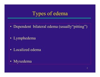
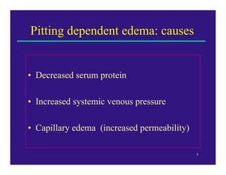
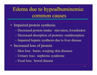
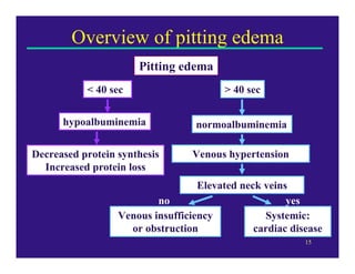
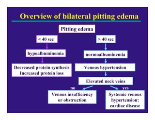
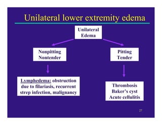
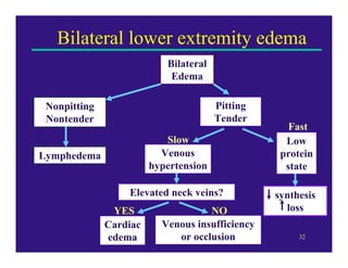
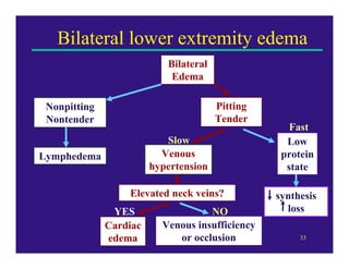
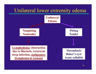
2. EDEMA
• Increased fluid in interstitial spaces
• Hydrothorax, hydropericardium, and
hydroperitoneum (ascites)
• Anasarca = severe and generalized edema
with profound sub-q tissue swelling .
NON-INFLAMMATORY CATEGORIES OF
EDEMA:
1. Incr hydrostatic pressure
2. Reduced plasma osmotic pressure
3. Lymphatic obstruction
4. Sodium Retention
3. FACTORS THAT GOVERN MOVEMENT OF WATER
B/C VASCULAR AND INTERSTITIAL SPACES:
Vascular hydrostatic pressure
Plasma colloid osmotic pressure
Incr capillary pressure or decr colloid osmotic
pressure incr interstitial fluid
Excess interstitial fluid is drained away by lymphatics
Lymphatic obstruction can cause edema
Edema fluid in hydrodynamic derangements is usually
a protein-poor transudate
Inflammatory edema is a ptn-rich exudate
4. INCREASED HYDROSTATIC PRESSURE:
Local increases in HP – may result from impaired venous outflow
Deep venous thrombosis in lower legs edema
GENERALIZED INCREASES IN HP SYSTEMIC EDEMA
• Occur most frequently in CHF
• CHF affects R vent cardiac fx
• Assoc w/ decr CO decr renal perfusion triggers renin-
angiotension-aldosterone axis Na and water retention
• Designed to incr intravascular volume improve CO restore renal
perfusion
• Heart cannot incr CO extra fluid load incr venous P incr
transudation edema
• Cycle of renal fluid restriction and worsening edema
• Edema of dependent parts of the body – prominent feature of CHF,
esp of R ventricle
• Constrictive pericarditis, ascites (liver cirrhosis), venous obstruction or
compression, arteriolar dilation
5. REDUCED PLASMA OSMOTIC PRESSURE:
• Results from excessive loss or decr production of albumin
• Albumin = serum ptn = most resp for maintaining colloid pressure
• Nephrotic Syndrome
• Leaky glomerular capillary wall, generalized edema
• Decr alb production b/c of diffuse liver pathology (cirrhosis), or from
ptn malnutrition
• Decr plasma osmotic pressure net movement of fluid into
interstitium decr in plasma V decr in renal perfusion
• Also have decr in CO b/c of decr in plasma V
• Decr in renal perfusion secondary aldosteronism Na and water
retention by kidneys
• Na and water retention cannot correct plasma V b/c still have low
serum ptns
• Initial edema ppt by hypoproteinemia is made worse by secondary
salt and water retention
• Edema caused by renal dysfx or nephrotic syndrome is generally
more severe, affects all parts of the body equally
• Liver cirrhosis, malnutrition, ptn-losing gastroenteropathy
6. LYMPHATIC OBSTRUCTION:
• Impaired lymphatic drainage and resulting lymphedema
is usually localized
• Can result from inflamm or neoplastic obstruction
• Parasitic infection filariases
• Often causes massive lymphatic and lymph node
fibrosis in inguinal region edema of external genitalia
and lower limbs
• Edema called elephantiasis
CANCER OF BREAST
• Can be tx by removal/irradiation of breast and axillary
lymph nodes resection of lymph nodes and scarring
severe edema of the arm
• Inflammatory, Neoplastic, Postsurgical, Postirradiation
7. Sodium and Water Retention:
• Either primary or secondary causes of edema
• Incr salt with obligate accompanying water:
• Incr intravascular fluid volume incr hydrostatic P
• Decr vascular colloid pressure
• Excessive salt intake w/ renal insufficiency, incr
tubular reabs of sodium (renal hypoperfusion, incr R-
A-Aldosterone secretion)
MORPHOLOGY OF EDEMA:
• Most easily recognized grossly
• By LM manifests as subtle cell swelling, w/ clearing
and separation of the ECM components
• Most commonly found in sub-q tissues, lungs, and
brain
8. SUBCUTANEOUS EDEMA:
• Can be diffuse or more conspicuous at sites of highest
hydrostatic pressures
• More conspicuous: called dependent edema,
distribution largely dependent on gravity
• Edema of dependent parts of the body – prominent
feature of CHF, esp of R ventricle
• Edema caused by renal dysfx or nephrotic syndrome is
generally more severe, affects all parts of the body
equally
• May initially be seen in tissues w/ soft tissue matrix; ex
– eyelids, causing periorbital edema
• Pitting edema – finger pressure on edematous areas
leaves a finger-shaped depression
9. PULMONARY EDEMA:
• Common clinical concern, most often in left
ventricular failure
• Also in renal failure, adult respiratory
distress syndrome, pulm infections, and
hypersensitivity rxns
• Lungs go to 2X to 3X their normal weight
• Sectioning: frothy, blood-tinged fluid –
represents a mixture of air, edema fluid, and
extravasated RBCs
10. EDEMA OF THE BRAIN:
• May be localized to sites of injury (abscess,
neoplasm), or may be generalized
(encephalitis, hypertensive crises, obstruction
to brain’s venous outflow)
• Trauma can cause local or generalized
edema, depending on nature and extent of
injury
• Generalized edema
• Brain is grossly swollen, narrowed sulci,
distended gyri
• Shows signs of flattening against skull
11. CLINICAL CORRELATION:
• Sub-q tissue edema - can impair wound
healing or clearance of infection
• Pulmonary edema – can cause death by
interfering w/ gas exchange; fluid in alveoli
create favorable place for bacterial infection
• Brain edema – serious, can be rapidly
fatal; brain herniation or brain stem vascular
supply can be compressed injure vent
centers
13. A. Intracellular Edema
a. depression of the metabolic
systems of the tissues
b. lack of adequate nutrition to the
cells
14. A. Extracellular Edema
a. abnormal leakage of fluid from the plasma to the
interstitial spaces across the capillaries
failure of the lymphatics to return fluid from the
interstitium back into the blood
The most common clinical cause of interstitial fluid
accumulation is excessive capillary fluid filtration
15. Factors That Can Increase Capillary Filtration
1. Filtration Coefficient – product of permeability and surface
area of the capillaries (Kf)
2. Capillary Hydrostatic Pressure (Pc)
3. Interstitial Fluid hydrostatic Pressure (Pif)
4. Capillary Plasma colloid Osmotic Pressure (iic)
5. Interstitial Fluid Colloid Osmotic Pressure (iiif)
Filtration = Kf x ( Pc – Pif – iic + iiif )
16. Major Factors that cause Increased
Capillary Filtration of Fluid and Protein into
the Interstitium:
1. Increased capillary Hydrostatic Pressure
2. Decreased Plasma Colloid Osmotic Pressure
3. Increased Capillary Permeability
17. Summary of Causes of Extracellular Edema
Increased capillary pressure
A. Excessive kidney retention of salt and water
1. Acute and chronic kidney failure
2. Mineralocorticoid excess
18. B. High venous pressure
1. Heart failure
2. Venous obstruction
3. Failure of venous pump
a) paralysis of muscles
b) immobilized parts of body
c) Failure of venous valves
19. C. Decreased arteriolar resistance
1. Excessive body heat
2. Insufficiency of sympathetic nervous system
3. Vasodilator drugs
20. II. Decreased plasma protein
A. Loss of proteins in urine (nephrotic syndrome)
B. Loss of protein from denuded skin areas
1. Burns
2. Wounds
C.Failure to produce proteins
1. Liver disease
2. Serious protein or caloric malnutrition
21. III. Increased capillary permeability
A. Immune reactions that cause release of histamine
B. Toxins
C. Bacterial infections
D. Vitamin deficiency, especially vitamin C
E. Prolonged ischemia
F. Burns
22. IV. Blockage of lymph return
A. Cancer
B. Infections (filarial nematodes)
C. Surgery
D. Congenital absence or abnormality of lymphatic
vessels
23. Safety Factors That Prevent Edema
1. The safety factors caused by low tissue compliance
( -3mmHg )
2. The safety factor caused by increased lymph flow ( 7 mmHg )
3. The safety factor caused by washdown of proteins from the
interstitial spaces ( 7 mmHg )
24. DAILY INTAKE AND OUTPUT OF WATER (in ml/day)________
Normal Prolonged
Heavy Exercise
Intake
Fluids ingested 2100 ?
From metabolism 200 200
Total intake ?
Output
Insensible – Skin 350 350
Insensible - Lungs 350 650
Sweat 100 5000
Feces 100 100
Urine 1400 500
Total output 2300 6600
25. Edema
Brad Anawalt, MD
VA Puget Sound
University of Washington
28. Edema due to hypoalbuminemia:
common causes
• Impaired protein synthesis
– Decreased protein intake: starvation, kwashiokor
– Decreased absorption of proteins: malabsorption
– Impaired hepatic synthesis due to liver disease
• Increased loss of protein
– Skin loss: burns, weeping skin diseases
– Urinary loss: nephrotic syndrome
– Fecal loss: bowel disease
4
30. Edema due to capillary permeability
• Vasculitis
• Idiopathic cyclic edema of women
– Varies with menstrual cycle
• Post-anoxic encephalopathy
6
31. Pitting recovery time
• Technique:
– Press firmly to bone
– Shine light and determine time to resolution of
shadow
• Interpretation
– Acute edema (< 3 months)
– < 40 seconds associated with low serum albumin
7
32. Rapid pitting recovery: < 40 seconds
• protein synthesis
– protein intake: dietary history
– absorption of proteins: diarrhea
– hepatic synthesis due to liver disease:
• History: alcohol, other hepatotoxins, hepatitis
• Physical findings: spider angiomata
• loss of protein
– Skin loss: skin lesions such as burns, ulcers
– Urinary loss: foamy urine with high protein
– Fecal loss: diarrhea, sticky, oily stools
8
33. Edema with rapid pitting recovery:
evaluation of etiology
• Impaired protein synthesis
– protein intake: starvation, kwashiokor
– absorption of proteins: malabsorption
– hepatic synthesis due to liver disease
• Increased loss of protein
– Skin loss: burns, weeping skin diseases
– Urinary loss: nephrotic syndrome
– Fecal loss: bowel disease 9
35. Venous hypertension & edema:
systemic vs regional
• Systemic venous hypertension
– Elevated neck veins
– Abdominojugular reflux and third heart sound
in heart failure
• Regional venous hypertension
– Neck veins not elevated
– No abdominal reflux, third heart sound
11
36. Regional venous hypertension:
Venous insufficiency vs obstruction
• Venous insufficiency
– Common
– Bilateral
– Chronic
• Associated with hemosiderin deposition
• Venous obstruction
– Often unilateral:
• Baker’s cyst, venous thrombosis
– Acute (< 3 months)
• Not associated with hemosiderosis 12
37. Characteristics of venous insufficiency
• Dependent edema: lower extremities, perineum
– May be asymmetric
– Often tender
– Usually chronic or recurrent
• Associated with hemosiderin deposition
– Ulceration often occurs
• Treatment
– Elevation
– Exercise to improve venous return
– Diuretics
– Compression +/- topical corticosteroids 13
38. Signs of systemic venous hypertension
• Elevated neck veins
– More than 3 cm above the angle of Lewis
• Angle of Lewis = sternal angle
– Abdominojugular reflux
• Suggests congestive heart failure
• Press for 10 seconds firmly on abdomen
• If neck veins fall after relief of pressure, then
suggests congestive heart failure
– Third heart sound
• Listen with bell of stethoscope
• Suggests congestive heart failure 14
39. Overview of pitting edema
Pitting edema
< 40 sec > 40 sec
hypoalbuminemia normoalbuminemia
Decreased protein synthesis Venous hypertension
Increased protein loss
Elevated neck veins
no yes
Venous insufficiency Systemic:
or obstruction cardiac disease
15
40. Treatment of fast-recovering
pitting edema
• hypoalbuminemic (<40 seconds)
– Treat malnutrition
– Treat underlying cause of malabsorption
– Treatment of edema due to cirrhosis
• Judicious use of diuretics and aldosterone
antagonist can alleviate edema
– Treat protein loss
• Skin or fecal loss: treat underlying disease
• Urinary loss: angiotensin-converting enzyme
inhibitor 16
41. Treatment of slow-recovering
pitting edema
• normoalbuminemic ( > 40 seconds)
– Treat congestive heart failure
• Bed rest and elevation of legs useful for acute
edema
• Loop diuretics, digoxin, angiotensin-converting
enzyme inhibitor, beta blocker if tolerated
– Treat venous insufficiency
• Diuretics, compression, leg-elevating exercises
– Treat underlying obstruction of veins
• Anticoagulants, leg elevation for thrombosis 17
42. Lymphedema: nonpitting edema
• Protein-rich edema due to abnormality
of lymphatic drainage
• Characteristics
– Nontender, painless
– Does not vary much during the day
– Ulceration rare
– Hyperkeratosis, thickening of skin
18
43. Lymphedema: causes
• Upper extremity
– breast cancer or surgery/radiation for breast
cancer
– Newborn baby, Turner’s syndrome (X0)
• Lower extremity
– Idiopathic: aplasia/dysplasia of lymphatics
• 3 types: congenital, praecox, form tarde
• Associated with yellow nails, pleural effusions
– Secondary
• Inflammatory
• Obstructive 19
44. Lymphedema: secondary causes
• Inflammatory
– Tropical: filariasis + recurrent strep infection
– Nontropical: recurrent streptococcal cellulitis
• Obstructive
– Unilateral in 95%
– Usually due to malignancy
– Prostate cancer most common in men
– Lymphoma most common in women
– Any pelvic tumor or major pelvic surgery 20
46. Lymphedema: treatment
• Fluid mobilization
– Diuretics
– Elevation
– Exercise
– Compressive, elastic stockings
– Massage
• Control of infection
– Treatment of dermatophytes
– Prophylaxis against streptococcal infections
• Amoxicillin, amoxicillin/clavulanate
22
47. Lymphedema: treatment #2
• Other therapies
– Coumadin
• Stimulates cutaneous macrophages and local
proteolysis
• Might be effective with topical administration
• Reduces edema ~55%
– Flavenoids
• Water soluble vitamin
– Surgery
• liposuction
23
48. Miscellaneous causes of edema
• Hot days: bilateral edema due to venous pooling +
compensatory salt and water retention (aldosterone)
• Localized edema
– Facial edema
• Trichinosis, hypothyroidism, allergies, nephrotic
syndrome
• Pretibial myxedema from Graves’ thyrotoxicosis
• Neurogenic edema
• Lipedema
– adiposity of the legs
• Pseudothrombophlebitis
– A form of unilateral edema with elevated venous
pressure due to a popliteal cyst 24
49. Summary of lower extremity edema
• Key questions:
• Are both legs edematous?
• Is it pitting edema?
• Is the edematous area tender or painful?
• Are the neck veins elevated? 25
50. Overview of bilateral pitting edema
Pitting edema
< 40 sec > 40 sec
hypoalbuminemia normoalbuminemia
Decreased protein synthesis Venous hypertension
Increased protein loss
Elevated neck veins
no yes
Venous insufficiency Systemic venous
or obstruction hypertension:
cardiac disease
26
52. Bilateral lower extremity edema
Bilateral
Edema
Nonpitting Pitting
Nontender Tender
Fast
Slow Low
Lymphedema Venous protein
hypertension state
Elevated neck veins? synthesis
YES NO loss
Cardiac Venous insufficiency
edema or occlusion 28
53. Case #1
A 55 year old man with a history of heavy alcohol use
complains of unilateral edema for one week. On exam he
has pitting edema of his left leg below the knee. His left
leg is tender. The skin of legs is not hyperpigmented.
The likely diagnosis is
a. Cirrhosis
b. Filariasis
c. Deep venous thrombosis
d. Prostate cancer
e. Congestive heart failure
29
54. Case #1 answer
A 45 year old man with a history of heavy alcohol use
complains of unilateral edema for one week. On exam he
has pitting edema of his left leg below the knee. His leg is
tende. The skin of legs is not hyperpigmented.
The diagnosis is likely to be deep venous thrombosis (c) or
cellulitis. The edema is unilateral (congestive heart failure
and cirrhosis are unlikely) and tender (lymphedema from
malignancy or filariasis is unlikely).
30
55. Case #2
A 45 year old man with a history of heavy alcohol use and
hepatitis complains of bilateral edema for two months.
On exam he has pitting edema of his legs below the knee.
Pitting resolves in 1 minute. His neck veins are elevated.
The likely diagnosis is
a. Cirrhosis
b. Filariasis
c. Inferior vena cava obstruction
d. Prostate cancer
e. Congestive heart failure
31
56. Bilateral lower extremity edema
Bilateral
Edema
Nonpitting Pitting
Nontender Tender
Fast
Slow Low
Lymphedema Venous protein
hypertension state
Elevated neck veins? synthesis
YES NO loss
Cardiac Venous insufficiency
edema or occlusion 32
57. Bilateral lower extremity edema
Bilateral
Edema
Nonpitting Pitting
Nontender Tender
Fast
Slow Low
Lymphedema Venous protein
hypertension state
Elevated neck veins? synthesis
YES NO loss
Cardiac Venous insufficiency
edema or occlusion 33
58. Case #2 answer
A 45 year old man with a history of heavy alcohol use and
hepatitis complains of bilateral edema for two months.
On exam he has pitting edema of his legs below the knee.
Pitting resolves in 1 minute. His neck veins are elevated.
The likely diagnosis is congestive heart failure (e). He has
bilateral edema with slow pitting (> 40 seconds) and neck
veins are elevated.
Bonus: What additional physical findings would be useful
to elicit?
34
59. Case #3
A 55 year old woman complains of right lower extremity
edema for one month. The leg was previously normal.
On exam she has nonpitting edema of the right leg. It is
nontender, and there is no hyperpigmentation.
The most likely diagnosis is
a. Baker’s cyst
b. Inferior vena cava obstruction
c. Chronic venous insufficiency
d. Lymphoma
e. Congestive heart failure
35
60. Case #3 answer
A 55 year old woman complains of right lower extremity
edema for one month. The leg was previously normal.
On exam she has nonpitting edema of the right leg. It is
nontender, and there is no hyperpigmentation.
The most likely diagnosis is lymphedema due to
lymphoma (d). She has nontender unilateral edema.
Therefore, causes of bilateral edema (congestive heart
failure and inferior vena cava obstruction) are unlikely.
A Baker’s cyst will cause tenderness and pitting edema.
Chronic venous insufficiency is unlikely given the acute
presentation and is usually associated with
hyperpigmentation.
36
62. Unilateral lower extremity edema
Unilateral
Edema
Nonpitting Pitting
Nontender Tender
Lymphedema: obstruction
due to filariasis, recurrent Thrombosis
strep infection, malignancy Baker’s cyst
(lymphoma in woman) Acute cellulitis
38
63. Edema references
Sapira JD. The Art & Science of Bedside Diagnosis.
Williams & Wilkins, Baltimore, 1990.
Rockson SG. Lymphedema. Am J Med. 2001;110:288-295.
Wiese J. The abdominojugular reflux sign. Am J Med.
2000;109:59-61.
McGee SR. Physical examination of venous pressure: a
critical review. Am Heart J. 1998;136:10-18.
Henry JA, et al. Assessment of hypoproteinaemic oedema: a
simple physical sign. Br Med J 1978;890.
39