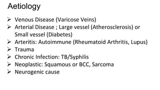
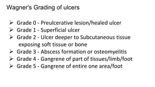
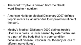
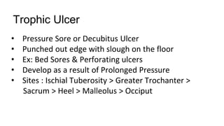
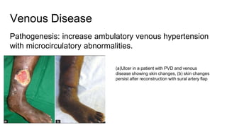
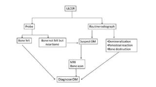
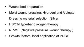
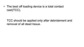
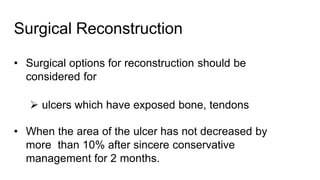
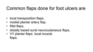
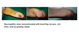
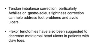
Trophic ulcer
Moderator: Dr Puneet Bhargava Sir
Presenter: Dr Gangadhar
Ulcer
• An ulcer is a break in the continuity of the covering
epithelium - skin or mucous membrane.
• It may either follow molecular death of the surface
epithelium or its traumatic removal.
Aetiology
 Venous Disease (Varicose Veins)
 Arterial Disease ; Large vessel (Atherosclerosis) or
Small vessel (Diabetes)
 Arteritis: Autoimmune (Rheumatoid Arthritis, Lupus)
 Trauma
 Chronic Infection: TB/Syphilis
 Neoplastic: Squamous or BCC, Sarcoma
 Neurogenic cause
Wagner's Grading of ulcers
 Grade 0 - Preulcerative lesion/healed ulcer
 Grade 1 - Superficial ulcer
 Grade 2 - Ulcer deeper to Subcutaneous tissue
exposing soft tissue or bone
 Grade 3 - Abscess formation or osteomyelitis
 Grade 4 - Gangrene of part of tissues/limb/foot
 Grade 5 - Gangrene of entire one area/foot
• The word ‘Trophic’ is derived from the Greek
word Trophe = nutrition.
• American Heritage Medical Dictionary 2007 defines
trophic ulcers as ‘an ulcer due to impaired nutrition of
the part’.
• Mosby's Medical Dictionary 2009 defines trophic
ulcer as ‘a pressure ulcer caused by external trauma
to a part of the body that is in poor condition
because of disease, vascular insufficiency or loss of
afferent nerve fibres’.
Classification of trophic ulcers
Trophic Ulcer
• Pressure Sore or Decubitus Ulcer
• Punched out edge with slough on the floor
• Ex: Bed Sores & Perforating ulcers
• Develop as a result of Prolonged Pressure
• Sites : Ischial Tuberosity > Greater Trochanter >
Sacrum > Heel > Malleolus > Occiput
Diabetic Ulcer
It may be caused due to
• Diabetic Neuropathy
• Diabetic Microangiopathy
• Increased Glucose : Increased Infection
• Foot ( Plantar ), Leg, Back, Scrotum, Perineum
• Ischemia, Septicemia, Osteomyelitis.
Venous Disease
Pathogenesis: increase ambulatory venous hypertension
with microcirculatory abnormalities.
(a)Ulcer in a patient with PVD and venous
disease showing skin changes, (b) skin changes
persist after reconstruction with sural artery flap
NEUROGENIC ULCER
• These include all ulcers in insensate hands and feet
in patients with neuropathy.
• Protective pain and pressure perception being
absent, they do not relieve pressure and hence the
repetitive trauma leads to skin breakdown and
ulceration.
EXAMINATIONS
INSPECTION
Location, size, shape, floor, edge, discharge, surrounding area.
PALPATION
Tenderness, local rise of temperature, bleeding on touch,
consistency of the ulcer, edge, surrounding area - oedema,
mobility.
REGIONAL LYMPH NODES
SENSATIONS
PULSATIONS
FUNCTION OF THE JOINT
SYSTEMIC EXAMINATION
Management
The key to successful management of a chronic ulcer
would be to correctly identify the etiology as well as the
local and systemic factors that could be contributing to its
non healing nature.
Investigations
• Complete blood picture: Hb%, TLC, DLC, ESR,
• Urine and blood examination to rule out diabetes
• Chest X-ray - PA.
• Pus for culture/sensitivity
• Lower limb angiography in cases of arterial diseases
• X-ray of the part to see for Osteomyelitis
• Biopsy: Non-healing/malignant ulcers
Treatment
• Address cause
• Correct deficiencies
• Control pain, infection
• Debridement, dressing
• Closure of defect
Debridement
• Surgical debridement should be aggressive to include
removal of all surrounding hard callus, hyperkeratotic
skin, all dead necrotic tissue, infected soft tissue and
bone.
• Activation of platelets for control of haemorrhage leads
to release of growth factors which begins the process
of healing.
(a) Neuropathic bone deep ulcer, (b) radiograph showing bone destruction
Osteomyelitis
• Wound bed preparation
• Moist wound dressing: Hydrogel and Alginate
Dressing material selection :Silver
• HBOT(Hyperbaric oxygen therapy)
• NPWT (Negative pressure wound therapy )
• Growth factors: local application of PDGF
Off Loading Measures
• Strict bed rest
• Use of crutches, Wheel chair , Walkers
• Pressure reducing measures like air cushion, waterbeds,
Plaster boot
• Removable contact casting, half shoes or specialised
footwear.
• The best off loading device is a total contact
cast(TCC).
TCC should be applied only after debridement and
removal of all dead tissue.
Surgical Reconstruction
• Surgical options for reconstruction should be
considered for
 ulcers which have exposed bone, tendons
• When the area of the ulcer has not decreased by
more than 10% after sincere conservative
management for 2 months.
Common flaps done for foot ulcers are
• local transposition flaps,
• medial plantar artery flap,
• fillet flaps,
• distally based sural neurocutaneous flaps,
• VY plantar flaps local muscle
flaps.
Neuropathic ulcer reconstructed with local flap (a) pre-, (b)
intra- and (c) postop views
• Tendon imbalance correction, particularly
Achilles or gastro-soleus tightness correction
can help address foot problems and avoid
ulcers.
• Flexor tenotomies have also been suggested to
decrease metatarsal head ulcers in patients with
claw toes.
Nerve decompression
• Nerve decompression as an adjunct therapy to medical
treatment should be used when there is clinical and / or
electrodiagnostic evidence of compression neuropathy.
• Prevention of limb loss in chronic diabetes mellitus, for
diminishment of pain and for restoration of sensory/
motor function.
Objective wound measurement
• Keeps the treating surgeon and the patient aware
of progress.
• Record keeping should be done by two methods-
• photographic record of the ulcers
• document the length, breadth and depth
measurements of the ulcer at weekly
intervals.
• It helps to objectively analyse healing and
motivates patients towards self-care.
Patient education and home care
1. Explanation in simple terms about their specific pathology.
2. Understanding that changing habits and making a few
lifestyle changes could go a long way to keep progression
of disease and its consequences in check, e.g. leg elevation
whenever possible, changing position to keep pressure off
one point.
3. Cessation of smoking.
4. Regular chiropodist care (foot and nail grooming).
5. Strict glycaemia control for diabetics.
6.Compression for venous diseases.
7.Daily end of day check of hands and feet for signs of
breakdown.
8.Self-monitoring of sole/fingertip temperature.
9.Specialized footwear for off-loading pressure.
10.Regular follow-up with physician even in periods of no
ulcer stage.
THANK YOU
Conclusion
Care of patients with trophic ulcers needs to be
multidisciplinary involving a large team which
includes Dermatologist.physician, general surgeon,
plastic surgeon, endocrinologist, vascular surgeon,
interventional radiologist, dietician, physiotherapist
and chiropodist.

trophic ulcer.pptx

  • 1.
    Trophic ulcer Moderator: DrPuneet Bhargava Sir Presenter: Dr Gangadhar
  • 2.
    Ulcer • An ulceris a break in the continuity of the covering epithelium - skin or mucous membrane. • It may either follow molecular death of the surface epithelium or its traumatic removal.
  • 3.
    Aetiology  Venous Disease(Varicose Veins)  Arterial Disease ; Large vessel (Atherosclerosis) or Small vessel (Diabetes)  Arteritis: Autoimmune (Rheumatoid Arthritis, Lupus)  Trauma  Chronic Infection: TB/Syphilis  Neoplastic: Squamous or BCC, Sarcoma  Neurogenic cause
  • 4.
    Wagner's Grading ofulcers  Grade 0 - Preulcerative lesion/healed ulcer  Grade 1 - Superficial ulcer  Grade 2 - Ulcer deeper to Subcutaneous tissue exposing soft tissue or bone  Grade 3 - Abscess formation or osteomyelitis  Grade 4 - Gangrene of part of tissues/limb/foot  Grade 5 - Gangrene of entire one area/foot
  • 5.
    • The word‘Trophic’ is derived from the Greek word Trophe = nutrition. • American Heritage Medical Dictionary 2007 defines trophic ulcers as ‘an ulcer due to impaired nutrition of the part’. • Mosby's Medical Dictionary 2009 defines trophic ulcer as ‘a pressure ulcer caused by external trauma to a part of the body that is in poor condition because of disease, vascular insufficiency or loss of afferent nerve fibres’.
  • 6.
  • 7.
    Trophic Ulcer • PressureSore or Decubitus Ulcer • Punched out edge with slough on the floor • Ex: Bed Sores & Perforating ulcers • Develop as a result of Prolonged Pressure • Sites : Ischial Tuberosity > Greater Trochanter > Sacrum > Heel > Malleolus > Occiput
  • 8.
    Diabetic Ulcer It maybe caused due to • Diabetic Neuropathy • Diabetic Microangiopathy • Increased Glucose : Increased Infection • Foot ( Plantar ), Leg, Back, Scrotum, Perineum • Ischemia, Septicemia, Osteomyelitis.
  • 10.
    Venous Disease Pathogenesis: increaseambulatory venous hypertension with microcirculatory abnormalities. (a)Ulcer in a patient with PVD and venous disease showing skin changes, (b) skin changes persist after reconstruction with sural artery flap
  • 11.
    NEUROGENIC ULCER • Theseinclude all ulcers in insensate hands and feet in patients with neuropathy. • Protective pain and pressure perception being absent, they do not relieve pressure and hence the repetitive trauma leads to skin breakdown and ulceration.
  • 13.
    EXAMINATIONS INSPECTION Location, size, shape,floor, edge, discharge, surrounding area. PALPATION Tenderness, local rise of temperature, bleeding on touch, consistency of the ulcer, edge, surrounding area - oedema, mobility. REGIONAL LYMPH NODES SENSATIONS PULSATIONS FUNCTION OF THE JOINT SYSTEMIC EXAMINATION
  • 14.
    Management The key tosuccessful management of a chronic ulcer would be to correctly identify the etiology as well as the local and systemic factors that could be contributing to its non healing nature.
  • 15.
    Investigations • Complete bloodpicture: Hb%, TLC, DLC, ESR, • Urine and blood examination to rule out diabetes • Chest X-ray - PA. • Pus for culture/sensitivity • Lower limb angiography in cases of arterial diseases • X-ray of the part to see for Osteomyelitis • Biopsy: Non-healing/malignant ulcers
  • 16.
    Treatment • Address cause •Correct deficiencies • Control pain, infection • Debridement, dressing • Closure of defect
  • 17.
    Debridement • Surgical debridementshould be aggressive to include removal of all surrounding hard callus, hyperkeratotic skin, all dead necrotic tissue, infected soft tissue and bone. • Activation of platelets for control of haemorrhage leads to release of growth factors which begins the process of healing.
  • 18.
    (a) Neuropathic bonedeep ulcer, (b) radiograph showing bone destruction Osteomyelitis
  • 20.
    • Wound bedpreparation • Moist wound dressing: Hydrogel and Alginate Dressing material selection :Silver • HBOT(Hyperbaric oxygen therapy) • NPWT (Negative pressure wound therapy ) • Growth factors: local application of PDGF
  • 21.
    Off Loading Measures •Strict bed rest • Use of crutches, Wheel chair , Walkers • Pressure reducing measures like air cushion, waterbeds, Plaster boot • Removable contact casting, half shoes or specialised footwear.
  • 22.
    • The bestoff loading device is a total contact cast(TCC). TCC should be applied only after debridement and removal of all dead tissue.
  • 23.
    Surgical Reconstruction • Surgicaloptions for reconstruction should be considered for  ulcers which have exposed bone, tendons • When the area of the ulcer has not decreased by more than 10% after sincere conservative management for 2 months.
  • 24.
    Common flaps donefor foot ulcers are • local transposition flaps, • medial plantar artery flap, • fillet flaps, • distally based sural neurocutaneous flaps, • VY plantar flaps local muscle flaps.
  • 25.
    Neuropathic ulcer reconstructedwith local flap (a) pre-, (b) intra- and (c) postop views
  • 26.
    • Tendon imbalancecorrection, particularly Achilles or gastro-soleus tightness correction can help address foot problems and avoid ulcers. • Flexor tenotomies have also been suggested to decrease metatarsal head ulcers in patients with claw toes.
  • 27.
    Nerve decompression • Nervedecompression as an adjunct therapy to medical treatment should be used when there is clinical and / or electrodiagnostic evidence of compression neuropathy. • Prevention of limb loss in chronic diabetes mellitus, for diminishment of pain and for restoration of sensory/ motor function.
  • 28.
    Objective wound measurement •Keeps the treating surgeon and the patient aware of progress. • Record keeping should be done by two methods- • photographic record of the ulcers • document the length, breadth and depth measurements of the ulcer at weekly intervals. • It helps to objectively analyse healing and motivates patients towards self-care.
  • 29.
    Patient education andhome care 1. Explanation in simple terms about their specific pathology. 2. Understanding that changing habits and making a few lifestyle changes could go a long way to keep progression of disease and its consequences in check, e.g. leg elevation whenever possible, changing position to keep pressure off one point. 3. Cessation of smoking. 4. Regular chiropodist care (foot and nail grooming).
  • 30.
    5. Strict glycaemiacontrol for diabetics. 6.Compression for venous diseases. 7.Daily end of day check of hands and feet for signs of breakdown. 8.Self-monitoring of sole/fingertip temperature. 9.Specialized footwear for off-loading pressure. 10.Regular follow-up with physician even in periods of no ulcer stage.
  • 32.
  • 33.
    Conclusion Care of patientswith trophic ulcers needs to be multidisciplinary involving a large team which includes Dermatologist.physician, general surgeon, plastic surgeon, endocrinologist, vascular surgeon, interventional radiologist, dietician, physiotherapist and chiropodist.