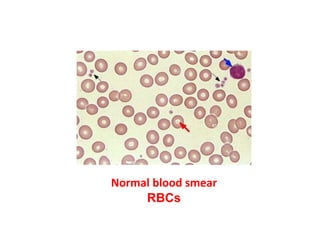
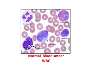
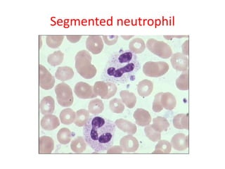
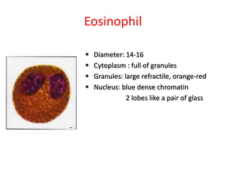
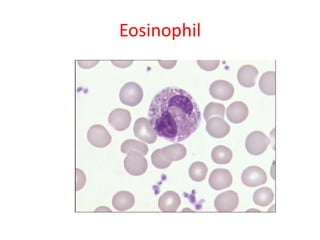
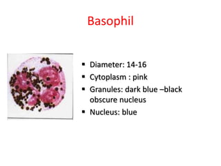
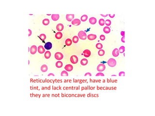
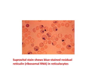
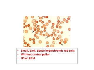
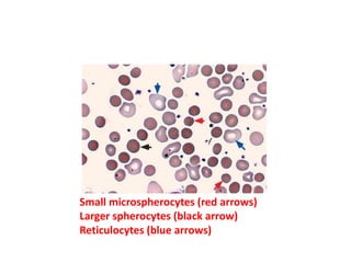
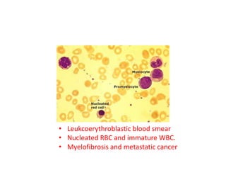
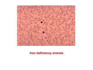
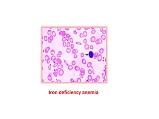
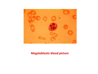
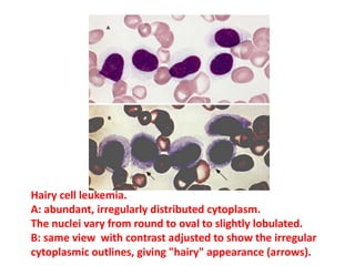
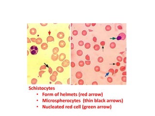
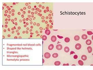
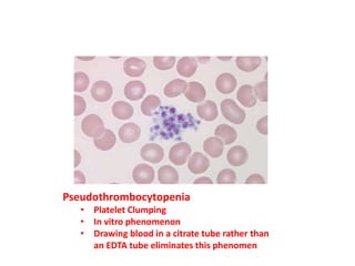
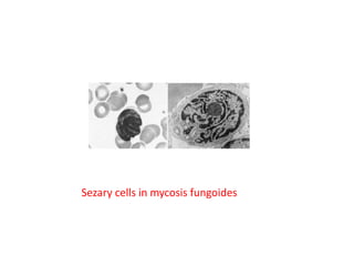
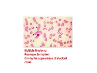
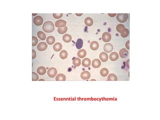
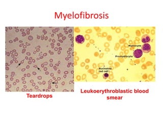
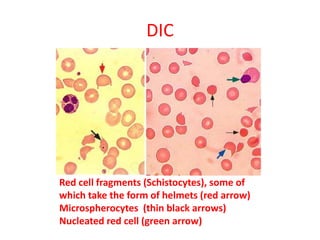
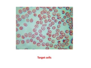
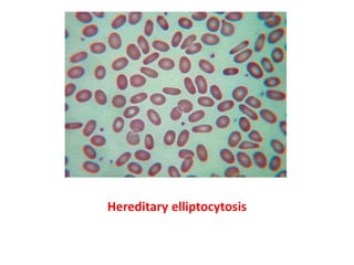
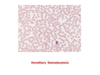
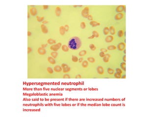
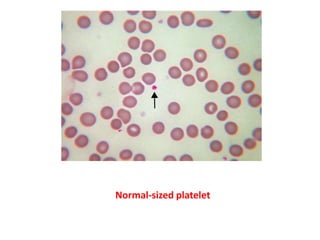
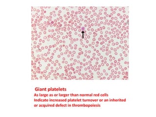
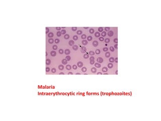
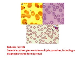
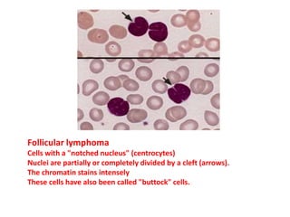
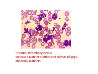
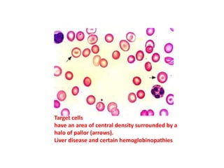
Evaluation of the
Peripheral Blood Smear
Ahmed Shorafa, MD
5/4/2015
Normal blood smear
RBCs
Normal blood smear
WBC
BAND SEGMENTED
LYMPHOCYTEBASOPHIL
EOSINOPHIL
MONOCYTE
Band neutrophil
 Diameter:12-16
 Cytoplasm : pink
 Granules
 Nucleus:
 dark purple blue dense
chromatin
Band neutrophil
Segmented neutrophil
 Diameter: 12-16
 Cytoplasm : pink
 Granules
 Nucleus:
 dark purple blue dense
chromatin
 2-5 lobes
Segmented neutrophil
Eosinophil
 Diameter: 14-16
 Cytoplasm : full of granules
 Granules: large refractile, orange-red
 Nucleus: blue dense chromatin
2 lobes like a pair of glass
Eosinophil
Basophil
 Diameter: 14-16
 Cytoplasm : pink
 Granules: dark blue –black
obscure nucleus
 Nucleus: blue
Basophil
Lymphocyte
 Diameter: small 7-9
large 12-16
 Cytoplasm: medium blue
 Granules: small agranular
large a few
primary granules
 Nucleus: dark blue round
dense chromatin
Lymphocyte
Monocyte
 Diameter: 14-20
 Cytoplasm : grey blue
 Granules: dust-like lilac color
granules
 Nucleus: blue
large irregularly shaped and
folded
Monocyte
Normal human promyelocyte
Larger than myeloblasts and myelocytes (>20 micron).
The nucleus and nuclear chromatin and nucleoli resemble myeloblasts but
the cardinal feature is the presence of many violet granules in the
cytoplasm with either a dense or coarse pattern often obscuring other cell
landmarks.
Normal human myeloblasts.
The size is medium (15 to 20 microns in diameter), the nucleus is
large and round with finely granular chromatin, nucleoli are
present, granules are absent, and cytoplasm is scanty.
The lymphocyte is a small cell with a heavily stained
condensed nucleus and a small rim of blue cytoplasm.
The monocyte has an irregular nucleus with grayish, blue
cytoplasm that has few granules.
The metamyelocyte has a kidney bean-shaped nucleus with
a granulated cytoplasm.
Reticulocytes are larger, have a blue
tint, and lack central pallor because
they are not biconcave discs
Supravital stain shows blue-stained residual
reticulin (ribosomal RNA) in reticulocytes
• Small, dark, dense hyperchromic red cells
• Without central pallor
• HS or AIHA
Small microspherocytes (red arrows)
Larger spherocytes (black arrow)
Reticulocytes (blue arrows)
• Leukcoerythroblastic blood smear
• Nucleated RBC and immature WBC.
• Myelofibrosis and metastatic cancer
Iron deficiency anemia
Megaloblastic blood picture
Marked macroovalocytosis
B12 deficiency
• Myeloblasts
• Large cells with high NC ratio
• Nucleoli
• Auer rod in the cytoplasm
• Chronic lymphocytic leukemia
• Almost mature appearing lymphocytes
• Sparse cytoplasm
• Round to slightly oval nuclei
• No evident nucleoli.
• Damaged lymphocytes ("smudge cells")
• CML
• Basophilia and granulocytosis
with neutrophils and
immature granulocytes
Chronic myelogenous leukemia
Three normal-appearing basophils.
These cells differ from a neutrophil by the presence of coarse,
dark blue-black granules.
Hairy cell leukemia.
A: abundant, irregularly distributed cytoplasm.
The nuclei vary from round to oval to slightly lobulated.
B: same view with contrast adjusted to show the irregular
cytoplasmic outlines, giving "hairy" appearance (arrows).
Schistocytes
• Form of helmets (red arrow)
• Microspherocytes (thin black arrows)
• Nucleated red cell (green arrow)
Schistocytes
• Fragmented red blood cells
• Shaped like helmets,
triangles.
• Microangiopathic
hemolytic process
Pseudothrombocytopenia
• Platelet Clumping
• In vitro phenomenon
• Drawing blood in a citrate tube rather than
an EDTA tube eliminates this phenomen
Pseudothrombocytopenia
•Sickle cell anemia
•Multiple spindly sickle cells (blue arrows)
•Nucleated red blood cell in the upper left
•Howell-Jolly body (black arrow), nuclear fragment normally removed
by the spleen.
•Target cells (red arrow)
bite-like deformity Heinz bodies
G-6-PD deficiency
• Sézary cells
• Lymphocytes with frequently convoluted
nuclei
Sezary cells in mycosis fungoides
Multiple Myeloma
Rouleaux formation
Giving the appearance of stacked
coins.
Essenntial thrombocythemia
Myelofibrosis
Leukoerythroblastic blood
smearTeardrops
Red cell fragments (Schistocytes), some of
which take the form of helmets (red arrow)
Microspherocytes (thin black arrows)
Nucleated red cell (green arrow)
DIC
t
Target cells
Hereditary elliptocytosis
Hereditary Stomatocytosis
Multiple myeloma
Basophilic stippling
Fine or coarse purplish blue dots dispersed through
the red cell
Very nonspecific feature occurring in thalassemia,
lead poisoning, and dyserythropoiesis in general
Basophilic stippling in several red cells from a patient with lead
poisoning.
The granules represent ribosomal precipitates.
A similar picture can be seen in a number of other conditions
including thalassemia, megaloblastic anemia, sickle cell
anemia, and sideroblastic anemia.
Red cell agglutination.
Irregular aggregates of red cells
Mycoplasma pneumoniae infection.
Infectious mononucleosis
Chronic cold hemagglutinin disease
Dِ hle’s body.
Pale blue-gray amorphous inclusion near
the cell membrane of a neutrophil.
Can result from infection and inflammation
Pelger-Huët anomaly
inherited, but similar Pelger neutrophils
are seen in myelodysplastic syndromes,
Pelger-Huet anomaly
Toxic granulation
Heavy staining of azurophilic granules of neutrophils.
When accompanied by neutrophil vacuolation, it is often indicative of
infection
Can also result from inflammation, tissue damage, and pregnancy
Hypersegmented neutrophil
More than five nuclear segments or lobes
Megaloblastic anemia
Also said to be present if there are increased numbers of
neutrophils with five lobes or if the median lobe count is
increased
Normal-sized platelet
Giant platelets
As large as or larger than normal red cells
Indicate increased platelet turnover or an inherited
or acquired defect in thrombopoiesis
Malaria
Intraerythrocytic ring forms (trophozoites)
Babesia microti
Several erythrocytes contain multiple parasites, including a
diagnostic tetrad form (arrow)
Follicular lymphoma
Cells with a "notched nucleus" (centrocytes)
Nuclei are partially or completely divided by a cleft (arrows).
The chromatin stains intensely
These cells have also been called "buttock" cells.
Essential thrombocythemia
increased platelet number and clumps of large,
abnormal platelets.
Target cells
have an area of central density surrounded by a
halo of pallor (arrows).
Liver disease and certain hemoglobinopathies
Splenic B-cell lymphoma
circulating villous lymphocytes
Echinocytes, also known as burr cells.
Echinocytes are often confused with acanthocytes.
However, the projections of the red cell membrane in
echinocytes are smaller and much more regular in shape
and distribution than in acanthocytes
Acanthocytes (spur cells).
These cells have prominent irregular protrusions scattered over
the surface in an asymmetrical pattern.
They are often confused with echinocytes in which the
protrusions are smaller and more regular
Neutrophils with toxic granulations, which are dark coarse
granules.
A Döhle body is also seen (arrow).
Right panel: A neutrophil with toxic granulations, vacuoles
(another toxic change), and a Döhle body (arrow).
Characteristic of toxic systemic illnesses.
Severe intravascular hemolysis due to sepsis with Clostridium perfringens.
Neutrophils show toxic changes, including toxic granulation and vacuoles.
There is an increased number of spherocytes (blue arrows) and
polychromatophilic red cells (ie, reticulocytes, red arrow).
The major finding on this slide is the large number of red blood cell ghosts
(black arrows), due to the intravascular lysis of red cells from the
phospholipase and other lytic enzymes elaborated by the Clostridial
organisms.
Howell-Jolly bodies in two red cells (black arrows), nuclear remnants
that are normally removed by the spleen. Thus, they are seen in
patients who have undergone splenectomy (as in this case) or have
functional asplenia (eg, sickle disease disease).
Target cells are also seen (blue arrows), another consequence of
splenectomy.
The peripheral blood smear on the left is from a 5-month-old boy with
eosinophilia of unknown cause. Eosinophil granules are normal in number and
size; nuclei are slightly hyperlobulated (ie, more than the usual two lobes).
Eosinophilia gradually regressed, with no residual organ dysfuction.
The peripheral blood smear on the right is from a 50-year-old male with
hypereosinophilic syndrome and a three year history of eosinophilia, bone marrow
failure and multiple end-organ involvement. The eosinophils show marked
hypogranulation.
Blood film

Blood film