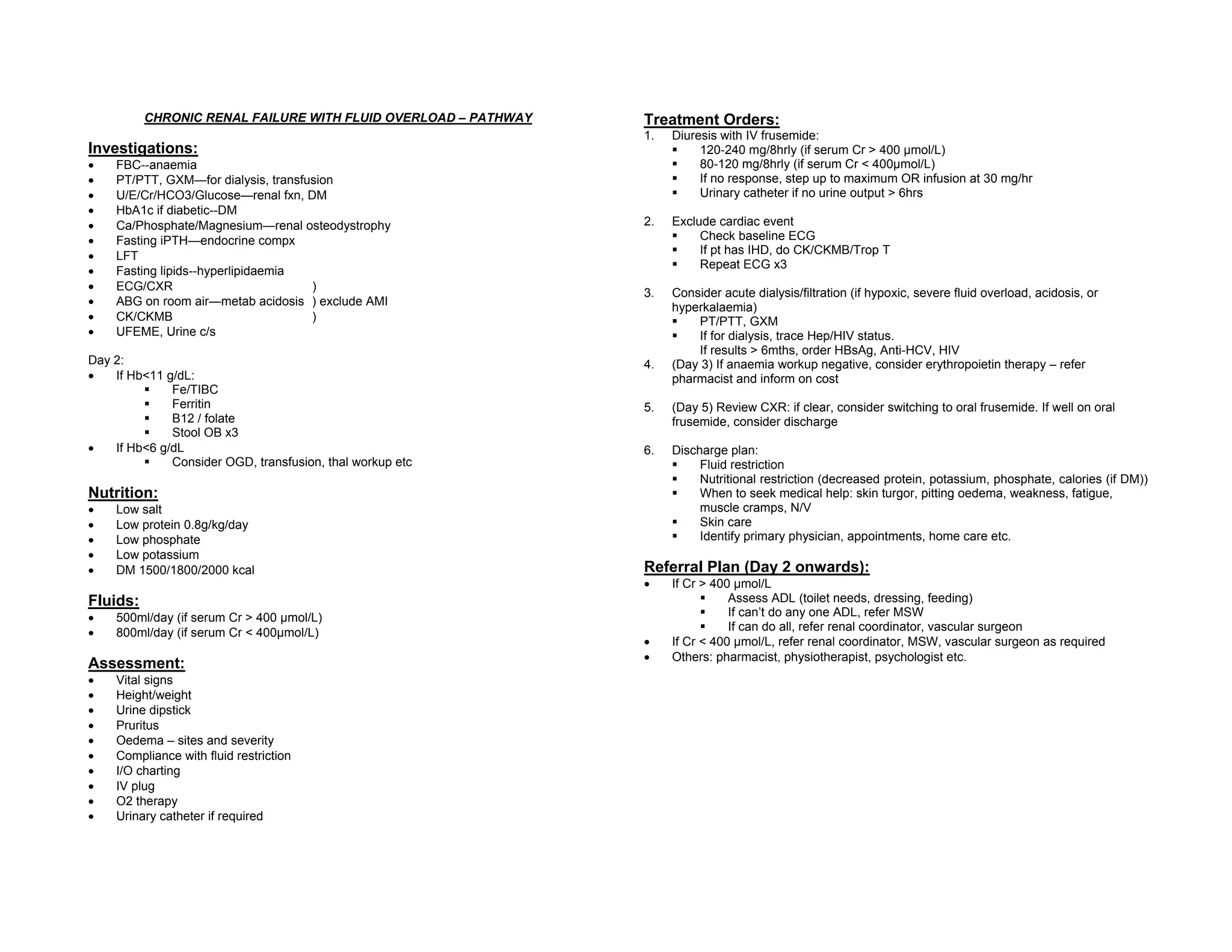
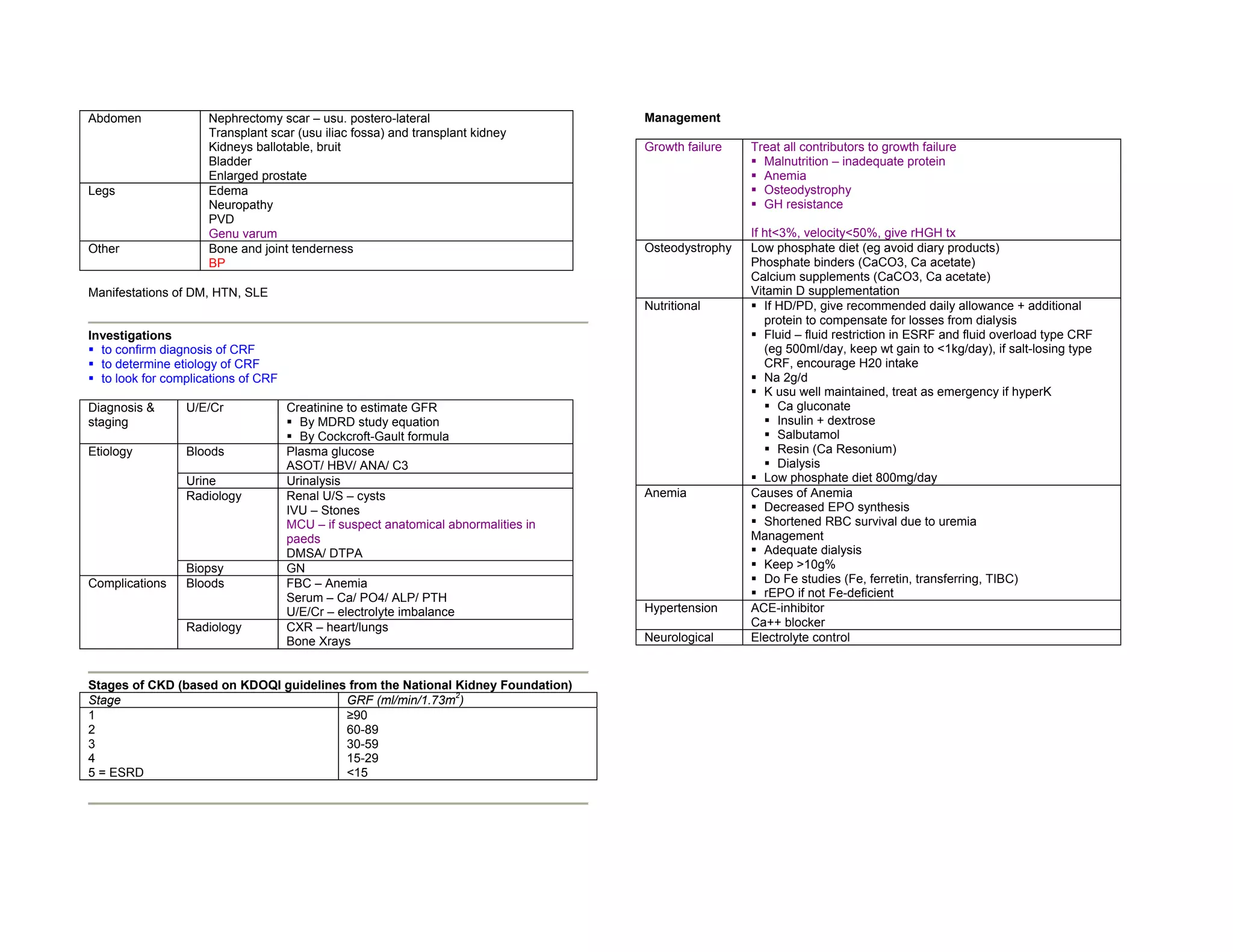
This document provides a concise long case approach for chronic renal failure. It outlines the key presenting complaints, history, physical exam findings, investigations, complications, and management strategies for chronic renal failure. The management involves addressing issues related to calcium/phosphate metabolism, anemia, osteodystrophy, fluid overload, and nutritional management. Treatment plans include diuresis, dialysis/filtration, erythropoietin therapy, calcium supplementation, and dietary restrictions. Referrals to other specialists are recommended based on the severity of renal failure and patient needs.
Chronic liver disease ( concise long case approach )Dr. Rubz
This document discusses the identification, diagnosis, and management of chronic liver disease (CLD). It identifies the main etiologies and risk factors for CLD, including infections like hepatitis B and C, alcohol, drugs, and metabolic disorders. It outlines the approach to diagnosing CLD based on symptoms, signs, laboratory tests, and imaging. Factors are assessed to determine if the CLD is compensated or decompensated. Management involves treating complications like ascites, bacterial peritonitis, variceal bleeding, and hepatic encephalopathy. Treatment may also include procedures like paracentesis or transplantation.
1. Mr. Al-Momtan, a 56-year-old male, presented with epigastric abdominal pain for 2 weeks which was worse after eating. Clinical exams and tests diagnosed him with a peptic ulcer disease.
2. He was prescribed a triple therapy of antibiotics and a PPI for 3 weeks to treat his condition.
3. Dyspepsia is a common gastrointestinal condition with many potential causes including non-ulcer dyspepsia, GERD, peptic ulcers, and H. pylori infection. Guidelines recommend lifestyle changes, antacids, and empirical PPI therapy as first-line treatment options.
Henoch–Schönlein purpura (HSP) is the most common vasculitis of childhood that presents with a tetrad of purpura, arthritis/arthralgia, abdominal pain, and renal involvement. It is characterized by IgA-containing immune complexes depositing in small vessels, skin, GI tract, joints, and kidneys. The diagnosis is based on purpura with lower limb predominance and at least one of the other criteria. Imaging and labs help assess organ involvement while biopsy confirms the diagnosis.
1. Polyuria is defined as urine output greater than 5 ml/kg/hr or 2 L/m2/day. Causes include increased fluid intake, increased urinary solute excretion, and impaired urinary concentration.
2. Evaluation of polyuria involves obtaining a detailed history, clinical examination, and investigations including a 24-hour urine output measurement and urine and serum analysis.
3. Distinguishing between central and nephrogenic diabetes insipidus involves tests like the water deprivation test and vasopressin response test.
This document provides definitions and diagnostic guidelines for recurrent abdominal pain (RAP) in children. It defines RAP as paroxysmal abdominal pain occurring between ages 4-16 years, affecting activities for at least 3 months. New definitions classify it as chronic abdominal pain without evidence of organic disease. RAP is a description, not a diagnosis, and can include various functional gastrointestinal disorders. Prevalence is reported as 10-15% of children. A thorough history and physical exam are important to identify alarm signals requiring further testing and to diagnose functional abdominal pain when organic causes are ruled out. Treatment involves reassurance, diet modification, pharmacotherapy like anticholinergics or TCAs, and psychological therapies like CBT.
This document discusses gastrointestinal bleeding in children. It notes that GI bleeding accounts for 10-20% of pediatric gastroenterology referrals and around 0.4% of PICU admissions are due to life-threatening GI bleeding. The presentation, classification, causes, diagnostic workup, and treatment of upper and lower GI bleeding in neonates, infants, and children are described in detail over multiple sections. Key points include distinguishing the source and severity of bleeding, identifying specific etiologies, and managing bleeding through supportive care, endoscopic procedures, medications, and surgery as needed.
Approach to child with generalized body swellingElhadi Hajow
Edema is characterized by swelling caused by excess fluid in the interstitial tissue. It can be localized or generalized. Common causes include cardiac, renal, or hepatic disease which decrease plasma oncotic pressure allowing fluid shift from vessels into tissue. A thorough history, physical exam, and lab tests are needed to determine the underlying cause and guide treatment such as diuretics, dietary changes, or treating the primary disease.
This document discusses chronic diarrhea in children, including definitions, causes, diagnosis, and treatment. It defines chronic diarrhea as diarrhea lasting 2-3 weeks or more. Common causes in infants include post-infectious lactase deficiency, celiac disease, cow's milk allergy, toddler's diarrhea, and infections like giardiasis. Diagnosis involves taking a thorough history and physical exam, with investigations tailored to the child's age and suspected causes, such as stool exams and tests for carbohydrate malabsorption.
This document discusses gastrointestinal bleeding in children beyond infancy. It notes that upper GI bleeding is common in pediatric ICU patients and is a frequent reason for referrals to pediatric gastroenterologists. The document then discusses the various etiologies of GI bleeding in children in detail, including gastrointestinal, systemic, genetic, and spurious causes. It provides information on diagnosing and grading GI bleeds and outlines management approaches including initial resuscitation, endoscopic therapies, pharmacotherapy, and in some cases surgery.
This document provides an overview of jaundice and its causes. It discusses the stages of jaundice progression based on bilirubin levels and the sites affected. Jaundice can be pre-hepatic due to hemolysis, hepatic due to liver disease, or post-hepatic/cholestatic due to biliary obstruction. A thorough history and physical exam are important to determine the underlying cause and type of jaundice. Lab tests and further imaging may then be used to make a specific diagnosis.
A 45-year-old male presented with jaundice, abdominal distension, and pain. Imaging found intrahepatic biliary radical dilatation and lymphadenopathy. Liver biopsy was recommended to determine the underlying cause of cholestatic jaundice and evaluate for possible malignancy given concerning findings on CT scan and clinical presentation.
This document provides information on the clinical management of a patient presenting with jaundice. It begins by defining jaundice and explaining bilirubin metabolism. Jaundice is classified by the type of circulating bilirubin (conjugated or unconjugated) and site of the problem (prehepatic, hepatocellular, or cholestatic/obstructive). The causes, clinical manifestations, appropriate laboratory tests, and imaging studies are described for each type of jaundice to aid in diagnosis and management. A thorough history, physical exam, and targeted lab and imaging workup are recommended to determine the underlying etiology causing a patient's jaundice.
A 30-year-old female presented with a 2-week history of fever, loss of appetite, weight loss, and fatigue. On examination, she was febrile and pale with cervical lymphadenopathy. Investigations revealed pancytopenia, elevated ESR and CRP, positive ANA and anti-DS DNA antibodies, low complement levels, and a positive bone marrow TB PCR. A lymph node biopsy showed necrotizing lymphadenitis. She developed hepatitis after starting anti-tuberculosis treatment, which was withdrawn. A Quantiferon test was ordered to further evaluate for tuberculosis. She is being started on treatment for systemic lupus erythematosus with isoniazid cover pending the Quant
WHO and UNICEF recommended management of Childhood Diarrhoea.
HLFPPT has been implementing Childhood Diarrhea management programmes with UNICEF and Micronutrient Initiative.
This document discusses gastrointestinal (GI) bleeding. It begins by stating that GI bleeding is a common gastrointestinal emergency, with 50% being upper GI bleeding and 40% being lower GI bleeding. Upper GI bleeding is more common and a major cause of morbidity and mortality. The document then discusses the classification, causes, risk factors, clinical presentation, diagnosis, management including endoscopy, and prevention of both upper and lower GI bleeding. It also covers obscure GI bleeding, differentiation of upper vs. lower GI bleeding, and references.
IBD Uicerative colitis versus crohn's disease.pptxPradeep Pande
This document provides tips for using a PowerPoint presentation on inflammatory bowel disease. It recommends actively engaging students by asking questions about blank slides before providing information on subsequent slides. This interactive approach can be used for self-study as well. The presentation covers topics like the definition of ulcerative colitis and Crohn's disease, their etiology, pathology, clinical features including symptoms, signs, and prognosis, as well as complications.
Henoch-Schönlein purpura (HSP) is a systemic vasculitis that commonly affects children. It is characterized by palpable purpura, arthritis or arthralgias, abdominal pain, and renal involvement. HSP is likely an immune-complex mediated disease caused by infections, drugs, or other antigens. It typically resolves on its own within 1-6 weeks but can occasionally lead to long-term kidney damage. Treatment involves symptom management, though steroids have not been proven effective.
Mrs. AR, a 62-year-old woman, presented with chest pain and was diagnosed with unstable angina. She had a history of hypertension. Physical examination and investigations found no signs of heart attack. She was treated with medications and discharged after three days in stable condition.
This document discusses electrolyte abnormalities in children. It provides information on the composition of body fluids, water balance, and maintenance fluid and electrolyte requirements in children. It then focuses on abnormalities in sodium, potassium, and other electrolytes. For each abnormality, it describes the causes, clinical features, evaluation, and treatment approaches. It emphasizes the importance of determining the underlying cause and correcting abnormalities slowly and carefully to avoid complications.
1. Findings in this x-ray: Bilateral coalesced opacities in upper and mid zones.
2. Two important conditions which produce similar findings: Pulmonary tuberculosis, pneumonia.
3. To differentiate radiologically: In tuberculosis, opacities are more dense and cavitation is seen. In pneumonia, opacities are less dense and consolidation is seen.
4. Hematological problems that can occur: Anemia due to chronic disease, thrombocytopenia.
This document provides an overview of approaches to evaluating and treating a patient presenting with jaundice. There are three main types of jaundice discussed: hemolytic, hepatic, and obstructive. For each type, the document outlines relevant clinical findings, laboratory investigations, and potential etiologies. Treatment options are also reviewed for obstructive jaundice, the most common cause being choledocholithiasis, which can be addressed through open or laparoscopic exploration/stone extraction or endoscopic papillotomy. Periampullary carcinoma is another potential etiology that may require curative surgery like the Whipple procedure or palliative interventions.
The document discusses the causes, evaluation, and differentiation of conjugated and unconjugated hyperbilirubinemia. Key causes of unconjugated hyperbilirubinemia include hemolysis and ineffective erythropoiesis. Causes of conjugated hyperbilirubinemia include genetic disorders like Dubin-Johnson syndrome and Rotor syndrome, as well as viral hepatitis, alcohol, drugs, toxins, and autoimmune conditions. Evaluation involves liver function tests and distinguishing between intrahepatic and extrahepatic cholestasis. A case example is presented with elevated direct bilirubin and aminotransferases suggestive of hepatitis.
This document discusses the evaluation and causes of chronic diarrhea. It begins by defining chronic diarrhea and outlining the normal stool production process. It then describes the main mechanisms that can cause diarrhea - osmotic, secretory, inflammatory, and dysmotility. Specific causes are discussed under each mechanism, including diseases, medications, toxins, and dietary factors. The document outlines the evaluation of a patient with chronic diarrhea, including history, physical exam, stool tests, imaging, and other lab tests. It provides guidance on testing for malabsorption and evaluating postsurgical causes of chronic diarrhea.
This document provides an approach to evaluating a patient presenting with jaundice. It defines jaundice and describes the production and metabolism of bilirubin. The causes of jaundice are divided into unconjugated and conjugated hyperbilirubinemia. A clinical history and physical exam are outlined to determine the underlying etiology. Key laboratory investigations including liver function tests and imaging studies are also reviewed to diagnose the cause of jaundice.
This document discusses Hirschsprung disease, a genetic birth defect where nerve cells are absent from parts of the colon. This causes blockages and requires surgery. The case study involves a 13-month-old boy with chronic constipation concerning for Hirschsprung's. Research shows patients have altered gut microbiomes with more inflammation-linked bacteria. Studies found probiotic supplementation reduced incidence and severity of a common post-surgery complication called enterocolitis. Breastfeeding was also protective through its effects on the microbiome.
This document discusses surgical jaundice, defined as jaundice that can be treated surgically, usually due to extrahepatic biliary obstruction. It covers the definition, causes, pathophysiology, clinical evaluation and treatment of surgical jaundice. The most common cause is gallstones, which can become lodged in the common bile duct. Physical examination may reveal jaundice and abdominal tenderness. Imaging studies can locate the obstruction and determine if it is intrahepatic or extrahepatic. Treatment involves addressing the underlying cause, often through surgery such as cholecystectomy for gallstones or bypass procedures for cancer.
Tuberculosis is caused by infection with Mycobacterium tuberculosis. It typically affects the lungs but can spread to other organs. Primary TB occurs after initial exposure and may result in an asymptomatic Ghon focus or spread to lymph nodes and other sites. Secondary TB occurs from reactivation of a dormant lesion, usually in the apices of the lungs. Diagnosis involves testing sputum, blood, or other fluids for acid-fast bacilli on smear or culture. Chest x-ray may show consolidations, cavities or fibrosis. Treatment involves a multi-drug regimen over 6-9 months to prevent resistance, with monitoring of side effects like hepatitis and optic neuritis. Contact tracing and screening of household members is
This document discusses various classes of antibiotics including their mechanisms of action, antimicrobial spectrum, therapeutic uses, and dosages. It covers penicillins, cephalosporins, macrolides, lincosamines, chloramphenicol, and aminoglycosides. For each class, it provides examples of specific antibiotics, the types of bacteria they are active against, common infections they are used to treat, and recommended dosages depending on the route of administration. Potential side effects are also outlined. The document provides a comprehensive overview of different antibiotic classes.
This document provides a regional overview of HIV/AIDS trends in Asia and the Pacific from 1990-2013. It summarizes that there are currently 4.8 million people living with HIV in the region, with new infections declining significantly since 2001 but remaining largely unchanged in the past 5 years. Treatment coverage has increased substantially, with 1.56 million people now on ART, however this is still only about one-third of those in need. The challenges ahead include addressing gaps in prevention for key populations and along the treatment cascade.
Gout is a mono or oligoarticular crystal arthropathy caused by uric acid crystals in the joints. It is associated with hyperuricemia and inflammation occurs when white blood cells phagocytose the urate crystals. It typically affects middle aged men or postmenopausal women. Clinically, it presents as acute arthritis, chronic tophaceous gout, urate urolithiasis or urate nephropathy. Diagnosis is confirmed by identifying needle-shaped urate crystals in joint fluid aspirate. Treatment involves bed rest, NSAIDs for acute attacks, and allopurinol or uricosurics for chronic management and prevention of further attacks and complications.
This document summarizes scleroderma, a chronic disorder characterized by diffuse skin and internal organ fibrosis. It affects women more than men and usually appears between ages 20-40. There are three main forms: limited cutaneous systemic sclerosis, diffuse cutaneous systemic sclerosis which involves systemic organ involvement, and morphea which rarely progresses. Symptoms can include thickened skin, Raynaud's phenomenon, esophageal difficulties, lung and heart involvement. Investigations are used to confirm the diagnosis and check for complications. Management focuses on supportive care, symptomatic treatment, vasodilators, immunosuppressants, and educating the patient.
Nephrotic syndrome is defined by hypoalbuminemia, proteinuria, and edema. It is caused by primary glomerular diseases like minimal change nephropathy or secondary diseases like diabetes. Complications include infections due to immunosuppression, thrombosis due to hypercoagulability, decreased blood volume, hyperlipidemia, hypocalcemia, and negative nitrogen balance. Investigations include blood tests to check for causes and urine tests to measure proteinuria. Management involves diet, diuretics, treating infections, hyperlipidemia, and hypertension, with corticosteroids often used for minimal change disease.
1. This document outlines the treatment plan and pathway for a patient presenting with chronic renal failure and fluid overload.
2. The plan involves diuresis with IV frusemide to reduce fluid, investigations to assess renal function and exclude other issues, and consideration of acute dialysis for severe cases.
3. Additional orders include monitoring for anemia, adjusting nutrition based on renal function, restricting fluid and protein intake, and making referrals to other services based on renal function and abilities.
This document discusses various causes of thrombocytopenia including bone marrow disorders, increased platelet consumption, and hypersplenism. It specifically describes idiopathic thrombocytopenic purpura (ITP) and thrombotic thrombocytopenic purpura (TTP). ITP is characterized by autoantibodies against platelets that cause their premature destruction. It commonly presents with purpura and mucosal bleeding. Treatment involves corticosteroids, IVIG, or splenectomy. TTP is a thrombotic microangiopathy associated with a deficiency in a protease that breaks down fibrin. It presents with a pentad of thrombocytopenia, microangiopathic hemolytic an
Anaemia is defined as a low haemoglobin level. It can be classified based on the mechanism (decreased red blood cell production or increased red blood cell loss) and mean corpuscular volume (MCV). Common symptoms include fatigue, dyspnea, and palpitations. Signs include pallor, jaundice, and heart murmurs. History should focus on iron loss, diet, medications, and family history. Investigations include full blood count, reticulocyte count, blood film, iron studies, folate, B12, and bone marrow biopsy if needed. The most common cause is iron deficiency due to blood loss. Treatment depends on the underlying cause.
Prostate cancer for public awareness by DR RUBZDr. Rubz
A presentation prepared for Charity Dinner with Fun Charity. All the profits of the event will go to FReHA (a NGO which supports women's and reproductive health.)
The document outlines the process for conducting a gynaecological history and physical examination. It details obtaining a patient's medical, menstrual, sexual and family history. The physical exam involves inspection and palpation of the abdomen and pelvis, as well as a speculum and bimanual digital examination of the external genitalia, vagina, cervix and uterus. The goal is to identify any masses, abnormalities, tenderness or discharge that could indicate medical issues.
1) This document provides guidance on evaluating and diagnosing the cause of proteinuria. It outlines a three step approach to determine the amount, condition, and type of protein being excreted.
2) Transient proteinuria is usually benign, while persistent proteinuria over 1g/day requires further investigation including renal biopsy to diagnose underlying renal disorders like glomerulonephritis.
3) Management involves controlling blood pressure, reducing cardiovascular risk factors, and treating any identified underlying conditions like diabetes or renal disease.
1) This document provides guidelines for the management of atrial fibrillation (AF), including evaluating the underlying causes, classifying the type of AF, assessing risk of complications, and approaches to rate control and rhythm control.
2) Rate control is recommended for most stable patients and involves pharmacological therapies like beta-blockers, calcium channel blockers, or digoxin, while assessing risk of thromboembolism and anticoagulating appropriately.
3) Rhythm control includes pharmacological or electrical cardioversion along with anticoagulation and maintenance medications to prevent AF recurrence, or non-pharmacological options like pacing or ablation for selected patients.
Viral meningitis is usually self-limiting and caused by viruses like mumps and herpes. Bacterial meningitis has a higher risk of mortality and morbidity and is commonly caused by bacteria like pneumococcus, meningococcus, and H. influenzae depending on age. Diagnosis involves CT scan, lumbar puncture, and CSF analysis looking at cell count, glucose, protein and culture. Treatment for bacterial meningitis involves IV antibiotics targeting the suspected bacteria and supportive care. Complications can include hydrocephalus.
1) The document outlines several emergency procedures including rapid sequence intubation, chest tube insertion, femoral line insertion, CVP line insertion, and peritoneal tap.
2) It provides detailed steps for each procedure including necessary equipment, patient positioning, anesthesia, insertion technique, and post-procedure care.
3) The procedures are commonly assessed in OSCE exams and require careful preparation, sterile technique, knowledge of anatomy, and verification of correct placement.
The document discusses various aspects of human sexuality including:
1) The normal sexual response cycle of desire, excitement and orgasm in men and women.
2) Several types of sexual dysfunctions that can occur including decreased libido, erectile dysfunction, delayed ejaculation and anorgasmia.
3) Paraphilias or abnormal sexual interests involving non-consenting partners or objects. Examples given include fetishism, voyeurism and pedophilia.
4) Gender identity disorders where one's identity does not match their biological sex.
Cardiac failure ( long case approach ) summaryDr. Rubz
Cardiac failure, or heart failure, is defined as a state where the heart is unable to maintain an adequate cardiac output or can only do so at the expense of an elevated filling pressure. It is diagnosed when a patient with heart disease develops symptoms of low cardiac output, pulmonary congestion, or systemic venous congestion. Compensatory changes in heart failure include chamber enlargement, myocardial hypertrophy, increased heart rate, activation of the renin-angiotensin-aldosterone system and antidiuretic hormone release. Precipitating factors include myocardial infarction, arrhythmias, infections, noncompliance with medication or diet, and medical conditions like anemia, hyperthyroidism, or sepsis.
1) This document provides an approach to managing patients presenting with chest pain and ST elevation on ECG with the aim of expediting diagnosis and treatment of acute myocardial infarction (AMI).
2) It outlines how to assess for AMI in ECGs with confounding patterns like left bundle branch block, ventricular paced rhythm, and left ventricular hypertrophy by applying Sgarbossa criteria to rule in AMI. Serial ECGs are used to look for dynamic changes if criteria are not met.
3) For uncomplicated ECGs, specific ECG features are examined to rule in AMI and serial ECGs are used to look for dynamic changes if initial features are not suspicious for AMI. The goal is
Chronic lower limb arterial disease is commonly caused by atherosclerosis and presents as intermittent claudication (IC) or critical limb ischemia (CLI). IC involves pain in the calf on walking that resolves with rest, while CLI includes tissue loss and gangrene. Treatment focuses on risk factor modification and exercise, with revascularization considered if best medical therapy fails. Endovascular procedures and bypass surgery aim to restore blood flow and relieve symptoms. Amputation may be necessary in advanced CLI if revascularization is not possible.
1) Dengue fever is caused by the dengue virus and transmitted by Aedes mosquitoes. It presents with fever, rash, and thrombocytopenia.
2) Dengue hemorrhagic fever is a severe form characterized by bleeding, low platelets, and fluid leakage causing shock.
3) Diagnosis is based on fever, thrombocytopenia, bleeding, and fluid shifts seen on hematocrit levels. Severe cases are classified as dengue shock syndrome.
Rheumatoid arthritis is a chronic inflammatory joint disease that commonly affects the small joints of the hands and feet in a symmetrical pattern. It is characterized by persistent synovitis that can lead to joint damage and deformity over time if left untreated. Diagnosis is based on meeting at least 4 out of 7 criteria defined by the American Rheumatism Association, including morning stiffness, arthritis of 3 or more joints, arthritis of hand joints, symmetrical arthritis, rheumatoid nodules, positive rheumatoid factor, and radiographic changes. Treatment involves disease-modifying drugs such as methotrexate, sulfasalazine, hydroxychloroquine, and biologics to reduce inflammation and prevent further joint damage.
This document provides guidance on evaluating patients presenting with arthritis. It discusses taking a rheumatologic history and performing a physical exam to determine if the arthritis is articular or non-articular, inflammatory or non-inflammatory, acute or chronic, and monoarticular or polyarticular. Key signs of inflammatory versus non-inflammatory arthritis are outlined. Common causes of mono/oligoarthritis like septic arthritis and gout are described. Approaches to polyarthritis and distinguishing rheumatoid arthritis from other conditions are also covered. Imaging and laboratory tests that can aid evaluation are mentioned.
Acromegaly is caused by excessive growth hormone (GH) secretion from the pituitary gland, usually presenting in adults aged 30-50 years old. Signs and symptoms include enlarged features of the face, hands, and feet; joint pain; headaches; and increased sweating. Treatment options include trans-sphenoidal surgery to remove the pituitary tumor, radiation therapy, and medication like somatostatin analogues to control GH levels. Complications can develop if acromegaly goes untreated and includes heart failure, arthritis, sleep apnea, and visual impairments.
This diagnostic tree provides guidance for evaluating and diagnosing cases of acute renal failure in dogs. It outlines key factors to consider in the animal's history and clinical signs. It then describes the initial diagnostic tests to perform and how to use their results to determine if the cause is intrinsic renal failure, prerenal azotemia, or postrenal azotemia. Additional diagnostics such as abdominal ultrasound, urine culture, renal biopsy, or radiographic tests are recommended as next steps to further localize the site of obstruction or rule out other underlying diseases. The tree also provides guidance on treatment based on the final diagnosis.
The document discusses a presentation given by Anwar Abd-Elfattah at the 18th Egyptian Society of Cardiothoracic Surgery conference on targeting post-ischemic reperfusion injury. It focuses on using selective adenosine deaminase inhibitors and nucleoside transport blockers to entrap intracellular adenosine and protect against reperfusion injury during cardiac surgery involving aortic cross-clamping. The presentation examines the role of the adenosine A1 receptor in this protective effect.
1. Genetic testing can be used to make diagnoses when clinical assessment alone is not conclusive, validate clinical diagnoses, choose appropriate therapies, establish genotype-phenotype correlations, and outline genetic disease heterogeneity.
2. Genetic screening can identify newborns at risk of treatable diseases, identify asymptomatic at-risk individuals, and recognize unaffected at-risk relatives through cascade screening.
3. Genetic counseling can be improved through risk assessment and identifying individuals who need periodic screening based on genetic testing results.
This document presents the case of a 7-year-old male child with umbilical hernia, noisy breathing, joint swelling, and short stature. On examination, the child had hepatosplenomegaly, murmur, and mild developmental delay. Investigations showed anemia, elevated alkaline phosphatase, and radiological findings suggestive of dysostosis multiplexa. Liver biopsy indicated a possible storage disorder. The document discusses evaluating splenomegaly through a thorough history, examination, and focused investigations to arrive at a diagnosis. A step-wise approach is outlined to systematically consider infectious, hematological, autoimmune, storage and other conditions.
Similar to Chronic renal failure concise long case approach & crf with fluid overload mx pathway (7)
HIV discrimination among health providers in Malaysia by Dr RubzDr. Rubz
Although doctors took oath that they will treat everyone the best they can and without judging anyone but discrimination still exist especially in HIV affected people. Due to this issue, Pertubuhan Advokasi Masyarakat Terpinggir Malaysia has taken a step to engage with doctors at government sector and desensitize them and find the line to stand together.
HIV/AIDS data Hub Asia Pacific -Malaysia 2014Dr. Rubz
This document provides a summary of HIV/AIDS data for Malaysia across multiple indicators:
- HIV prevalence is highest among key populations like people who inject drugs, female sex workers, and men who have sex with men. Condom use and safe injection practices have increased over time but remain below optimal levels.
- The number of reported HIV infections and AIDS-related deaths has declined in recent years. Most HIV transmissions are through heterosexual contact and injecting drug use.
- Vulnerability remains high as many key populations lack comprehensive HIV knowledge and access to prevention programs, testing, and treatment.
- Government spending on HIV has increased but more funding needs to be directed towards programs for key populations at higher
1. The document summarizes Malaysia's HIV/AIDS situation and recommendations for interventions. It reports that while HIV transmission is declining overall, sexual transmission now accounts for over 50% of new cases, especially among heterosexuals and men who have sex with men. 2. Key recommendations include expanding access to antiretroviral treatment, strengthening prevention services for at-risk groups, and implementing proven interventions like condom promotion and harm reduction programs. 3. Achieving the UNAIDS 90-90-90 targets of diagnosing 90% of HIV cases, treating 90% of those diagnosed, and virally suppressing 90% of those treated is an important goal.
The document announces an e-mail auction to raise funds for the United Learning Centre, which provides education and meals to 140 refugee children in Malaysia. The auction includes donations of a Rado watch, porcelain vases, paintings, a lamp, and other items. Proceeds will help the learning center continue offering refugee children education, nutrition, and boarding for those whose parents work far away. The auction encourages supporting this charity auction to help children in need.
Testicular cancer for public awareness by Dr RubzDr. Rubz
A presentation prepared for Charity Dinner with Fun Charity. All the profits of the event will go to FReHA (a NGO which supports women's and reproductive health.)
Breast Cancer for public awareness by Dr RubzDr. Rubz
This document provides information from a presentation on breast cancer given by Dr. Ruby Bazeer. It discusses the anatomy of the breast and lymphatic system. Breast cancer is the most common cancer in women, with over 1.5 million new cases diagnosed annually. While breast cancer can be fatal if not detected early, it is curable when found early through methods like breast self-exams, clinical exams, ultrasound and mammography. The document outlines risk factors, signs and symptoms, screening recommendations, cancer stages and types of treatment for breast cancer. It aims to educate about this disease and the importance of early detection.
This is the first phase (qualitative) of the current project we are working on with the supervision of University Malaya and Yale School of Medicine.It will be publish as IBBS 2013 by end of the year. This slide is just a rough picture of what we are doing at the moment. This is copyright protected!
This document appears to be a set of slides for a lecture or teaching session on rapidly interpreting electrocardiograms (ECGs) given by Dr. James Smitt of Monash University on July 25, 2013 for third year medical students. The slides provide instruction on efficiently analyzing ECG readings to identify potential cardiac issues or abnormalities.
The document summarizes key details about the inguinal canal and inguinal hernias. It describes the anatomy of the inguinal canal including its entrance, exit, roof, floor, and walls. It then discusses direct and indirect inguinal hernias, their causes, signs and symptoms, examination findings, and surgical repair techniques like Lichtenstein and Shouldice repairs. Femoral hernias are also briefly covered.
The document describes the anatomy and physiology of the breast as well as common breast conditions. It discusses the structure of the breast including lobes, lobules, ducts, and surrounding tissues. It then covers common benign and malignant breast diseases like fibroadenomas, cysts, mastitis, and ductal carcinoma in situ. The document concludes with descriptions of clinical exam findings, imaging tests, biopsy procedures, and management of various breast abnormalities.
This document discusses techniques for breast examination and signs of breast cancer. It describes various types of lumps, skin changes, and nipple disorders that may indicate breast cancer, including hard or soft lumps, skin dimpling or redness, nipple inversion or discharge. It also summarizes ductal carcinoma in situ, invasive ductal carcinoma, invasive lobular carcinoma, and how cancer can spread through lymph or blood vessels. Risk factors like genetics, lifestyle, and environment that may contribute to breast cancer development are outlined. Diagrams depict breast anatomy and different stages of cancer progression.
The document discusses various conditions that can affect the male genital tract including hydrocele, hematocele, spermatocele, varicocele, testicular tumors, testicular torsion, epididymo-orchitis, and undescended testis. It provides information on the presentation, risk factors, investigations, management, and complications of each condition. The document is a reference for doctors on evaluating and treating various scrotal and testicular issues.
This document discusses different types of hernias, including ventral, incisional, and Spigelian hernias. It defines a hernia as an abnormal protrusion of an organ outside its normal cavity. It classifies hernias based on their location, such as inguinal or femoral. Incisional hernias occur through a previous surgical wound. Signs and symptoms vary from a painless lump to a painful, swollen protrusion. Management typically involves surgical repair to excise the hernia sac and close the defect.
1. Orchitis and epididymo-orchitis are usually caused by blood-borne infections like Chlamydia, gonorrhea, or E. coli. They present with acute pain and swelling of the testes or epididymis.
2. Undescended testes occur in 1% of boys after 1 year of age and can lead to infertility if not treated. Risk factors include prematurity and family history. Treatment is orchidopexy to bring the testes into the scrotum.
3. Testicular torsion occurs when the spermatic cord twists, cutting off blood supply to the testes. It requires urgent surgery to untwist the cord or
Malaria is the most commonly imported tropical disease in the UK, with 1,500-2,000 cases reported annually. Three-quarters of cases are caused by Plasmodium falciparum, which can rapidly cause severe multi-organ disease if not treated promptly. Diagnosis relies on examination of blood films by an expert microscopist to detect parasites, though rapid diagnostic tests can also identify P. falciparum. Treatment depends on the Plasmodium species and severity of illness. Uncomplicated non-falciparum malaria is usually treated with chloroquine, while uncomplicated P. falciparum is treated with atovaquone-proguanil, quinine,
This document summarizes different types of shock including hypovolemic, cardiogenic, neurogenic, septic, anaphylactic, and obstructive shock. It provides details on definitions, signs and symptoms, investigations, and management for each type of shock. General management includes maintaining the airway, providing oxygen, establishing intravenous access, monitoring vital signs, and administering fluids with or without vasopressors depending on the type and severity of shock. Specific investigations and treatments are outlined for each shock type.
This document provides a summary of the physical findings associated with Acromegaly. It describes the characteristic features seen in the hands, face, neck, thorax, abdomen, skeletal system, and common lab abnormalities. The hands are large with broad palms and spatulate fingers, and may have hyperhidrosis, carpal tunnel syndrome, or osteoarthrosis. The face has prominent supraorbital ridges, large nose and lips, prognathism, interdental spacing, macroglossia, and possible optic atrophy or bitemporal hemianopia. Other findings include goiter in the neck, skin tags or acanthosis nigricans in the axilla, gynecomastia or cardiomegal
This document summarizes the physical exam findings that suggest Cushing's disease. When lying down, signs on the face include a moon face, hirsutism, acne, plethora from telangiectasia, and oral thrush. The shoulders may show supraclavicular fat pads and the abdomen may be obese with thin skin, purple striae, and bruising and wasting of the limbs. When standing up, the shoulders may have an interscapular buffalo hump and the spine may show kyphoscoliosis, vertebral fractures or osteoporosis. Long-term steroid use can cause similar findings and systemic exams include checking the urine for glucose, visual fields for pituitary tumors, and
Short case approach to parkinson's dz summaryDr. Rubz
This document provides guidance on evaluating a patient for Parkinson's disease and related conditions. It describes key signs and symptoms to assess including mask-like facies, tremors, rigidity, bradykinesia, and postural instability. It also lists tests to help differentiate between Parkinson's disease and similar conditions like progressive supranuclear palsy or multiple system atrophy. Causes of true Parkinsonism are identified as well as differential diagnoses to consider.
Chronic renal failure concise long case approach & crf with fluid overload mx pathway
1. Chronic Renal Failure GONAH + skin + neuro + GIT
Concise Long Case Approach Growth (paeds) Height/ weight
Osteodystrophy Bone pain
History Fractures
Arthritis
Presenting complaint Proximal myopathy
“I have renal failure” Nutrition Protein intake
complications Water restriction (glomerular) or excess (interstitial)
unrelated problem Electrolytes
Anemia Pallor, lethargy, fatigue
Past history SOB
first diagnosis when Hypertension treatment
presenting complaint Skin Sallow
o enquire about urinary symptoms Pruritis
frothy urine GIT NVD
0
hematuria Neuro Seizures – 2 electrolyte disturbances
oliguria/anuria/polyuria/nocturia Encephalopathy
Peripheral neuropathy
etiology:
Complications of treatment
Commonest Other Dialysis – bleeding/ infections/ occlusion
DM Renal Immunosuppressive drugs – cyclosporine/ azothioprine/ prednisolone
ask for past history Renovascular
ask for polyuria/polydipsia/polyphagia Interstitial nephritis e.g. drugs Functional – days off work/school, change of job, financial
HTN Cystic kidney diseases – family history The standard remaining history
ask NSAIDs—analgesic nephropathy
GN Pyelonephritis e.g. told of kidney
VITAMIN infection? Fever? Physical examination
Vascular – HSP – rashes/joint/abdpain Stones – loin to groin pain, previous
Infectious – strep sore throat/ HBV stones
General Ht/Wt
Toxin – gold/ penicillamine Anatomical – e.g. VUR, BPH
Cachexia
Autoimmune – SLE symptoms o
Myoclonus 2 uremia
Metabolic – DM Extrarenal o
0 Cusingoid appearance 2 steroids
1 causes SLE
Skin Sallow
systemic sclerosis
Scratch marks
myeloma
Hands/ Arms Asterixis
Leuconychia
Investigations done
Lindsay’s ½ and ½ nails (proximal white distal brown)
U/S – anatomical malformations
Palmar crease pallor
Biopsy - GN
AV fistula – thrill present is important sign of patency
Myopathy
Management
medications Face/ chest Fundoscopy – HTN/DM changes
dialysis Anemia
AV grafts Central line
Transplant/ waiting list? Tanner Staging
Rickety rosary ribs
Disease progression and Complications of disease Heart Pericardial rub
CCF
DO YOU STILL PASS URINE NOW? (GIVES AN ESTIMATION OF SEVERITY OF Bruit suggest vascular cause of CRF
RENAL FAILURE) Lungs Creps
2. Abdomen Nephrectomy scar – usu. postero-lateral Management
Transplant scar (usu iliac fossa) and transplant kidney
Kidneys ballotable, bruit Growth failure Treat all contributors to growth failure
Bladder Malnutrition – inadequate protein
Enlarged prostate Anemia
Legs Edema Osteodystrophy
Neuropathy GH resistance
PVD
Genu varum If ht<3%, velocity<50%, give rHGH tx
Other Bone and joint tenderness Osteodystrophy Low phosphate diet (eg avoid diary products)
BP Phosphate binders (CaCO3, Ca acetate)
Calcium supplements (CaCO3, Ca acetate)
Manifestations of DM, HTN, SLE Vitamin D supplementation
Nutritional If HD/PD, give recommended daily allowance + additional
protein to compensate for losses from dialysis
Investigations Fluid – fluid restriction in ESRF and fluid overload type CRF
to confirm diagnosis of CRF (eg 500ml/day, keep wt gain to <1kg/day), if salt-losing type
to determine etiology of CRF CRF, encourage H20 intake
to look for complications of CRF Na 2g/d
K usu well maintained, treat as emergency if hyperK
Diagnosis & U/E/Cr Creatinine to estimate GFR Ca gluconate
staging By MDRD study equation Insulin + dextrose
By Cockcroft-Gault formula Salbutamol
Etiology Bloods Plasma glucose Resin (Ca Resonium)
ASOT/ HBV/ ANA/ C3 Dialysis
Urine Urinalysis Low phosphate diet 800mg/day
Radiology Renal U/S – cysts Anemia Causes of Anemia
IVU – Stones Decreased EPO synthesis
MCU – if suspect anatomical abnormalities in Shortened RBC survival due to uremia
paeds Management
DMSA/ DTPA Adequate dialysis
Biopsy GN Keep >10g%
Complications Bloods FBC – Anemia Do Fe studies (Fe, ferretin, transferring, TIBC)
Serum – Ca/ PO4/ ALP/ PTH rEPO if not Fe-deficient
U/E/Cr – electrolyte imbalance Hypertension ACE-inhibitor
Radiology CXR – heart/lungs Ca++ blocker
Bone Xrays Neurological Electrolyte control
Stages of CKD (based on KDOQI guidelines from the National Kidney Foundation)
2
Stage GRF (ml/min/1.73m )
1 ≥90
2 60-89
3 30-59
4 15-29
5 = ESRD <15
3. Issues: CKD Vit D products (eg calcitriol)
A) Ca/PO4 metabolism in CKD - give if iPTH >21 or 3x normal upper limit
- contraindications: Ca- PO4 >55, PO4 >2.0 or iPTH <15 (as further suppression of iPTH
by Vit D will cause impaired bone remodeling and higher risk of fractures
Decreased renal Decreased Acidosis secondary to - monitoring of iPTH – ideally, yearly in stage 3, half yrly in stage 4, 4-6 mthly in stage 5
excretion of PO4 renal mass
+
decreased H excretion 4) Secondary hyperparathyroidism due to hyperplasia of parathyroid gland
iPTH usually >100
perform U/S or Sestamibi scan to locate PTH glds, as there may be >4. Sestamibi scan
preferred
Decreased Rx: surgical removal +/- partial reimplantation of portion of PTH gland
hydroxylation of Vit D 5) Treating hypocalcaemia
Decreased GI IV Ca Gluconate contains 2.4mmol/10ml, IV CaCl2 contains 7 mmol/10ml of calcium
Ca absorption ions
Higher risk of phlebitis with CaCl2, and also, more severe in event of extravasation.
Always give CaCl2 intra-hemodialysis, via central line, or at least via large bore cannula
Decreased if possible, unless in event of emergency.
serum Ca
Increased bone B) Anaemia in CKD
Increased iPTH osteoclastic activity Causes LVH and increase risk of cardiovascular disease
secretion Ideal target 11-11.5 g/dL, however locally, usually aim for 8g/dL in view of Rx costs.
(Note: Hb > 12.5 also a/w increased mortality)
iPTH/PO4 Work up:
iPTH and PO4 levels start rising o Fe panel – Ferritin <200 + Tsat <20% = Fe deficiency in hemodialysis
exponentially from CKD stage 3 (in peritoneal dialysis, limit for ferritin is <100)
increased PO4 levels correlates to greater Tsat = transferrin saturation = Fe ÷ transferring x100%
cardiovascular risk o Vit B12/folate
o PBF
o OGD
o +/- stool OB
Mx 1 2 3 4 5 Mx
1) Diet – first line Fe Fumarate – 400-800mg/day, give on empty stomach (eg ON)
- limit PO4 intake to 800mg/day (dietician r/v) IV venofer 100mg x 8-10 doses. CI: Ferritin >500 or Tsat >50%
2) Phosphate binders Erythropoeitin
i) CaCO3 - higher elemental Ca load, & better tasting. Therefore better for o Start if Hb <10-11. Starting dose 100unit/kg/wk (eg 6000U/wk).
calcium supplementation Monitor for 3-4 wks before increasing dose as necessary.
ii) Ca Acetate – higher phosphate binding capacity, but large and bad tasting. o Max 30,000 U/wk
Better for managing high phosphate levels o If poor response, consider possible causes
iii) MgSO4 – bad tasting, risk of Mg toxicity. Common SE of diarrhea Inflammation/infection (raised ferritin)
iv) Al(OH)3 – risk of aluminium toxicity (eg poor BP ctrl, encephalopathy), not for Malnutrition (low albumin)
use for >6wks. Secondary hyperparathyroidism
v) Seralamer – amino acid polymer. Gd for ctrl of acidosis as well, but expensive Malignancy
vi) Dialysis Aluminium toxicity
Give CaCO3/ Ca acetate between meals if trying to supplement Ca. Give with meals if ACE/ARB
trying to bind phosphate. Insufficient dialysis
Calcium-phosphate binding product = [Ca] x [PO4] Pure red cell aplasia (rare)
- if >55 (calculations in mmol), OR if [Ca] 2.50 mmol/L, avoid Ca based phosphate
binders in view of risk of metastatic calcium product deposition
Targets: CKD Stage 3-4 keep [PO4] <1.5; Stage 5 keep [PO4] <1.8
Low phosphate levels also indicate likely malnutrition, as PO4 is found in high protein
Digitally signed by DR WANA HLA SHWE
DN: cn=DR WANA HLA SHWE, c=MY,
food o=UCSI University, School of Medicine, KT-
Campus, Terengganu, ou=Internal Medicine
3) Calcium supplementation Group, email=wunna.hlashwe@gmail.com
Reason: This document is for UCSI year 4
CaCO3/ Ca acetate between meals students.
Date: 2009.02.22 15:45:33 +08'00'
4. CHRONIC RENAL FAILURE WITH FLUID OVERLOAD – PATHWAY Treatment Orders:
1. Diuresis with IV frusemide:
Investigations: 120-240 mg/8hrly (if serum Cr > 400 μmol/L)
• FBC--anaemia 80-120 mg/8hrly (if serum Cr < 400μmol/L)
• PT/PTT, GXM—for dialysis, transfusion If no response, step up to maximum OR infusion at 30 mg/hr
• U/E/Cr/HCO3/Glucose—renal fxn, DM Urinary catheter if no urine output > 6hrs
• HbA1c if diabetic--DM
• Ca/Phosphate/Magnesium—renal osteodystrophy 2. Exclude cardiac event
• Fasting iPTH—endocrine compx Check baseline ECG
• LFT If pt has IHD, do CK/CKMB/Trop T
Repeat ECG x3
• Fasting lipids--hyperlipidaemia
• ECG/CXR )
3. Consider acute dialysis/filtration (if hypoxic, severe fluid overload, acidosis, or
• ABG on room air—metab acidosis ) exclude AMI hyperkalaemia)
• CK/CKMB ) PT/PTT, GXM
• UFEME, Urine c/s If for dialysis, trace Hep/HIV status.
If results > 6mths, order HBsAg, Anti-HCV, HIV
Day 2: 4. (Day 3) If anaemia workup negative, consider erythropoietin therapy – refer
• If Hb<11 g/dL: pharmacist and inform on cost
Fe/TIBC
Ferritin 5. (Day 5) Review CXR: if clear, consider switching to oral frusemide. If well on oral
B12 / folate frusemide, consider discharge
Stool OB x3
• If Hb<6 g/dL 6. Discharge plan:
Consider OGD, transfusion, thal workup etc Fluid restriction
Nutritional restriction (decreased protein, potassium, phosphate, calories (if DM))
Nutrition: When to seek medical help: skin turgor, pitting oedema, weakness, fatigue,
• Low salt muscle cramps, N/V
• Low protein 0.8g/kg/day Skin care
• Low phosphate Identify primary physician, appointments, home care etc.
• Low potassium
• DM 1500/1800/2000 kcal Referral Plan (Day 2 onwards):
• If Cr > 400 μmol/L
Fluids: Assess ADL (toilet needs, dressing, feeding)
• 500ml/day (if serum Cr > 400 μmol/L) If can’t do any one ADL, refer MSW
• 800ml/day (if serum Cr < 400μmol/L) If can do all, refer renal coordinator, vascular surgeon
• If Cr < 400 μmol/L, refer renal coordinator, MSW, vascular surgeon as required
• Others: pharmacist, physiotherapist, psychologist etc.
Assessment:
• Vital signs
• Height/weight
• Urine dipstick
• Pruritus
• Oedema – sites and severity Digitally signed by DR WANA HLA
• Compliance with fluid restriction SHWE
• I/O charting DN: cn=DR WANA HLA SHWE, c=MY,
o=UCSI University, School of Medicine,
• IV plug KT-Campus, Terengganu, ou=Internal
• O2 therapy Medicine Group, email=wunna.
hlashwe@gmail.com
• Urinary catheter if required Reason: This document is for UCSI year
4 students.
Date: 2009.02.22 15:43:25 +08'00'