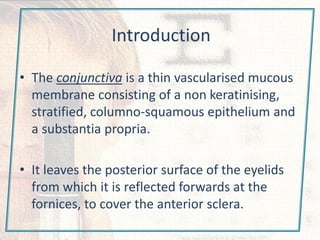
Introduction
• The conjunctiva is a thin vascularised mucous
membrane consisting of a non keratinising,
stratified, columno-squamous epithelium and
a substantia propria.
• It leaves the posterior surface of the eyelids
from which it is reflected forwards at the
fornices, to cover the anterior sclera.
Subdivisions
• 1. palpebral.
it is firmly adherent to
the tarsal plate.
• 2. fornicial.
loose, redundant and
swells easily.
• 3. bulbar.
covers ant sclera.
loosely attached to
underlying Tenon
except at limbus.
Histology
• 1. conjunctival epithelium
2-5 cells layer
- epithelial basal wing and
superficial polyhedral cells
- others :
• - melanocytes (in the basal
layer)
• - Langerhans antigen
presenting cells also (in the
basal layer)
• - goblet cells more numerous
in the fornices and plica (infro
nasaly)
• 2. the stroma ( substantia
propria)
- richly vascularized connective
tissue.
Note:-
Adenoid superfacial layer
doesn’t develop until age
of 3 months.
Conjunctivitis
• Conjunctivitis is an inflammatory process involving the
surface of the eye and characterized by vascular dilation,
cellular infiltration, and exudation.
• Two forms of the disorder are distinguished:
• 1. Acute conjunctivitis. Onset is abrupt and initially
unilateral with inflammation of the second eye within one
week. Duration is less than four weeks.
• 2. Chronic conjunctivitis. Duration is longer than three to
four weeks.
Conjunctivitis
• The most common ophthalmic
symptom is one of "red sticky
eyes" secondary to
conjunctivitis.
• Types of conjunctivitis
-Bacterial
-Chlamydial
-Viral
-Allergic/Hypersensitivity
-Toxic
-Other
Others:
• Blepharo-conjunctivitis.
• Rosacea kerato-
conjunctivitis
• Dry eye syndromes.
• Fungal conjunctivitis.
• Parasitic conjunctivitis.
• Ligneous conjunctivitis.
• Granulomatous
conjunctivitis.
• Systemic disease.
Conjunctivitis may be a feature
of a number of systemic
disorders including rheumatoid
arthritis, Wegener's
granulomatosis, Stevens-
Johnson syndrome, Lyell's
syndrome (toxic epidermal
necrolysis), scleroderma and
linear IgA disease.
Clinical evaluation
• Differential diagnosis determined from:
a. symptoms (eg. pain, Itching).
b. discharge.
c. conjunctival reaction.
d. membranes
e. lymphadenopathy.
Discharge
• It is exudate with epith debris, mucus and tears.
1. Watery (serous exudate+tears):
acute viral, allergic.
2. Mucoid :
vernal conj & keratoconj sicca.
3. purulent: severe acute bacterial.
4. mucopurulent:
mild bacterial, chlamydial.
Conjunctival reaction
• 1. Injection: more in fornices.
• 2. subconjunctival
hemorrhage
usually viral (mainly
picorna) highly
infectious but self-
limiting.
also strep. Pnemonia &
H. aegypticus do.
• 3. Oedema (chemosis)
In severely inflamed conj due to protien rich fluid
• 4. Scarring
trachoma, ocular cicatricial pemphygoid, atopic
conjunctivitis & prolonged use of topical medications.
6. Papillary reaction
• Non specific.
• Hyperplastic conjunctival epithelium with central
vessels & diffuse infiltrate of chronic inflamatory
cells.
• Can develop only in palpebral conj & bulbar at
limbus.
• Mostly seen in upper palpebral conj as fine mosaic-
like elevated polygonal hyperemic areas separated
by palor channels.
• Causes: chronic blepharitis, allergic & bact
conjunctivitis, contact lenses and superior
limbic keratoconjunctivitis.
• Note: the appearance of normal sup edge of tarsal plate( inf
when inverted) may mimic papillae & follicles so should not
be used as a clinical sign.
Membranes
• 1. pseudo
membranes:
coagulated exudate
adherent w inflamed
conj, easily peeled of
leaving intact
epithelium.
Causes: adenoviral,
gonococcal, ligneous
conj & Steven Johnson
syndrome.
• 2. true
membrane:
infiltrate
superfecial
layer of epith
conj.
if removed
bleeding occurs.
causes: strep
pypgenes and
diphtheria.
Lymphadenopathy
• Lymph drainage of the conj is to the
preauricular and sub mandibular regions
corresponding to the drainage of eye lids.
• Causes: viral, chlamydial, gonococcal and
perinaud oculoglandular syndrome.
Lab investigations
• Indications:
- sever purulent conjunctivitis.
- follicular conjunctivitis.
to R/O chlamydial.
- conjunctival inflammations
that are insufficient ti achieve diagnosis.
- neonatal conjunctivitis.
• Cultures. These are usually made on
blood (aerobic bacteria, fungi) and
chocolate agar (neisseria, haemophilus),
thioglycolate and meat broths
(anaerobes) or on other special media
when necessary. Sabouraud's agar and
brain-heart broth may be used to grow
fungi.
• Others: cytologic investigations, detection
of viral or chlamydial antigens, impression
cytology and PCR.
Simple bacterial conjunctivitis
• The commonest causative bacteria are
Staphylococcus aureus (in children and adults),
Streptococcus pneumonia and Haemophilus
influenzas (especially in children) and others
include Streptococcus viridans and pyogenes.
• It is a common self limiting condition mostly
affecting children.
• Spread by direct contact with infected secretions.
-Usually the onset is relatively acute.
-The patient usually has discomfort and a purulent
discharge in one eye that characteristically spreads to
the other eye. The eye may be difficult to open in the
morning because the discharge sticks the lashes
together. There may be a history of contact with a
person with similar symptoms.
- Discharge initially watery
mimiking viral.
History:
• —The vision should be normal after the discharge has been blinked clear
of the cornea. The discharge usually is mucopurulent and there is uniform
engorgement of all the conjunctival blood vessels. When fluorescein
drops are instilled in the eye there is no staining of the cornea.
• Injection maximal at fornices and less at limbus.
- Valvety, beefy red tarsal conjunctiva.
- Superfacial punctate epithelial erosions.
Examination
Management
• even without treatment, simple types resolves
within 10-14 days .
• Topical antibiotic eye drops (for example,
chloramphenicol) should be instilled every two hours
for the first 24 hours to hasten recovery, decreasing
to four times a day for one week. Chloramphenicol
ointment applied at night may also increase comfort
and reduce the stickiness of the eyelids in the
morning. Patients should be advised about general
hygiene measures; for example, not sharing face
towels.
Gonococcal keratoconjunctivitis
• Caused by gram –ve diplococcus
Neisseria gonorrhoeae.
• It is capable of invading intact
corneal epith.
• Gonococcal conjunctivitis:-
- acute profuse purulent
discharge.
- edematous tender eye
lids, hyperemia, chemosis
and pseudo membranes.
- lymphadenopathy.
• Gonococcal keratitis:
- if conjunctivitis not
treated well, keratitis
occurs as follows:
1- marginal ulceration.
2 - coalescence to form a
pereferal ring ulcer.
3 - central ulceration
that may rapidly
lead to perforation and
endopthalmitis.
• Treatment:
1. admit, take cultures and remove discharge.
2. systemic
cefotaxime 1gm I.V b.d
( only 1 day if only conj involved).
3. topical gentamycin or bacitracin at very
frequent intervals.
Note: recognize and treat any associated chlamydial
infection.
Ophthalmia neonatorum
• is conjunctivitis occuring within the first three weeks of
life.
• The most serious cause is infection with Neisseria
gonorrhoeae,but in the UK it is caused more commonly
by Chlamydia trachomatis (neonatal inclusion
conjunctivitis - NIC) and by staphylococci or
pneumococci. Herpes simplex type 2 virus is a relatively
rare cause.
• Follicles and lymphadenopathy do not occur
• has an early onset from between 1 and 3 days post
partum.
Management
• Specimens should be obtained from the child, the
mother and her sexual partners).
• frequent irrigation.
• systemic and local antibiotics is appropriate for
bacterial conditions and aciclovir is used in
herpetic infection.
• Inclusion conjunctivitis is treated with systemic
and topical erythromycin.
• In all cases, the mother and her sexual partner
require appropriate treatment.
Adenoviral conjunctivitis
• varies from very mild to very severe.
• It is an occupational hazard of
opthalmologists.
• It is highly contagious and transmitted via
respiratory or ocular secretions &
dissimination by towels and equipments
as tonometer.
• Incubation period 4-10 days.
Adenoviral conjunctivitis.
• Adenoviral conjunctivitis can be classified into 3 types:
• -pharyngoconjunctival fever (PCF),
type 3, 4or 7 & may be 5
fever and general malaise
usually bilateral,
• -epidemic keratoconjunctivitis (EKC).
adenovirus type 8 or 19
absence of major systemic symptoms.
usually unilateral
• -non-specific follicular conjunctivitis (NFC).
mild adenoviral conjunctivitis, which is usually self-limiting
and requires no treatment.
Diagnosis is clinical, but can be aided by serology, or ELISA.
• Viral keratitis
• Stage 1:
- within 7-10 days
- punctate epith keratitis.
• Stage 2:
- focal white sub epithelial opacity, represents
immune response.
• Stage 3:
- anterior stromal infiltrates fades over months to
years.
• Clinical presentation
• acute watering, redness,
discomfort & photophobia.
Both eyes.
• usually lasts longer than
bacterial conjunctivitis and
may go on for many weeks;
patients need to be
informed of this.
• Photophobia and
discomfort may be severe if
the patient goes on to
develop discrete corneal
opacities.
• Examination
• bilateral diffuse
conjunctival injection.
• clear discharge.
• Small white lymphoid
aggregations may be
present on the conjunctiva
(follicles).
• Subconjunctival
hemorrhage.
• pre-auricular or
submandibular
lymphadenopathy
Management
• Viral conjunctivitis is generally a self limiting condition,
So only symptomatic, they mostly resolves spontaneously
within 2 weeks.
• but antibiotic eye drops (for example, chloramphenicol)
provide symptomatic relief and help prevent secondary
bacterial infection. Viral conjunctivitis is extremely contagious,
and strict hygiene measures are important for both the
patient and the doctor; for example, washing of hands and
sterilizing of instruments. The period of infection is often
longer than with bacterial pathogens and patients should be
warned that symptoms may be present for several weeks. In
some patients the infection may have a chronic, protracted
course and steroid eye drops may be indicated if the corneal
lesions and symptoms are persistent.
Conjuctival diseases

Conjuctival diseases

  • 2.
    Introduction • The conjunctivais a thin vascularised mucous membrane consisting of a non keratinising, stratified, columno-squamous epithelium and a substantia propria. • It leaves the posterior surface of the eyelids from which it is reflected forwards at the fornices, to cover the anterior sclera.
  • 3.
    Subdivisions • 1. palpebral. itis firmly adherent to the tarsal plate. • 2. fornicial. loose, redundant and swells easily. • 3. bulbar. covers ant sclera. loosely attached to underlying Tenon except at limbus.
  • 4.
    Histology • 1. conjunctivalepithelium 2-5 cells layer - epithelial basal wing and superficial polyhedral cells - others : • - melanocytes (in the basal layer) • - Langerhans antigen presenting cells also (in the basal layer) • - goblet cells more numerous in the fornices and plica (infro nasaly) • 2. the stroma ( substantia propria) - richly vascularized connective tissue. Note:- Adenoid superfacial layer doesn’t develop until age of 3 months.
  • 5.
    Conjunctivitis • Conjunctivitis isan inflammatory process involving the surface of the eye and characterized by vascular dilation, cellular infiltration, and exudation. • Two forms of the disorder are distinguished: • 1. Acute conjunctivitis. Onset is abrupt and initially unilateral with inflammation of the second eye within one week. Duration is less than four weeks. • 2. Chronic conjunctivitis. Duration is longer than three to four weeks.
  • 6.
    Conjunctivitis • The mostcommon ophthalmic symptom is one of "red sticky eyes" secondary to conjunctivitis. • Types of conjunctivitis -Bacterial -Chlamydial -Viral -Allergic/Hypersensitivity -Toxic -Other Others: • Blepharo-conjunctivitis. • Rosacea kerato- conjunctivitis • Dry eye syndromes. • Fungal conjunctivitis. • Parasitic conjunctivitis. • Ligneous conjunctivitis. • Granulomatous conjunctivitis. • Systemic disease. Conjunctivitis may be a feature of a number of systemic disorders including rheumatoid arthritis, Wegener's granulomatosis, Stevens- Johnson syndrome, Lyell's syndrome (toxic epidermal necrolysis), scleroderma and linear IgA disease.
  • 7.
    Clinical evaluation • Differentialdiagnosis determined from: a. symptoms (eg. pain, Itching). b. discharge. c. conjunctival reaction. d. membranes e. lymphadenopathy.
  • 8.
    Discharge • It isexudate with epith debris, mucus and tears. 1. Watery (serous exudate+tears): acute viral, allergic. 2. Mucoid : vernal conj & keratoconj sicca. 3. purulent: severe acute bacterial. 4. mucopurulent: mild bacterial, chlamydial.
  • 10.
    Conjunctival reaction • 1.Injection: more in fornices.
  • 12.
    • 2. subconjunctival hemorrhage usuallyviral (mainly picorna) highly infectious but self- limiting. also strep. Pnemonia & H. aegypticus do.
  • 13.
    • 3. Oedema(chemosis) In severely inflamed conj due to protien rich fluid
  • 14.
    • 4. Scarring trachoma,ocular cicatricial pemphygoid, atopic conjunctivitis & prolonged use of topical medications.
  • 16.
    6. Papillary reaction •Non specific. • Hyperplastic conjunctival epithelium with central vessels & diffuse infiltrate of chronic inflamatory cells. • Can develop only in palpebral conj & bulbar at limbus. • Mostly seen in upper palpebral conj as fine mosaic- like elevated polygonal hyperemic areas separated by palor channels. • Causes: chronic blepharitis, allergic & bact conjunctivitis, contact lenses and superior limbic keratoconjunctivitis.
  • 18.
    • Note: theappearance of normal sup edge of tarsal plate( inf when inverted) may mimic papillae & follicles so should not be used as a clinical sign.
  • 19.
    Membranes • 1. pseudo membranes: coagulatedexudate adherent w inflamed conj, easily peeled of leaving intact epithelium. Causes: adenoviral, gonococcal, ligneous conj & Steven Johnson syndrome.
  • 20.
    • 2. true membrane: infiltrate superfecial layerof epith conj. if removed bleeding occurs. causes: strep pypgenes and diphtheria.
  • 21.
    Lymphadenopathy • Lymph drainageof the conj is to the preauricular and sub mandibular regions corresponding to the drainage of eye lids. • Causes: viral, chlamydial, gonococcal and perinaud oculoglandular syndrome.
  • 22.
    Lab investigations • Indications: -sever purulent conjunctivitis. - follicular conjunctivitis. to R/O chlamydial. - conjunctival inflammations that are insufficient ti achieve diagnosis. - neonatal conjunctivitis.
  • 23.
    • Cultures. Theseare usually made on blood (aerobic bacteria, fungi) and chocolate agar (neisseria, haemophilus), thioglycolate and meat broths (anaerobes) or on other special media when necessary. Sabouraud's agar and brain-heart broth may be used to grow fungi. • Others: cytologic investigations, detection of viral or chlamydial antigens, impression cytology and PCR.
  • 26.
    Simple bacterial conjunctivitis •The commonest causative bacteria are Staphylococcus aureus (in children and adults), Streptococcus pneumonia and Haemophilus influenzas (especially in children) and others include Streptococcus viridans and pyogenes. • It is a common self limiting condition mostly affecting children. • Spread by direct contact with infected secretions.
  • 27.
    -Usually the onsetis relatively acute. -The patient usually has discomfort and a purulent discharge in one eye that characteristically spreads to the other eye. The eye may be difficult to open in the morning because the discharge sticks the lashes together. There may be a history of contact with a person with similar symptoms. - Discharge initially watery mimiking viral. History:
  • 28.
    • —The visionshould be normal after the discharge has been blinked clear of the cornea. The discharge usually is mucopurulent and there is uniform engorgement of all the conjunctival blood vessels. When fluorescein drops are instilled in the eye there is no staining of the cornea. • Injection maximal at fornices and less at limbus. - Valvety, beefy red tarsal conjunctiva. - Superfacial punctate epithelial erosions. Examination
  • 29.
    Management • even withouttreatment, simple types resolves within 10-14 days . • Topical antibiotic eye drops (for example, chloramphenicol) should be instilled every two hours for the first 24 hours to hasten recovery, decreasing to four times a day for one week. Chloramphenicol ointment applied at night may also increase comfort and reduce the stickiness of the eyelids in the morning. Patients should be advised about general hygiene measures; for example, not sharing face towels.
  • 30.
    Gonococcal keratoconjunctivitis • Causedby gram –ve diplococcus Neisseria gonorrhoeae. • It is capable of invading intact corneal epith. • Gonococcal conjunctivitis:- - acute profuse purulent discharge. - edematous tender eye lids, hyperemia, chemosis and pseudo membranes. - lymphadenopathy.
  • 31.
    • Gonococcal keratitis: -if conjunctivitis not treated well, keratitis occurs as follows: 1- marginal ulceration. 2 - coalescence to form a pereferal ring ulcer. 3 - central ulceration that may rapidly lead to perforation and endopthalmitis.
  • 32.
    • Treatment: 1. admit,take cultures and remove discharge. 2. systemic cefotaxime 1gm I.V b.d ( only 1 day if only conj involved). 3. topical gentamycin or bacitracin at very frequent intervals. Note: recognize and treat any associated chlamydial infection.
  • 34.
    Ophthalmia neonatorum • isconjunctivitis occuring within the first three weeks of life. • The most serious cause is infection with Neisseria gonorrhoeae,but in the UK it is caused more commonly by Chlamydia trachomatis (neonatal inclusion conjunctivitis - NIC) and by staphylococci or pneumococci. Herpes simplex type 2 virus is a relatively rare cause. • Follicles and lymphadenopathy do not occur • has an early onset from between 1 and 3 days post partum.
  • 37.
    Management • Specimens shouldbe obtained from the child, the mother and her sexual partners). • frequent irrigation. • systemic and local antibiotics is appropriate for bacterial conditions and aciclovir is used in herpetic infection. • Inclusion conjunctivitis is treated with systemic and topical erythromycin. • In all cases, the mother and her sexual partner require appropriate treatment.
  • 39.
    Adenoviral conjunctivitis • variesfrom very mild to very severe. • It is an occupational hazard of opthalmologists. • It is highly contagious and transmitted via respiratory or ocular secretions & dissimination by towels and equipments as tonometer. • Incubation period 4-10 days.
  • 40.
    Adenoviral conjunctivitis. • Adenoviralconjunctivitis can be classified into 3 types: • -pharyngoconjunctival fever (PCF), type 3, 4or 7 & may be 5 fever and general malaise usually bilateral, • -epidemic keratoconjunctivitis (EKC). adenovirus type 8 or 19 absence of major systemic symptoms. usually unilateral • -non-specific follicular conjunctivitis (NFC). mild adenoviral conjunctivitis, which is usually self-limiting and requires no treatment. Diagnosis is clinical, but can be aided by serology, or ELISA.
  • 41.
    • Viral keratitis •Stage 1: - within 7-10 days - punctate epith keratitis. • Stage 2: - focal white sub epithelial opacity, represents immune response. • Stage 3: - anterior stromal infiltrates fades over months to years.
  • 43.
    • Clinical presentation •acute watering, redness, discomfort & photophobia. Both eyes. • usually lasts longer than bacterial conjunctivitis and may go on for many weeks; patients need to be informed of this. • Photophobia and discomfort may be severe if the patient goes on to develop discrete corneal opacities. • Examination • bilateral diffuse conjunctival injection. • clear discharge. • Small white lymphoid aggregations may be present on the conjunctiva (follicles). • Subconjunctival hemorrhage. • pre-auricular or submandibular lymphadenopathy
  • 45.
    Management • Viral conjunctivitisis generally a self limiting condition, So only symptomatic, they mostly resolves spontaneously within 2 weeks. • but antibiotic eye drops (for example, chloramphenicol) provide symptomatic relief and help prevent secondary bacterial infection. Viral conjunctivitis is extremely contagious, and strict hygiene measures are important for both the patient and the doctor; for example, washing of hands and sterilizing of instruments. The period of infection is often longer than with bacterial pathogens and patients should be warned that symptoms may be present for several weeks. In some patients the infection may have a chronic, protracted course and steroid eye drops may be indicated if the corneal lesions and symptoms are persistent.