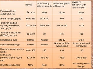
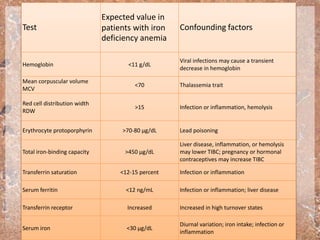
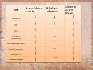
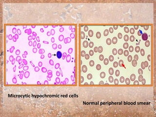
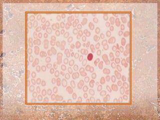
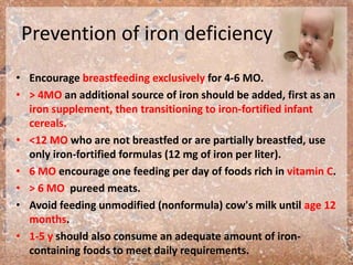
Done by:
Mohammed A Qazzaz
Objective
• INTRODUCTION
• PREVALENCE
• IRON BALANCE
• REQUIREMENTS
• Causes
• CLINICAL MANIFESTATIONS
• Diagnosis
• Treatment
• Prevention of iron deficiency
INTRODUCTION
• Iron deficiency (ID) is the most common nutritional
deficiency in children.
• WHO estimates that anemia affects one quarter of the
world's population and is concentrated within pre-school age
children and women.
• Iron deficiency anemia (IDA) is a microcytic, hypochromic,
and hypoproductive state.
PREVALENCE
• Iron deficiency anaemia is an important public health
problem in the Eastern Mediterranean Region.
• It is estimated that more than one third of the
population in the Region is anaemic.
• Pregnant women and young children are most at risk
– 50% of pregnant women and
– 63% of children under-5 have iron deficiency anaemia.
• 9 % incidence of iron deficiency and a 2 % incidence of
anemia among American females between the ages 12
and 15 years.
IRON BALANCE
• 75% bound in heme proteins (hemoglobin and myoglobin).
• In normal subjects - small amount of iron enters and leaves the
body on a daily basis.
• Iron balance is achieved primarily by mechanisms affecting
intestinal absorption and transport.
• In infants and children, 30% of daily iron needs must come
from diet.
• Intestinal iron absorption is a function of three principal factors:
– body iron stores (transferrin and ferritin)
– erythropoietic rate
– bioavailability of dietary iron.
• Iron absorption also is increased when there is increased
erythropoiesis and reticulocytosis or ineffective erythropoiesis, as
in beta thalassemia.
• Heme dietary sources have a higher bioavailability of iron than do
non-heme sources (30 versus 10 percent)
• Ascorbic acid enhances the absorption of non-animal sources of
iron.
• Tannates (teas), bran foods rich in phosphates, and phytates
(plant fiber, especially in seeds and grains) inhibit iron absorption.
REQUIREMENTS
• Breast milk contains only 0.3 to 1.0 mg/L iron,
but has a high bioavailability (50 percent)
• Iron-containing formulas with 12 mg/L iron
have only 4 to 6 percent bioavailability.
• Full-term: 1 mg/kg (maximum 15 mg)
• Children 1 to 3 years old: 7 mg/day
• Children 4 to 8 years old: 10 mg/day
• Children 9 to 13 years old: 8 mg/day
Perinatal risk factors
• At birth- iron stores ~75 mg/kg, mean hemoglobin
concentrations are 15 t 17 g/dL.
• First three to six months of life, by reducing the iron
stores at birth or through other mechanisms:
– Maternal iron deficiency
– Prematurity
– Fetal-maternal hemorrhage (FMH)
– Twin-twin transfusion syndrome (TTS)
– Other perinatal hemorrhagic events
– Insufficient dietary intake of iron during early infancy
Dietary factors
• Insufficient iron intake
• Decreased absorption due to poor dietary
sources of iron
• Introduction of unmodified cow's milk
(non-formula cow’s milk) before 12 months
of age
• Occult blood loss secondary to cow's milk
protein-induced colitis
Gastrointestinal disease
• Gastrointestinal malabsorption of iron:
– Active celiac disease
– Crohn’s disease
– Giardiasis
– Resection of the proximal small intestine.
• Conditions that cause gastrointestinal blood loss:
– Cow’s milk protein-induced colitis
– Inflammatory bowel disease
– chronic use of aspirin or nonsteroidal
antiinflammatory drugs, are also associated with iron
deficiency.
CLINICAL MANIFESTATIONS
• Iron deficiency anemia (IDA) is a microcytic, hypochromic,
and hypoproductive state.
• The most common presentation of IDA is an otherwise
asymptomatic, well nourished infant or child who has a mild
to moderate microcytic, hypochromic anemia
• Much less frequent are infants with severe anemia, who
present with:
– Lethargy
– Pallor
– Irritability
– Cardiomegaly
– Poor feeding
– Tachypnea.
• A number of abnormalities of epithelial tissues are
described in association with iron deficiency anemia.
These include:
– Esophageal webbing
– Koilonychias
– Glossitis
– Angular stomatitis
– Gastric atroph
• Neurodevelopmental
– Impaired psychomotor and/or mental development.
– cognitive impairment can occur in adolescents.
– negatively impact infant social-emotional behavior
– may contribute to the development of attention deficit
hyperactivity disorder.
• Exercise capacity
• Pica and pagophagia
• Thrombosis — cerebral vein thrombosis
• Birth -Mean Hb = elevated, but highly
variable
• 2 mos –“physiologic” anemia
–-2 SD Hb = 9.4 g/dL
• 6 mos to 24 mos
–-2.5 SD Hb 11.0 g/dL
• American Academy of Pediatrics
–Hb <11.0, Hct < 33% defines anemia
• For infants up to 24 months
• The most cost effective strategy is a therapeutic trial of
iron .
• Ferrous sulfate this is given at 3 mg/kg of elemental
iron, given once or twice daily between meals (ie, 3 to 6
mg/kg/day total).
• If four weeks of this treatment produces a hemoglobin
rise of greater than 1 gm/dL, this confirms the diagnosis
of iron deficiency.
• 2 years and adolescence, we suggest slightly more
evaluation.
• This is because IDA is somewhat less common in
otherwise-healthy children than in infants.
• Therefore, in addition to evaluating CBC (with indices
for MCV and RDW), we suggest performing a
reticulocyte count and reviewing a blood smear, and
screening several stools for occult blood.
Normal
Fe deficiency
without anemia
Fe deficiency with
mild anemia
Severe Fe
deficiency with
severe anemia
Marrow reticulo-
endothelial iron
2+ to 3+ None None None
Serum iron (SI), µg/dL 60 to 150 60 to 150 <60 <40
Total iron binding
capacity (transferrin,
TIBC), µg/dL
300 to 360 300 to 390 350 to 400 >410
Transferrin saturation
(SI/TIBC), percent
20 to 50 30 <15 <10
Hemoglobin, g/dL Normal Normal 9 to 12 6 to 7
Red cell morphology Normal Normal
Normal or slight
hypochromia
Hypochromia and
microcytosis
Plasma or serum ferritin,
ng/mL
40 to 200 <40 <20 <10
Erythrocyte
protoporphyrin, ng/mL
RBC
30 to 70 30 to 70 >100 100 to 200
Other tissue changes None None None
Nail and epithelial
changes
Test
Expected value in
patients with iron
deficiency anemia
Confounding factors
Hemoglobin <11 g/dL
Viral infections may cause a transient
decrease in hemoglobin
Mean corpuscular volume
MCV
<70 Thalassemia trait
Red cell distribution width
RDW
>15 Infection or inflammation, hemolysis
Erythrocyte protoporphyrin >70-80 µg/dL Lead poisoning
Total iron-binding capacity >450 µg/dL
Liver disease, inflammation, or hemolysis
may lower TIBC; pregnancy or hormonal
contraceptives may increase TIBC
Transferrin saturation <12-15 percent Infection or inflammation
Serum ferritin <12 ng/mL Infection or inflammation; liver disease
Transferrin receptor Increased Increased in high turnover states
Serum iron <30 µg/dL
Diurnal variation; iron intake; infection or
inflammation
Test
Iron deficiency
anemia
Alpha/beta
thalassemia
Anemia of
chronic
disease
Hemoglobin
MCV
RDW
Erythrocyte
protoporphyrin
Total iron-binding capacity
Transferrin saturation
Serum ferritin
Transferrin receptor
Microcytic hypochromic red cells
Normal peripheral blood smear
Treatment
• Oral iron therapy is started at a dose of 3 mg/kg of elemental
iron, given once or twice daily. It should be given 30 to 45
minutes before meals or two hours after meals, and only with
juice or water, rather than with food or milk.
• <12 months:
– iron-fortified formula
– A cow’s milk-based formula
– Unmodified cow’s milk (non-formula cow’s milk) should not be given
to infants.
• >12 months of age, intake of cow's milk should be limited to
less than 20 oz per day and bottle feeding should be
discontinued.
• CBC is reevaluated in 4 weeks when the child is
healthy. If the hemoglobin (Hgb) has increased
by 1 g/dL, therapy is continued and a CBC is
retested every 2 to 3 months until the Hgb
reaches the age-adjusted normal range.
• Oral iron is continued for an additional two
months after the Hgb reaches the normal range
for age.
Prevention of iron deficiency
• Encourage breastfeeding exclusively for 4-6 MO.
• > 4MO an additional source of iron should be added, first as an
iron supplement, then transitioning to iron-fortified infant
cereals.
• <12 MO who are not breastfed or are partially breastfed, use
only iron-fortified formulas (12 mg of iron per liter).
• 6 MO encourage one feeding per day of foods rich in vitamin C.
• > 6 MO pureed meats.
• Avoid feeding unmodified (nonformula) cow's milk until age 12
months.
• 1-5 y should also consume an adequate amount of iron-
containing foods to meet daily requirements.
Thank
you

Iron deficiency in children

  • 1.
  • 2.
    Objective • INTRODUCTION • PREVALENCE •IRON BALANCE • REQUIREMENTS • Causes • CLINICAL MANIFESTATIONS • Diagnosis • Treatment • Prevention of iron deficiency
  • 3.
    INTRODUCTION • Iron deficiency(ID) is the most common nutritional deficiency in children. • WHO estimates that anemia affects one quarter of the world's population and is concentrated within pre-school age children and women. • Iron deficiency anemia (IDA) is a microcytic, hypochromic, and hypoproductive state.
  • 4.
    PREVALENCE • Iron deficiencyanaemia is an important public health problem in the Eastern Mediterranean Region. • It is estimated that more than one third of the population in the Region is anaemic. • Pregnant women and young children are most at risk – 50% of pregnant women and – 63% of children under-5 have iron deficiency anaemia. • 9 % incidence of iron deficiency and a 2 % incidence of anemia among American females between the ages 12 and 15 years.
  • 5.
    IRON BALANCE • 75%bound in heme proteins (hemoglobin and myoglobin). • In normal subjects - small amount of iron enters and leaves the body on a daily basis. • Iron balance is achieved primarily by mechanisms affecting intestinal absorption and transport. • In infants and children, 30% of daily iron needs must come from diet.
  • 7.
    • Intestinal ironabsorption is a function of three principal factors: – body iron stores (transferrin and ferritin) – erythropoietic rate – bioavailability of dietary iron. • Iron absorption also is increased when there is increased erythropoiesis and reticulocytosis or ineffective erythropoiesis, as in beta thalassemia. • Heme dietary sources have a higher bioavailability of iron than do non-heme sources (30 versus 10 percent) • Ascorbic acid enhances the absorption of non-animal sources of iron. • Tannates (teas), bran foods rich in phosphates, and phytates (plant fiber, especially in seeds and grains) inhibit iron absorption.
  • 9.
    REQUIREMENTS • Breast milkcontains only 0.3 to 1.0 mg/L iron, but has a high bioavailability (50 percent) • Iron-containing formulas with 12 mg/L iron have only 4 to 6 percent bioavailability. • Full-term: 1 mg/kg (maximum 15 mg) • Children 1 to 3 years old: 7 mg/day • Children 4 to 8 years old: 10 mg/day • Children 9 to 13 years old: 8 mg/day
  • 11.
    Perinatal risk factors •At birth- iron stores ~75 mg/kg, mean hemoglobin concentrations are 15 t 17 g/dL. • First three to six months of life, by reducing the iron stores at birth or through other mechanisms: – Maternal iron deficiency – Prematurity – Fetal-maternal hemorrhage (FMH) – Twin-twin transfusion syndrome (TTS) – Other perinatal hemorrhagic events – Insufficient dietary intake of iron during early infancy
  • 12.
    Dietary factors • Insufficientiron intake • Decreased absorption due to poor dietary sources of iron • Introduction of unmodified cow's milk (non-formula cow’s milk) before 12 months of age • Occult blood loss secondary to cow's milk protein-induced colitis
  • 13.
    Gastrointestinal disease • Gastrointestinalmalabsorption of iron: – Active celiac disease – Crohn’s disease – Giardiasis – Resection of the proximal small intestine. • Conditions that cause gastrointestinal blood loss: – Cow’s milk protein-induced colitis – Inflammatory bowel disease – chronic use of aspirin or nonsteroidal antiinflammatory drugs, are also associated with iron deficiency.
  • 14.
    CLINICAL MANIFESTATIONS • Irondeficiency anemia (IDA) is a microcytic, hypochromic, and hypoproductive state. • The most common presentation of IDA is an otherwise asymptomatic, well nourished infant or child who has a mild to moderate microcytic, hypochromic anemia • Much less frequent are infants with severe anemia, who present with: – Lethargy – Pallor – Irritability – Cardiomegaly – Poor feeding – Tachypnea.
  • 15.
    • A numberof abnormalities of epithelial tissues are described in association with iron deficiency anemia. These include: – Esophageal webbing – Koilonychias – Glossitis – Angular stomatitis – Gastric atroph
  • 20.
    • Neurodevelopmental – Impairedpsychomotor and/or mental development. – cognitive impairment can occur in adolescents. – negatively impact infant social-emotional behavior – may contribute to the development of attention deficit hyperactivity disorder. • Exercise capacity • Pica and pagophagia • Thrombosis — cerebral vein thrombosis
  • 22.
    • Birth -MeanHb = elevated, but highly variable • 2 mos –“physiologic” anemia –-2 SD Hb = 9.4 g/dL • 6 mos to 24 mos –-2.5 SD Hb 11.0 g/dL • American Academy of Pediatrics –Hb <11.0, Hct < 33% defines anemia
  • 23.
    • For infantsup to 24 months • The most cost effective strategy is a therapeutic trial of iron . • Ferrous sulfate this is given at 3 mg/kg of elemental iron, given once or twice daily between meals (ie, 3 to 6 mg/kg/day total). • If four weeks of this treatment produces a hemoglobin rise of greater than 1 gm/dL, this confirms the diagnosis of iron deficiency.
  • 24.
    • 2 yearsand adolescence, we suggest slightly more evaluation. • This is because IDA is somewhat less common in otherwise-healthy children than in infants. • Therefore, in addition to evaluating CBC (with indices for MCV and RDW), we suggest performing a reticulocyte count and reviewing a blood smear, and screening several stools for occult blood.
  • 25.
    Normal Fe deficiency without anemia Fedeficiency with mild anemia Severe Fe deficiency with severe anemia Marrow reticulo- endothelial iron 2+ to 3+ None None None Serum iron (SI), µg/dL 60 to 150 60 to 150 <60 <40 Total iron binding capacity (transferrin, TIBC), µg/dL 300 to 360 300 to 390 350 to 400 >410 Transferrin saturation (SI/TIBC), percent 20 to 50 30 <15 <10 Hemoglobin, g/dL Normal Normal 9 to 12 6 to 7 Red cell morphology Normal Normal Normal or slight hypochromia Hypochromia and microcytosis Plasma or serum ferritin, ng/mL 40 to 200 <40 <20 <10 Erythrocyte protoporphyrin, ng/mL RBC 30 to 70 30 to 70 >100 100 to 200 Other tissue changes None None None Nail and epithelial changes
  • 26.
    Test Expected value in patientswith iron deficiency anemia Confounding factors Hemoglobin <11 g/dL Viral infections may cause a transient decrease in hemoglobin Mean corpuscular volume MCV <70 Thalassemia trait Red cell distribution width RDW >15 Infection or inflammation, hemolysis Erythrocyte protoporphyrin >70-80 µg/dL Lead poisoning Total iron-binding capacity >450 µg/dL Liver disease, inflammation, or hemolysis may lower TIBC; pregnancy or hormonal contraceptives may increase TIBC Transferrin saturation <12-15 percent Infection or inflammation Serum ferritin <12 ng/mL Infection or inflammation; liver disease Transferrin receptor Increased Increased in high turnover states Serum iron <30 µg/dL Diurnal variation; iron intake; infection or inflammation
  • 27.
  • 28.
    Microcytic hypochromic redcells Normal peripheral blood smear
  • 31.
    Treatment • Oral irontherapy is started at a dose of 3 mg/kg of elemental iron, given once or twice daily. It should be given 30 to 45 minutes before meals or two hours after meals, and only with juice or water, rather than with food or milk. • <12 months: – iron-fortified formula – A cow’s milk-based formula – Unmodified cow’s milk (non-formula cow’s milk) should not be given to infants. • >12 months of age, intake of cow's milk should be limited to less than 20 oz per day and bottle feeding should be discontinued.
  • 32.
    • CBC isreevaluated in 4 weeks when the child is healthy. If the hemoglobin (Hgb) has increased by 1 g/dL, therapy is continued and a CBC is retested every 2 to 3 months until the Hgb reaches the age-adjusted normal range. • Oral iron is continued for an additional two months after the Hgb reaches the normal range for age.
  • 33.
    Prevention of irondeficiency • Encourage breastfeeding exclusively for 4-6 MO. • > 4MO an additional source of iron should be added, first as an iron supplement, then transitioning to iron-fortified infant cereals. • <12 MO who are not breastfed or are partially breastfed, use only iron-fortified formulas (12 mg of iron per liter). • 6 MO encourage one feeding per day of foods rich in vitamin C. • > 6 MO pureed meats. • Avoid feeding unmodified (nonformula) cow's milk until age 12 months. • 1-5 y should also consume an adequate amount of iron- containing foods to meet daily requirements.
  • 34.

Editor's Notes

  • #6 The remainder is bound in the storage proteins ferritin and hemosiderin, with a small portion (3 percent) bound in critical enzyme systems, such as catalase and cytochromes. because of the growth spurt and increase in body (muscle) mass.
  • #8 Thus, iron deficiency may be a particular problem in vegetarian children.
  • #10 Requirements for absorbed iron in otherwise uncomplicated states approximate 0.5 to 0.8 mg/day. However, only a fraction of dietary iron is absorbed, and the dietary iron requirement is considerably higher and depends on the bioavailability of the iron in the food. As an example
  • #13 Unmodified cow's milk increases intestinal blood loss in infants as compared to formula feeding or breast feeding.
  • #14 Duodenum
  • #21 Moderately severe IDA is associated with decreased work capacity in part because iron is an essential cofactor for enzyme-driven aerobic metabolism. Decreased iron stores, in the absence of anemia, is associated with decreased exercise performance in laboratory animals.
  • #24 is an inexpensive and convenient form of iron; Once the response is confirmed, iron supplementation and monitoring should be continued for several months after the hemoglobin reaches the age-adjusted normal range.
  • #25 before embarking on a therapeutic trial of iron If the results of these tests are consistent with IDA (hypochromic microcytic anemia) and there is no evidence of gastrointestinal blood loss or lead toxicity, the child is treated with iron, and further evaluation is performed only if the response is inadequate.
  • #27 An elevated red cell distribution width (RDW) is the earliest hematologic manifestation of iron deficiency
  • #32 if the infant is not breastfed or only partially breastfed. is acceptable if there is no evidence of cow’s milk protein-induced colitis.
  • #34 The heme iron in meats is more bioavailable than nonheme iron and also increases the absorption of nonheme iron.