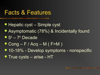
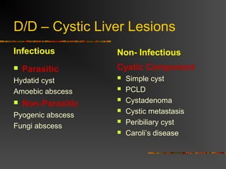
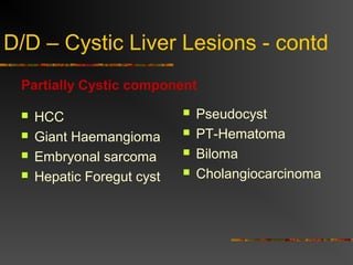
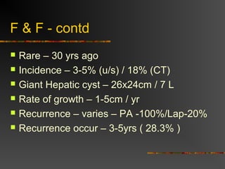
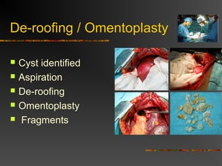
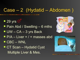
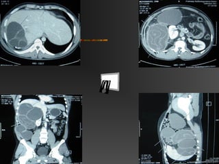
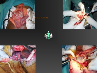
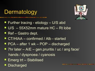
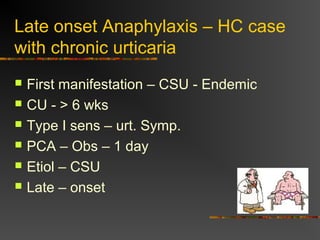
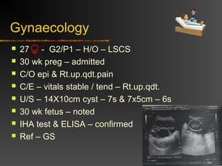
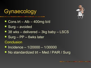
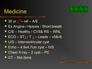
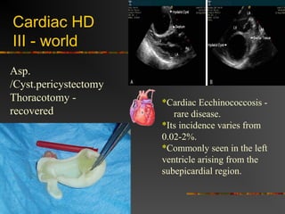
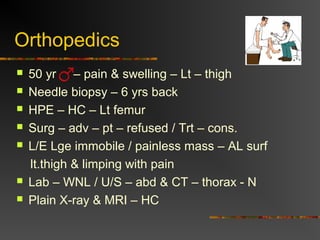
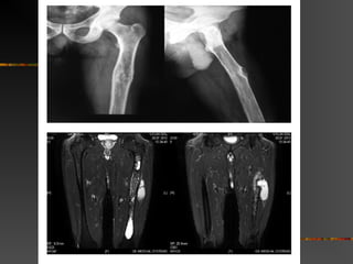
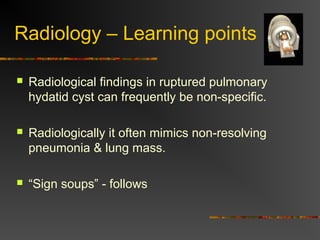
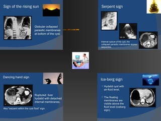
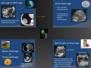
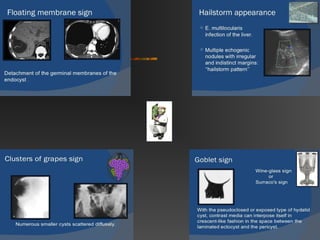
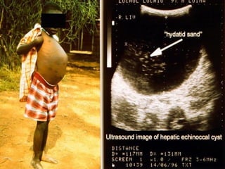
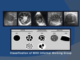
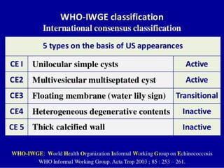
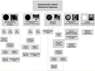
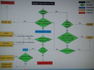
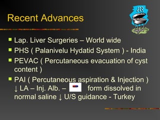
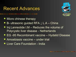
LIVER CYSTS 
Prof.Dr.U.Murali.M.S;MBA 
Dept.Of Surgery 
D Y Patil Medical College 
Mauritius.
“ Is Life worth living ? 
It all depends on the Liver ! ” 
- William James 
(1842-1910 )
Contents of Discussion 
 Facts & Salient Features – Liver Cysts 
 My Personal Experience – Hydatid Cyst 
 Around Each Dept – Interesting case reports 
 Recent Advances 
 Key points
Salient Features 
 Anatomy – Segmental Anatomy 
 Common – Rt side – “stream-line” 
 Segment – VII - common 
 Hepatomegaly – Sign – U.problem 
 Cysts > 10cm – CB communication 
 Most series in the literature are relatively 
small, reporting fewer than 50 patients each.
Couinaud Classification 
•Divides the liver into 8 functionally independent segments. 
•Each segment has its own vascular inflow, outflow and biliary drainage
Facts & Features 
 Hepatic cyst – Simple cyst 
 Asymptomatic (78%) & Incidentally found 
 5th – 7th Decade 
 Cong – F / Acq – M ( F>M ) 
 10-18% - Develop symptoms - nonspecific 
 True cysts – arise - HT
D/D – Cystic Liver Lesions 
Infectious 
 Parasitic 
Hydatid cyst 
Amoebic abscess 
 Non-Parasitic 
Pyogenic abscess 
Fungi abscess 
Non- Infectious 
Cystic Component 
 Simple cyst 
 PCLD 
 Cystadenoma 
 Cystic metastasis 
 Peribiliary cyst 
 Caroli’s disease
D/D – Cystic Liver Lesions - contd 
Partially Cystic component 
 HCC 
 Giant Haemangioma 
 Embryonal sarcoma 
 Hepatic Foregut cyst 
 Pseudocyst 
 PT-Hematoma 
 Biloma 
 Cholangiocarcinoma
F & F - contd 
 Rare – 30 yrs ago 
 Incidence – 3-5% (u/s) / 18% (CT) 
 Giant Hepatic cyst – 26x24cm / 7 L 
 Rate of growth – 1-5cm / yr 
 Recurrence – varies – PA -100%/Lap-20% 
 Recurrence occur – 3-5yrs ( 28.3% )
F & F - contd 
 Hydatid cyst > E.histolytica 
 Disease – worldwide 
 Endemic – M/ME/I/SA/C 
 Eradicated - Iceland 
 Annual incidence – 1-220/100,000 
 New era – Mgt on HC 
 Lge & recur – Rt.post.lobe 
 Recurrence – 2.2-22%
Giant Hepatic Cyst – 26x24cm
Personal Experience 
 Case 1 – Hydatid Cyst – Liver 
 Case 2 – Recurrent Hydatid - Abdomen
Case – 1 (Hydatid – Liver ) 
 55 yrs 
 Pain Abd / Swelling – 1 mth 
 No LOW / Fever 
 P/A – Hepatomegaly 
 CBC – WNL 
 CT Scan – Hydatid Cyst
De-roofing / Omentoplasty 
 Cyst identified 
 Aspiration 
 De-roofing 
 Omentoplasty 
 Fragments
Case – 2 (Hydatid – Abdomen ) 
 29 yrs 
 Pain Abd / Swelling – 6 mths 
 UW – CA – 3 yrs Back 
 P/A – Liver + / + masses abd 
 CBC – WNL 
 CT Scan – Hydatid Cyst 
Multiple Liver & Mes.
Around the Depts -
Anaesthesia 
 62 - admitted – removal – hydatid cyst liver 
 IO – hypotension / tachycardia & ST segment 
elevation 
 No erythema / bronchospasm / desaturation 
 Anas. Agents stopped – O2 – increased 
 View- recr.hypotension – shifted –ICU 
 Acute MI – ruled out – resusitated 
 Anaphylaxis – well managed
Anaesthesia 
 Stabilized – back – cyst removed 
 HPE – confirmed 
 Conclusion – 
 Atypical IO Anaphylactic shock with ECG changes 
sec – non- ruptured hapatic hydatid cyst. 
( Anaphylaxis sec – non-ruptured cyst – uncommon )
Dermatology 
 21 yrs - 3mths - urticaria 
 Trt – Ah b/d – OP 
 Lost wt 4-5kg – 6mths 
 C/E – WNL 
 Lab – WBC ↑ / ESR – 45mm/hr / CRP-14.3mg/dl 
 Chest X-ray – Cavity – upper lobes 
 AFB – Sputum +ve 
 Other parameters - WNL
Dermatology 
 ATT – Started 
(a) 
 Post Att – Wt + But UC contd- 
 Rpt X-ray – No lesions 
(b)
Dermatology 
 Further tracing - etiology – U/S abd 
 U/S – 55X52mm mature HC – Rt lobe 
 Ref – Gastro dept. 
 CT/HIAA – confirmed / Alb - started 
 PCA – after 1 wk – POP – discharged 
 7hr later – A/E – gen.pruritis / uc / ang face/ 
hands / dyspnoea / cyanosis 
 Emerg trt – Stabilised 
 Discharged
Late onset Anaphylaxis – HC case 
with chronic urticaria 
 First manifestation – CSU - Endemic 
 CU - > 6 wks 
 Type I sens – urt. Symp. 
 PCA – Obs – 1 day 
 Etiol – CSU 
 Late – onset
Gynaecology 
 27 - G2/P1 – H/O – LSCS 
 30 wk preg – admitted 
 C/O epi & Rt.up.qdt.pain 
 C/E – vitals stable / tend – Rt.up.qdt. 
 U/S – 14X10cm cyst – 7s & 7x5cm – 6s 
 30 wk fetus – noted 
 IHA test & ELISA – confirmed 
 Ref – GS
Gynaecology 
 Cons.trt – Alb – 400mg b/d 
 Surg – avoided 
 38 wks – delivered – 3kg baby – LSCS 
 Surg – PP – 6wks later 
Conclusion 
 Incidence – 1/20000 – 1/30000 
 No standardized trt – Med / PAIR / Surg
Medicine 
 30 yr – ref – A/E 
 Ex.Angina / Hptysis / Short.breath 
 C/E – Healthy / CVS& RS – WNL 
 ECG – ST↓ / T ↓ – Leads – v5&v6 
 U/S – Interventricular cyst 
 Echo – 4.9x4.7cm cyst – IVS 
 Chest X-ray – 2 cyst – PC 
 CT – Not done
Cardiac HD 
III - world 
*Cardiac Ecchinococcosis - 
rare disease. 
*Its incidence varies from 
0.02-2%. 
*Commonly seen in the left 
ventricle arising from the 
subepicardial region. 
Asp. 
/Cyst.pericystectomy 
Thoracotomy - 
recovered
Orthopedics 
 50 yr – pain & swelling – Lt – thigh 
 Needle biopsy – 6 yrs back 
 HPE – HC – Lt femur 
 Surg – adv – pt – refused / Trt – cons. 
 L/E Lge immobile / painless mass – AL surf 
lt.thigh & limping with pain 
 Lab – WNL / U/S – abd & CT – thorax - N 
 Plain X-ray & MRI – HC
Orthopedics 
 Surg – Debridement 
 E-O portion of the cyst was dissected and excised 
10 minutes after injection of 50 mL 10% povidone-iodine 
solution into the cyst 
 Lt k.jt was opened .Intercondylar notch was drilled 
upto distal femoral metaphysis. An 8 mm ET tube 
was inserted into the intercondylar notch, and its 
cuff has been inflated to prevent any leakage.
Orthopedics 
 P-iodine soln. was injected - intubation tube into 
the medullary cavity. After 10 mts, all daughter 
vesicles and germinative membranes aspirated 
safely from the distal end through the intubation 
tube, which has been placed previously, while 
aspirating simultaneous drilling and broaching the 
medullary cavity of the left femur from proximal to 
distal was performed.
Orthopedics 
 Drains were removed - 3rd POD. HPE - was 
reported as echinococcal hydatid cysts of femur. 
Pt – discharged with crutches. 
 10 days - discharge, the patient came back with a 
spontaneous pathological fracture of the left femur 
Thorough debridement – done. NEO hydatid 
disease was observed intraoperatively.
Orthopedics 
 The IMC was filled with vancomycin beads 
& closed, and the patient was put into skeletal 
traction from tuberosity of tibia. 
 When the infection has ceased, the pathological 
fracture has been treated using a custom-made 
interlocking intramedullary nail.
HD - Femur with an Extraosseous Extent 
-pathological Fracture 
 Bone hydatidosis is about 1–2.5% of all human 
hydatid disease 
 Spine is the most common location (50%) 
 D/D – Bone tumors & TB / Fungal 
 Bone hydatidosis - often asymptomatic & is 
usually detected only after a pathologic fracture, 
secondary infection, or neurovascular symptoms 
caused by compression
HD - Femur with an Extraosseous Extent 
-pathological Fracture 
 The forming defect after curettage or excision is 
a major problem. The dead space can be filled 
with bone grafts, bone cement, or 
endoprostheses. 
 Bone grafts can be invaded by the parasites, 
and recurrence is often common. 
 A stepped surgical care should be planned.
Paediatrics 
 6 yr boy – attended – AE 
 Prod.cough / dyspnoea 
 P/H – LOA & weakness 
 C/E - ↓ air entry Rt + wheeze 
 Trt – Bdtrs. – no response 
 Chest X-ray – cyst – middle lobe - Rt 
 CECT – “ Warerlilly sign ” + pleural eff. 
 Trt – Alb + Radical cystectomy 
 Fully recovered
Waterlilly 
sign 
Endocyst collapses & 
Floats – cystic fluid
Radiology – Learning points 
 Radiological findings in ruptured pulmonary 
hydatid cyst can frequently be non-specific. 
 Radiologically it often mimics non-resolving 
pneumonia & lung mass. 
 “Sign soups” - follows
Recent Advances 
 Lap. Liver Surgeries – World wide 
 PHS ( Palanivelu Hydatid System ) - India 
 PEVAC ( Percutaneous evacuation of cyst 
content ) 
 PAI ( Percutaneous aspiration & Injection ) 
↓ LA – Inj. Alb. – form dissolved in 
normal saline ↓ U/S guidance - Turkey
Recent Advances 
 Micro chinese therapy 
 B- ultrasonic guided RFA ↓ L.A – China 
 Inj.Lanreotide I.M – Reduces the volume of 
Polycystic liver disease - Netherlands 
 EG -95 Recombinant vaccine – Hydatid Disease 
 Amoebiasis vaccine – under trial 
 Liver Care Foundation - India
Key points 
 Most app. Method – Lack consensus 
 Scolicidal Agents – No consensus 
 Recurrence disease – 5 yrs 
 Hydatid disease – Diverse forms 
 Serodiagnostic test – High sens & spec 
 Clinically – Difficult – Complicated cysts 
 CEUS – Acc. excludes cystic neoplasms.
Liver cyst - Facts & Interesting Case Reports

Liver cyst - Facts & Interesting Case Reports

  • 1.
    LIVER CYSTS Prof.Dr.U.Murali.M.S;MBA Dept.Of Surgery D Y Patil Medical College Mauritius.
  • 2.
    “ Is Lifeworth living ? It all depends on the Liver ! ” - William James (1842-1910 )
  • 3.
    Contents of Discussion  Facts & Salient Features – Liver Cysts  My Personal Experience – Hydatid Cyst  Around Each Dept – Interesting case reports  Recent Advances  Key points
  • 4.
    Salient Features Anatomy – Segmental Anatomy  Common – Rt side – “stream-line”  Segment – VII - common  Hepatomegaly – Sign – U.problem  Cysts > 10cm – CB communication  Most series in the literature are relatively small, reporting fewer than 50 patients each.
  • 5.
    Couinaud Classification •Dividesthe liver into 8 functionally independent segments. •Each segment has its own vascular inflow, outflow and biliary drainage
  • 6.
    Facts & Features  Hepatic cyst – Simple cyst  Asymptomatic (78%) & Incidentally found  5th – 7th Decade  Cong – F / Acq – M ( F>M )  10-18% - Develop symptoms - nonspecific  True cysts – arise - HT
  • 7.
    D/D – CysticLiver Lesions Infectious  Parasitic Hydatid cyst Amoebic abscess  Non-Parasitic Pyogenic abscess Fungi abscess Non- Infectious Cystic Component  Simple cyst  PCLD  Cystadenoma  Cystic metastasis  Peribiliary cyst  Caroli’s disease
  • 8.
    D/D – CysticLiver Lesions - contd Partially Cystic component  HCC  Giant Haemangioma  Embryonal sarcoma  Hepatic Foregut cyst  Pseudocyst  PT-Hematoma  Biloma  Cholangiocarcinoma
  • 9.
    F & F- contd  Rare – 30 yrs ago  Incidence – 3-5% (u/s) / 18% (CT)  Giant Hepatic cyst – 26x24cm / 7 L  Rate of growth – 1-5cm / yr  Recurrence – varies – PA -100%/Lap-20%  Recurrence occur – 3-5yrs ( 28.3% )
  • 10.
    F & F- contd  Hydatid cyst > E.histolytica  Disease – worldwide  Endemic – M/ME/I/SA/C  Eradicated - Iceland  Annual incidence – 1-220/100,000  New era – Mgt on HC  Lge & recur – Rt.post.lobe  Recurrence – 2.2-22%
  • 11.
    Giant Hepatic Cyst– 26x24cm
  • 12.
    Personal Experience Case 1 – Hydatid Cyst – Liver  Case 2 – Recurrent Hydatid - Abdomen
  • 13.
    Case – 1(Hydatid – Liver )  55 yrs  Pain Abd / Swelling – 1 mth  No LOW / Fever  P/A – Hepatomegaly  CBC – WNL  CT Scan – Hydatid Cyst
  • 14.
    De-roofing / Omentoplasty  Cyst identified  Aspiration  De-roofing  Omentoplasty  Fragments
  • 15.
    Case – 2(Hydatid – Abdomen )  29 yrs  Pain Abd / Swelling – 6 mths  UW – CA – 3 yrs Back  P/A – Liver + / + masses abd  CBC – WNL  CT Scan – Hydatid Cyst Multiple Liver & Mes.
  • 18.
  • 19.
    Anaesthesia  62- admitted – removal – hydatid cyst liver  IO – hypotension / tachycardia & ST segment elevation  No erythema / bronchospasm / desaturation  Anas. Agents stopped – O2 – increased  View- recr.hypotension – shifted –ICU  Acute MI – ruled out – resusitated  Anaphylaxis – well managed
  • 20.
    Anaesthesia  Stabilized– back – cyst removed  HPE – confirmed  Conclusion –  Atypical IO Anaphylactic shock with ECG changes sec – non- ruptured hapatic hydatid cyst. ( Anaphylaxis sec – non-ruptured cyst – uncommon )
  • 21.
    Dermatology  21yrs - 3mths - urticaria  Trt – Ah b/d – OP  Lost wt 4-5kg – 6mths  C/E – WNL  Lab – WBC ↑ / ESR – 45mm/hr / CRP-14.3mg/dl  Chest X-ray – Cavity – upper lobes  AFB – Sputum +ve  Other parameters - WNL
  • 22.
    Dermatology  ATT– Started (a)  Post Att – Wt + But UC contd-  Rpt X-ray – No lesions (b)
  • 23.
    Dermatology  Furthertracing - etiology – U/S abd  U/S – 55X52mm mature HC – Rt lobe  Ref – Gastro dept.  CT/HIAA – confirmed / Alb - started  PCA – after 1 wk – POP – discharged  7hr later – A/E – gen.pruritis / uc / ang face/ hands / dyspnoea / cyanosis  Emerg trt – Stabilised  Discharged
  • 24.
    Late onset Anaphylaxis– HC case with chronic urticaria  First manifestation – CSU - Endemic  CU - > 6 wks  Type I sens – urt. Symp.  PCA – Obs – 1 day  Etiol – CSU  Late – onset
  • 25.
    Gynaecology  27- G2/P1 – H/O – LSCS  30 wk preg – admitted  C/O epi & Rt.up.qdt.pain  C/E – vitals stable / tend – Rt.up.qdt.  U/S – 14X10cm cyst – 7s & 7x5cm – 6s  30 wk fetus – noted  IHA test & ELISA – confirmed  Ref – GS
  • 26.
    Gynaecology  Cons.trt– Alb – 400mg b/d  Surg – avoided  38 wks – delivered – 3kg baby – LSCS  Surg – PP – 6wks later Conclusion  Incidence – 1/20000 – 1/30000  No standardized trt – Med / PAIR / Surg
  • 27.
    Medicine  30yr – ref – A/E  Ex.Angina / Hptysis / Short.breath  C/E – Healthy / CVS& RS – WNL  ECG – ST↓ / T ↓ – Leads – v5&v6  U/S – Interventricular cyst  Echo – 4.9x4.7cm cyst – IVS  Chest X-ray – 2 cyst – PC  CT – Not done
  • 28.
    Cardiac HD III- world *Cardiac Ecchinococcosis - rare disease. *Its incidence varies from 0.02-2%. *Commonly seen in the left ventricle arising from the subepicardial region. Asp. /Cyst.pericystectomy Thoracotomy - recovered
  • 29.
    Orthopedics  50yr – pain & swelling – Lt – thigh  Needle biopsy – 6 yrs back  HPE – HC – Lt femur  Surg – adv – pt – refused / Trt – cons.  L/E Lge immobile / painless mass – AL surf lt.thigh & limping with pain  Lab – WNL / U/S – abd & CT – thorax - N  Plain X-ray & MRI – HC
  • 31.
    Orthopedics  Surg– Debridement  E-O portion of the cyst was dissected and excised 10 minutes after injection of 50 mL 10% povidone-iodine solution into the cyst  Lt k.jt was opened .Intercondylar notch was drilled upto distal femoral metaphysis. An 8 mm ET tube was inserted into the intercondylar notch, and its cuff has been inflated to prevent any leakage.
  • 32.
    Orthopedics  P-iodinesoln. was injected - intubation tube into the medullary cavity. After 10 mts, all daughter vesicles and germinative membranes aspirated safely from the distal end through the intubation tube, which has been placed previously, while aspirating simultaneous drilling and broaching the medullary cavity of the left femur from proximal to distal was performed.
  • 33.
    Orthopedics  Drainswere removed - 3rd POD. HPE - was reported as echinococcal hydatid cysts of femur. Pt – discharged with crutches.  10 days - discharge, the patient came back with a spontaneous pathological fracture of the left femur Thorough debridement – done. NEO hydatid disease was observed intraoperatively.
  • 34.
    Orthopedics  TheIMC was filled with vancomycin beads & closed, and the patient was put into skeletal traction from tuberosity of tibia.  When the infection has ceased, the pathological fracture has been treated using a custom-made interlocking intramedullary nail.
  • 35.
    HD - Femurwith an Extraosseous Extent -pathological Fracture  Bone hydatidosis is about 1–2.5% of all human hydatid disease  Spine is the most common location (50%)  D/D – Bone tumors & TB / Fungal  Bone hydatidosis - often asymptomatic & is usually detected only after a pathologic fracture, secondary infection, or neurovascular symptoms caused by compression
  • 36.
    HD - Femurwith an Extraosseous Extent -pathological Fracture  The forming defect after curettage or excision is a major problem. The dead space can be filled with bone grafts, bone cement, or endoprostheses.  Bone grafts can be invaded by the parasites, and recurrence is often common.  A stepped surgical care should be planned.
  • 37.
    Paediatrics  6yr boy – attended – AE  Prod.cough / dyspnoea  P/H – LOA & weakness  C/E - ↓ air entry Rt + wheeze  Trt – Bdtrs. – no response  Chest X-ray – cyst – middle lobe - Rt  CECT – “ Warerlilly sign ” + pleural eff.  Trt – Alb + Radical cystectomy  Fully recovered
  • 38.
    Waterlilly sign Endocystcollapses & Floats – cystic fluid
  • 39.
    Radiology – Learningpoints  Radiological findings in ruptured pulmonary hydatid cyst can frequently be non-specific.  Radiologically it often mimics non-resolving pneumonia & lung mass.  “Sign soups” - follows
  • 49.
    Recent Advances Lap. Liver Surgeries – World wide  PHS ( Palanivelu Hydatid System ) - India  PEVAC ( Percutaneous evacuation of cyst content )  PAI ( Percutaneous aspiration & Injection ) ↓ LA – Inj. Alb. – form dissolved in normal saline ↓ U/S guidance - Turkey
  • 50.
    Recent Advances Micro chinese therapy  B- ultrasonic guided RFA ↓ L.A – China  Inj.Lanreotide I.M – Reduces the volume of Polycystic liver disease - Netherlands  EG -95 Recombinant vaccine – Hydatid Disease  Amoebiasis vaccine – under trial  Liver Care Foundation - India
  • 51.
    Key points Most app. Method – Lack consensus  Scolicidal Agents – No consensus  Recurrence disease – 5 yrs  Hydatid disease – Diverse forms  Serodiagnostic test – High sens & spec  Clinically – Difficult – Complicated cysts  CEUS – Acc. excludes cystic neoplasms.