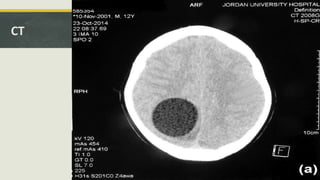
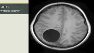
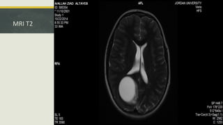
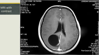
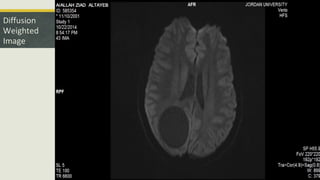
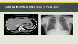
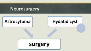
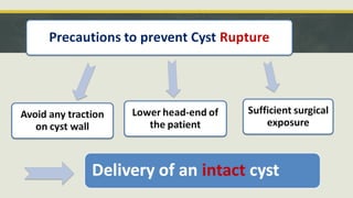
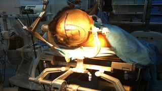
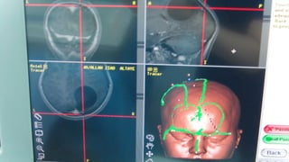
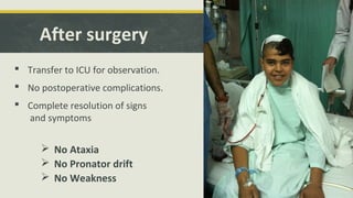
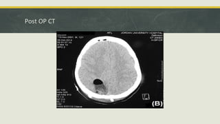
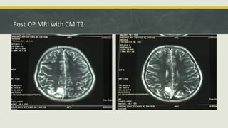
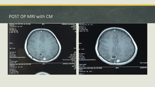
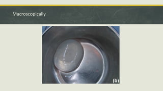
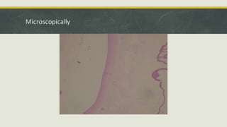
Isolated intracranial Hydatid
Cyst – Multidisplinary Approach
sa•Ahmed Altibi
•Heba Daradkeh
•Ibrahim Nour
Case Presentation
saJordan University Hospital
Department of Neurosurgery
History
A 13 year old male patient, previously healthy, living in Madaba.
Presented to our pediatrics neurology clinic on the 22nd
of October, 2014.
Complaining of
HEADACHE OF ONE MONTH DURATION
History
The headache was :
- Gradual in onset.
- Initially right-sided then later became diffuse.
- Dull in nature.
He had that headache almost daily throughout the month and
it was more severe upon waking up in the morning.
History
 It was partially relieved with simple analgesia (Paracetamol
and Iboprufen) but did not have specific exacerbating factors.
 The severity of the headache was progressively increasing,
reaching its peak a couple of days prior to his presentation.
History
 The headache was sometimes associated with:
-Nausea without vomiting
-Blurring of vision
-weakness in his left upper and lower limbs, associated with numbness in the
upper limb, he described difficulty carrying objects with his left upper limb. Those
developed only a couple of days before his presentation.
History
NO history of :
-Fever
-Seizures
-Loss of consciousness
-trauma
-significant weight loss, poor appetite, or night sweats.
History
Past medical history: free
Past surgical history: free
No significant relevant family history
Social history:
He is the youngest in 6 brothers, lives in a rural area.
Physical Examination
General :
-In a good general condition
-Fully awake and oriented to place, time & person.
-He did look in pain !
Physical Examination
Neurological examination
-No meningeal signs
-Examination of the cranial nerves was normal
-The visual filed by confrontation test was normal
-Fundoscopic examination at the clinic showed
MILD BILATERAL PAPILLEDEMA
Physical Examination
Neurological examination ctd.
- He had an obvious left-sided PRONATOR DRIFT
- WEAKNESS in the left upper and lower limbs (4/5)
- SENSORY ATAXIA with a clear Positive Romberg’s Sign
- Deep tendon reflexes were normal
- Babinski & Hoffman’s signs were absent
Summary
a13 year old boy who presented with signs and symptoms of increased ICP :
progressive headache, postive pronator drift and mild bilateral papillaedema and
left sided weakness
Labs: CBC, Electrolytes
 Electrolytes were normal
 CBC:
 Hb :12.8
 WBC: 7.1 x 10⁹
 Neutrophils: 35%
 lymphocytes: 40%
 Eosinophils : 11%
 Basophils: 1.92%
 Monocytes: 5.68%
CT
MRI T1
without contrast
MRI T2
MRI with
contrast
Diffusion
Weighted
Image
Quick Summary
 So this is an Intra axial supratentorial parieto-occipital cystic lesion with focal
area of enhancement within its wall, no restricted diffusion, and with some
vasogenic edema.
List of differentials
Is it a Pilocytic astrocytoma?
Is it an abscess?
 1. Not an abscess because the DWI was free from any restricted diffusion.
These are the images of the child’s liver and lungs:
Neurosurgery
We were consulted by pediatrics team
Physical findings were consistent with their findings
Ordered imaging were reviewed
Neurosurgery
Preparation for surgery
 Patients was started on Mebendizole
3 days before the operation.
Hypertonic saline was prepared to
continuously irrigate and possibly
inject the cyst if needed.
Anesthesia - Preoperative
 ASA score: I
 Malampati class 1
 GA
 ASA Standard Monitoring:
 Oxygenation: Pulse Oximetery
 Ventilation
 Circulation: ECG & BP
 Temperature
Anesthesia
 Precaution
 Canulae (Gauge 16)
 Central line and arterial line
 Adrenaline 50 mic
 Hydrocortisone
 Antihistamine
Imaging:
Imaging:
After surgery
 Transfer to ICU for observation.
 No postoperative complications.
 Complete resolution of signs
and symptoms
 No Ataxia
 No Pronator drift
 No Weakness
Post OP CT
Post OP MRI with CM T2
POST OP MRI with CM
 I recommend an MRI for follow up, especially that this is a child and I would want
to decrease the amount of radiation he would be exposed to, also an MRI is
superior in providing better characterization of the soft tissue.
Pathology
Macroscopically
Microscopically
Hydatid Disease
Overview
Definition
“Hydatid Disease Is a parasitic infection by the tapeworm echinococcus.
Four species of Echinococcus produce infection in humans;
1. E. granulosus causing cystic echinococcosis (CE)
2.E. multilocularis causing alveolar echinococcosis (AE),
3. E. vogeli causing polycystic echinococcosis
4.E. oligarthrus being an extremely rare cause of human echinococcosis.
1&2 being the most common”. 8
Reproduced from: Centers for Disease Control and Prevention. DPDx: Echinococcosis. Available at:http://www.cdc.gov/dpdx/echinococcosis/index.html.
Notes on Lifecycle
 Definitive host  dogs or related species
 Intermediate host  sheep, goats, camels, horses, cattle, and swine
 Humans are incidental hosts; they do not play a role in the transmission cycle. 8
 Eggs are highly resistant and can remain infective for a year in a moist
environment at low temperature. 8
 Infectious eggs re passed to humans via fecal-oral transmission. This may occur
via environmental contamination of water and cultivated vegetables or contact
between infected domestic dogs and humans (often in children). 8
 The same life cycle occurs with E. multilocularis (1.2 to 3.7 mm), with the
following differences: the definitive hosts are foxes, and to a lesser extent dogs,
cats, coyotes, and wolves; the intermediate host are small rodents; and larval
growth (in the liver) remains indefinitely in the proliferative stage, resulting in
invasion of the surrounding tissues. 8
Epidemiology
 Cystic hydatidosis is a significant public health problem in South America, the
Middle East and eastern Mediterranean, some sub-Saharan African countries,
western China, and the former Soviet Union. 8
 Echinococcosis is one of the major zoonotic parasitic diseases in the Middle East
and Arabic North Africa from Morocco to Egypt.
 It is hyper endemic in Iran, Turkey, Iraq, Jordan, Morocco, Libya, Tunisia, and
Algeria, and endemic in Egypt. 10
 Both cystic and alveolar echinococcosis has been reported from these areas.
However, cystic echinococcosis is more prevalent. 10
Clinical Presentation
 Initially asymptomatic and may remain so for many years.
 The clinical presentation depends upon the site of the cysts and their size.
 Hydatid cysts may be found in almost any site of the body, either from primary
inoculation or via secondary spread.
 the liver (63%), lungs (25%), muscles (5%), bones (3%), kidneys (2%), brain (1%),
and spleen (1%). 9
Cerebral Hydatid disease (Neurohydatidosis)
 Epidemiology
 Cerebral hydatid disease is a rare parasitic infestation and accounts for 1-2 % of
all cystic echinococcosis. 2
 “Intracranial involvement is more commonly seen in pediatric population, and
80% of patients in the recent series were children.” 7
 “This high incidence in children is probably related to patent ductus arteriosus.” 7
Clinical Presentation of Neurohydatidosis
Symptoms and signs include:
 focal neurological deficits
 headaches
 increased intracranial pressure
 hydrocephalus
 papilledema and loss of vision
 altered mental status
 seizures (rare)
Clinical Presentation, ctd
 Intracranial cysts are more frequently settled in the supratentorial compartment
in the vascular territory of middle cerebral artery 1
and the parietal lobe is the
most preferable site.
 The other less common sites reported are skull, cavernous sinus, eyeball, pons,
skull, extradural, cerebellum and ventricles.
 Intracranial hydatid cysts are commonly solitary. Multiple cysts are rare. 7
Types of intracranial hydatid cysts
 Primary hydatid cysts
 occur as direct invasion of larva that managed and filtered via liver and lung to the
brain
 usually solitary but may be multiple
 is fertile
 Secondary hydatid cysts
 occur as a result of rupture of primary cysts in others organs then reaching by
embolization to the brain
 usually multiple
 infertile
 do not have brood capsule or scolices
Treatment and prognosis of neurohydatidosis
 Management is surgical, with removal of the entire cyst without rupture using
Dowling’s maneuver (instilling warm saline between the cyst wall and the
brain) 5
.
 In some cases where it is felt that removing the cyst intact (without cyst rupture
during surgery) is unlikely to succeed, the cyst can be removed after puncture
and aspiration of its contents.
 Serology and histopathology of the excised cyst will confirm the diagnosis
of neurohydatidosis.
 Rupture of the cyst can result in recurrence in the subarachnoid space, both
intracranially and of the spine 6
.
References
 1. Arora SK, Aggarwal A, Datta V. Giant primary cerebral hydatid cyst: A rare cause of childhood seizure. J Pediatr Neurosci. 2014;9 (1): 73-5.
 2. Polat P, Kantarci M, Alper F et-al. Hydatid disease from head to toe. Radiographics. 2003;23 (2): 475-94.
 3. Senturk S, Oguz KK, Soylemezoglu F et-al. Cerebral alveolar echinoccosis mimicking primary brain tumor. AJNR Am J Neuroradiol. 2006;27 (2):
 4. Binesh F, Mehrabanian M, Navabii H. Primary brain hydatosis. BMJ Case Rep. 2011;2011 (mar05 1): .
 5. Ulutas M, Cinar K, Secer M. Removal of large hydatid cysts with balloon-assisted modification of Dowling's method: technical report. Acta Neurochir (Wien). 2015;157 (7): 1221-
4.
 6. Izci Y, Tüzün Y, Seçer HI et-al. Cerebral hydatid cysts: technique and pitfalls of surgical management. Neurosurg Focus. 2008;24 (6): E15.
 7. Basarslan, S. K., Gocmez, C., Kamasak, K., & Ceviz, A. (2015). The Gigant primary cerebral hydatid cyst with no marked manifestation: a case report and review of literature.
European Review for Medical and Pharmacological Sciences, 19(8), 1327–9. Retrieved from http://www.ncbi.nlm.nih.gov/pubmed/25967703

8. Pedro L Moro, MD, MPH (2016). Epidemiology and control of echinococcosis. In: UpToDate, Post, TW (Ed), UpToDate, Waltham, MA, 2016.
 9. Pedro L Moro, MD, MPH (2016). Clinical manifestations and diagnosis of echinococcosis. In: UpToDate, Post, TW (Ed), UpToDate, Waltham, MA, 2016.
 10. Sadjjadi, S. M. (2006). Present situation of echinococcosis in the Middle East and Arabic North Africa. Parasitology International, 55 Suppl, S197–202.
Isolated Intracranial Hydatid Cyst - Multidisplinary Approach

Isolated Intracranial Hydatid Cyst - Multidisplinary Approach

  • 1.
    Isolated intracranial Hydatid Cyst– Multidisplinary Approach sa•Ahmed Altibi •Heba Daradkeh •Ibrahim Nour
  • 2.
    Case Presentation saJordan UniversityHospital Department of Neurosurgery
  • 3.
    History A 13 yearold male patient, previously healthy, living in Madaba. Presented to our pediatrics neurology clinic on the 22nd of October, 2014. Complaining of HEADACHE OF ONE MONTH DURATION
  • 4.
    History The headache was: - Gradual in onset. - Initially right-sided then later became diffuse. - Dull in nature. He had that headache almost daily throughout the month and it was more severe upon waking up in the morning.
  • 5.
    History  It waspartially relieved with simple analgesia (Paracetamol and Iboprufen) but did not have specific exacerbating factors.  The severity of the headache was progressively increasing, reaching its peak a couple of days prior to his presentation.
  • 6.
    History  The headachewas sometimes associated with: -Nausea without vomiting -Blurring of vision -weakness in his left upper and lower limbs, associated with numbness in the upper limb, he described difficulty carrying objects with his left upper limb. Those developed only a couple of days before his presentation.
  • 7.
    History NO history of: -Fever -Seizures -Loss of consciousness -trauma -significant weight loss, poor appetite, or night sweats.
  • 8.
    History Past medical history:free Past surgical history: free No significant relevant family history Social history: He is the youngest in 6 brothers, lives in a rural area.
  • 9.
    Physical Examination General : -Ina good general condition -Fully awake and oriented to place, time & person. -He did look in pain !
  • 10.
    Physical Examination Neurological examination -Nomeningeal signs -Examination of the cranial nerves was normal -The visual filed by confrontation test was normal -Fundoscopic examination at the clinic showed MILD BILATERAL PAPILLEDEMA
  • 11.
    Physical Examination Neurological examinationctd. - He had an obvious left-sided PRONATOR DRIFT - WEAKNESS in the left upper and lower limbs (4/5) - SENSORY ATAXIA with a clear Positive Romberg’s Sign - Deep tendon reflexes were normal - Babinski & Hoffman’s signs were absent
  • 12.
    Summary a13 year oldboy who presented with signs and symptoms of increased ICP : progressive headache, postive pronator drift and mild bilateral papillaedema and left sided weakness
  • 14.
    Labs: CBC, Electrolytes Electrolytes were normal  CBC:  Hb :12.8  WBC: 7.1 x 10⁹  Neutrophils: 35%  lymphocytes: 40%  Eosinophils : 11%  Basophils: 1.92%  Monocytes: 5.68%
  • 15.
  • 16.
  • 17.
  • 18.
  • 19.
  • 20.
    Quick Summary  Sothis is an Intra axial supratentorial parieto-occipital cystic lesion with focal area of enhancement within its wall, no restricted diffusion, and with some vasogenic edema.
  • 21.
  • 22.
    Is it aPilocytic astrocytoma?
  • 23.
    Is it anabscess?  1. Not an abscess because the DWI was free from any restricted diffusion.
  • 24.
    These are theimages of the child’s liver and lungs:
  • 25.
    Neurosurgery We were consultedby pediatrics team Physical findings were consistent with their findings Ordered imaging were reviewed
  • 26.
  • 27.
    Preparation for surgery Patients was started on Mebendizole 3 days before the operation. Hypertonic saline was prepared to continuously irrigate and possibly inject the cyst if needed.
  • 29.
    Anesthesia - Preoperative ASA score: I  Malampati class 1  GA  ASA Standard Monitoring:  Oxygenation: Pulse Oximetery  Ventilation  Circulation: ECG & BP  Temperature
  • 30.
    Anesthesia  Precaution  Canulae(Gauge 16)  Central line and arterial line  Adrenaline 50 mic  Hydrocortisone  Antihistamine
  • 31.
  • 32.
  • 36.
    After surgery  Transferto ICU for observation.  No postoperative complications.  Complete resolution of signs and symptoms  No Ataxia  No Pronator drift  No Weakness
  • 38.
  • 39.
    Post OP MRIwith CM T2
  • 40.
    POST OP MRIwith CM
  • 41.
     I recommendan MRI for follow up, especially that this is a child and I would want to decrease the amount of radiation he would be exposed to, also an MRI is superior in providing better characterization of the soft tissue.
  • 42.
  • 43.
  • 44.
  • 47.
  • 48.
    Definition “Hydatid Disease Isa parasitic infection by the tapeworm echinococcus. Four species of Echinococcus produce infection in humans; 1. E. granulosus causing cystic echinococcosis (CE) 2.E. multilocularis causing alveolar echinococcosis (AE), 3. E. vogeli causing polycystic echinococcosis 4.E. oligarthrus being an extremely rare cause of human echinococcosis. 1&2 being the most common”. 8
  • 49.
    Reproduced from: Centersfor Disease Control and Prevention. DPDx: Echinococcosis. Available at:http://www.cdc.gov/dpdx/echinococcosis/index.html.
  • 50.
    Notes on Lifecycle Definitive host  dogs or related species  Intermediate host  sheep, goats, camels, horses, cattle, and swine  Humans are incidental hosts; they do not play a role in the transmission cycle. 8
  • 51.
     Eggs arehighly resistant and can remain infective for a year in a moist environment at low temperature. 8  Infectious eggs re passed to humans via fecal-oral transmission. This may occur via environmental contamination of water and cultivated vegetables or contact between infected domestic dogs and humans (often in children). 8
  • 52.
     The samelife cycle occurs with E. multilocularis (1.2 to 3.7 mm), with the following differences: the definitive hosts are foxes, and to a lesser extent dogs, cats, coyotes, and wolves; the intermediate host are small rodents; and larval growth (in the liver) remains indefinitely in the proliferative stage, resulting in invasion of the surrounding tissues. 8
  • 53.
    Epidemiology  Cystic hydatidosisis a significant public health problem in South America, the Middle East and eastern Mediterranean, some sub-Saharan African countries, western China, and the former Soviet Union. 8  Echinococcosis is one of the major zoonotic parasitic diseases in the Middle East and Arabic North Africa from Morocco to Egypt.  It is hyper endemic in Iran, Turkey, Iraq, Jordan, Morocco, Libya, Tunisia, and Algeria, and endemic in Egypt. 10  Both cystic and alveolar echinococcosis has been reported from these areas. However, cystic echinococcosis is more prevalent. 10
  • 54.
    Clinical Presentation  Initiallyasymptomatic and may remain so for many years.  The clinical presentation depends upon the site of the cysts and their size.  Hydatid cysts may be found in almost any site of the body, either from primary inoculation or via secondary spread.  the liver (63%), lungs (25%), muscles (5%), bones (3%), kidneys (2%), brain (1%), and spleen (1%). 9
  • 55.
    Cerebral Hydatid disease(Neurohydatidosis)  Epidemiology  Cerebral hydatid disease is a rare parasitic infestation and accounts for 1-2 % of all cystic echinococcosis. 2  “Intracranial involvement is more commonly seen in pediatric population, and 80% of patients in the recent series were children.” 7  “This high incidence in children is probably related to patent ductus arteriosus.” 7
  • 56.
    Clinical Presentation ofNeurohydatidosis Symptoms and signs include:  focal neurological deficits  headaches  increased intracranial pressure  hydrocephalus  papilledema and loss of vision  altered mental status  seizures (rare)
  • 57.
    Clinical Presentation, ctd Intracranial cysts are more frequently settled in the supratentorial compartment in the vascular territory of middle cerebral artery 1 and the parietal lobe is the most preferable site.  The other less common sites reported are skull, cavernous sinus, eyeball, pons, skull, extradural, cerebellum and ventricles.  Intracranial hydatid cysts are commonly solitary. Multiple cysts are rare. 7
  • 58.
    Types of intracranialhydatid cysts  Primary hydatid cysts  occur as direct invasion of larva that managed and filtered via liver and lung to the brain  usually solitary but may be multiple  is fertile  Secondary hydatid cysts  occur as a result of rupture of primary cysts in others organs then reaching by embolization to the brain  usually multiple  infertile  do not have brood capsule or scolices
  • 59.
    Treatment and prognosisof neurohydatidosis  Management is surgical, with removal of the entire cyst without rupture using Dowling’s maneuver (instilling warm saline between the cyst wall and the brain) 5 .  In some cases where it is felt that removing the cyst intact (without cyst rupture during surgery) is unlikely to succeed, the cyst can be removed after puncture and aspiration of its contents.  Serology and histopathology of the excised cyst will confirm the diagnosis of neurohydatidosis.  Rupture of the cyst can result in recurrence in the subarachnoid space, both intracranially and of the spine 6 .
  • 60.
    References  1. AroraSK, Aggarwal A, Datta V. Giant primary cerebral hydatid cyst: A rare cause of childhood seizure. J Pediatr Neurosci. 2014;9 (1): 73-5.  2. Polat P, Kantarci M, Alper F et-al. Hydatid disease from head to toe. Radiographics. 2003;23 (2): 475-94.  3. Senturk S, Oguz KK, Soylemezoglu F et-al. Cerebral alveolar echinoccosis mimicking primary brain tumor. AJNR Am J Neuroradiol. 2006;27 (2):  4. Binesh F, Mehrabanian M, Navabii H. Primary brain hydatosis. BMJ Case Rep. 2011;2011 (mar05 1): .  5. Ulutas M, Cinar K, Secer M. Removal of large hydatid cysts with balloon-assisted modification of Dowling's method: technical report. Acta Neurochir (Wien). 2015;157 (7): 1221- 4.  6. Izci Y, Tüzün Y, Seçer HI et-al. Cerebral hydatid cysts: technique and pitfalls of surgical management. Neurosurg Focus. 2008;24 (6): E15.  7. Basarslan, S. K., Gocmez, C., Kamasak, K., & Ceviz, A. (2015). The Gigant primary cerebral hydatid cyst with no marked manifestation: a case report and review of literature. European Review for Medical and Pharmacological Sciences, 19(8), 1327–9. Retrieved from http://www.ncbi.nlm.nih.gov/pubmed/25967703  8. Pedro L Moro, MD, MPH (2016). Epidemiology and control of echinococcosis. In: UpToDate, Post, TW (Ed), UpToDate, Waltham, MA, 2016.  9. Pedro L Moro, MD, MPH (2016). Clinical manifestations and diagnosis of echinococcosis. In: UpToDate, Post, TW (Ed), UpToDate, Waltham, MA, 2016.  10. Sadjjadi, S. M. (2006). Present situation of echinococcosis in the Middle East and Arabic North Africa. Parasitology International, 55 Suppl, S197–202.

Editor's Notes

  • #7 And this what brought him to us
  • #10 Clinically the pt was in a good condition!!!??. He was fully awake and oriented to place time & person. He did look in pain though! Upon examining him , he did not have meningismus (mening signs?). Cranial nerves’ examination was completely normal. Performing the confrontation test the visual field was intact. Visual acuity was intact too (by finger counting) --to investigate blurring of vision--- Ophthalmoscopy was performed in the clinic and it showed bilat papilloedema. The pt had obvious left-sided pronator drift with minimal weakness in the left upper and lower limbs (4/5) DTRs were normal He did not have babinski nor hoffman’s signs The pt showed positive Romberg signs Cerebellar signs to wrap things up this pt presented with h/a, signs of elevated ICP and focal neuro deficits which necessitated admition to the hosp for investigations. The pt was admitted on the same day. We ordered a brain MRI but it couldn’t be done as early as needed, so a CT scan was done initially instead, to help us know what was going on.And the following labs were ordered: - CBC
  • #12 Upon examining the upper limbs The pt had obvious left-sided pronator drift with minimal weakness in the left upper and lower limbs (4/5) DTRs were normal He did not have babinski nor hoffman’s signs But the pt showed positive Romberg signs Cerebellar signs?! --- to wrap things up this pt presented with h/a, signs of elevated ICP and focal neuro deficits which necessitated admition to the hosp for investigations.
  • #15 The pt was admitted on the same day. And the following labs were ordered: WBCs count was normal but surprisingly there was marked eosoniphila We also ordered a brain MRI but it couldn’t be done as early as needed, so a CT scan was done initially instead, to help us know what was going on.
  • #16 well-defined CSF density lesion right parieto-occipital lobe hypodense with some mass effect on the midline which appears slightly bulging to the left (pushing the posterior interhemispheric fissure) effacement of the cortical sulci with minor compression of the posterior body of the right lateral ventricle, but no overt midline shift no significant perilesional hypo density on the adjacent brain parenchyma to suggest vasogenic edema at this particular cut there are no foci calcification at the lesion or at its free of any septations or daughter cysts. it is thought to be intraaxial. For further characterization of this lesion by an MRI is needed.
  • #17 Axial T1 sequence without contrast Intra axial lesion in the right parietal lobe that confirms the cystic nature of this lesion because of its signal characteristics which is very similar to that of the CSF. well defined. some hypo-intensity within the adjacent brain parenchyma in the parasagittal occipital lobe which suggests a vasogenic edema no apparent soft tissue component.
  • #18 Emphasizes the vasogenic edema in these images.
  • #19 There is a linear, mural enhancement on the medial aspect of its parasagittal border. no other areas of abnormal enhancement in other parts of the brain parenchyma, either in the ventricular system or the subarachnoid space
  • #20 no evidence of any restricted diffusion. thus ruling out the possibility that it may be an abscess. Its an intra-axial lesion. so we needed to give contrast to give more diagnostic character into this lesion.
  • #23 the adjacent ventricle it is more prominent than the other ventricle, thus opposing the theory of mass effect, and suggests volume loss due to the insult.
  • #24 Showing the clear restricted diffusion within it. (in image D) Enhancement in its periphery all through (image B), not just a focality of enhancement like our patient. More extensive amounts of edema is shown here. (image A)
  • #39 This image shows a craniotomy defect in the right side with adjacent postoperative changes including; subgaleal changes. some pneumocephaly. a resection cavity within the right parietal lobe; it does haves some air-fluid level, and some hyper-density which suggest a bit of a hemorrhage. the size of the resection cavity is much smaller than the original size of the lesion.
  • #40 These are axial T2 sequence cuts; which show a very small cystic cavity at the location of the excised cyst with a tract, a bit of edema some encephalomalacia within the adjacent brain parenchyma. no mass effect is found all the shifted structures were restored to their original position. Now regarding this cystic cavity, we presume that it’s a post-surgical change that is followed with this post op T1 with contrast images which show some peripheral enhancement which was not present initially.
  • #41 This is a Post op T1 with contrast images which show very clear peripheral enhancement, vs the original lesion than only showed some focal enhancement, thus suggesting that this is a cystic cavity.
  • #44 The specimen was fixed in formalin labeled with the patient's name It consisted of an intact cyst measuring 5*5 cm. The surface of the cyst was transparent. The cyst contained a clear fluid.
  • #50 Causal agents: Human echinococcosis (hydatidosis, or hydatid disease) is caused by the larval stages of cestodes (tapeworms) of the genus Echinococcus. Echinococcus granulosuscauses cystic echinococcosis, the form most frequently encountered; E. multilocularis causes alveolar echinococcosis; E. vogeli causes polycystic echinococcosis; and E. oligarthrus is an extremely rare cause of human echinococcosis.The adult Echinococcus granulosus (3 to 6 mm long) (1) resides in the small bowel of the definitive hosts, dogs, or other canids. Gravid proglottids release eggs (2) that are passed in the feces. After ingestion by a suitable intermediate host (under natural conditions: sheep, goat, swine, cattle, horses, camel), the egg hatches in the small bowel and releases an oncosphere (3) that penetrates the intestinal wall and migrates through the circulatory system into various organs, especially the liver and lungs. In these organs, the oncosphere develops into a cyst (4) that enlarges gradually, producing protoscolices and daughter cysts that fill the cyst interior. The definitive host becomes infected by ingesting the cyst-containing organs of the infected intermediate host. After ingestion, the protoscolices (5)evaginate, attach to the intestinal mucosa (6), and develop into adult stages (1) in 32 to 80 days. The same life cycle occurs with E. multilocularis (1.2 to 3.7 mm), with the following differences: the definitive hosts are foxes, and to a lesser extent dogs, cats, coyotes, and wolves; the intermediate host are small rodents; and larval growth (in the liver) remains indefinitely in the proliferative stage, resulting in invasion of the surrounding tissues. WithE. vogeli (up to 5.6 mm long), the definitive hosts are bush dogs and dogs; the intermediate hosts are rodents; and the larval stage (in the liver, lungs, and other organs) develops both externally and internally, resulting in multiple vesicles. E. oligarthrus (up to 2.9 mm long) has a life cycle that involves wild felids as definitive hosts and rodents as intermediate hosts. Humans become infected by ingesting eggs (Δ), with resulting release of oncospheres(◊) in the intestine and the development of cysts (*) in various organs.E. granulosus occurs practically worldwide, and more frequently in rural, grazing areas where dogs ingest organs from infected animals. E. multilocularis occurs in the northern hemisphere, including central Europe and the northern parts of Europe, Asia, and North America. E. vogeli and E. oligarthrus occur in Central and South America.
  • #55 Echinococcus granulosus infection is initially asymptomatic and may remain so for many years. Many infections are acquired in childhood but do not cause clinical manifestations until adulthood. The clinical presentation of E. granulosus infection depends upon the site of the cysts and their size. Small and/or calcified cysts may remain asymptomatic indefinitely. Hydatid cysts may be found in almost any site of the body, either from primary inoculation or via secondary spread.