Hydatid cyst
•Download as PPTX, PDF•
46 likes•3,087 views

hydatid cyst, pathophysiology, rupture of cyst, complications, investigation, treatment surgical and medical, garbi and who classification

Recommended
More Related Content
What's hot
What's hot (20)
Similar to Hydatid cyst
Similar to Hydatid cyst (20)
More from Rojan Adhikari
More from Rojan Adhikari (19)
Recently uploaded
Recently uploaded (20)
Hydatid cyst
- 1. HYDATID CYST Dr Rojan Adhikari FCPS Resident General Surgery Kathmandu Model Hospital
- 2. Hydatid cyst • Echinococcosis (hydatid disease) is a zoonosis caused by the larval stage of Echinococcus. • Species: granulosus , multilocularis, ligartus, vogeli • cestodes (flat worms)
- 3. Epidemology • The first case was observed in North America in 1808 and published in 1822. • E. granulosus is commonly seen in the Mediterranean, South America, the Middle East, Australia, and New Zealand, and is the most common type of hydatid disease in humans • In humans, 50–75% of the cysts occur in the liver, 25% are located in the lungs, and 5–10% distribute along the arterial system
- 4. Life cycle • Definitve host- Dog and some other carnivore • intermediate host – most commonly sheep, • Humans are the accidental intermediate host • The adult worm of the parasite lives in the proximal small bowel of the definitive host attached by hooklets to the mucosa
- 6. Life Cycle After ingesion by intermittent host ovum loses the protective chitinous layer and is digested in the duodenum. The released hexacanth embryo (oncosphere) passes through the intestinal wall into the portal circulation and develops into cysts within the liver. The definitive host eats the viscera of the intermediate host and the cycle is completed.
- 7. Pathology • By 21 days becomes visible with with naked eye • Host tissue response- covers parasite in fibous tissue • Parasite responds by forming inert chitinous material
- 8. • There are three known forms of echinococcosis in humans: (i) cystic echinococcosis (hydatid disease) caused by Echinococcus granulosus, (ii) alveolar echinococcosis (alveolar hydatid disease) caused by Echinococcus multilocularis, and (iii) polycystic echinococcosis caused by Echinococcus vogeli
- 9. • Hydatid cyst – Unilocular – Increases size about 1 to 1.5mm/month – Fluid is under pressure – Liters of fluid
- 10. Pericyst • thin, indistinct fibrous tissue layer representing an adventitial reaction to the parasitic infection. • acts as a mechanical support for the hydatid cyst • metabolic interface between the host and the parasite. • As the cyst grows, bile ducts and blood vessels stretch and become incorporated within this structure, which explains the biliary and hemorrhagic complications of cyst growth and difficulties with resection. • Over time, the pericyst calcifies.
- 11. The fully developed wall of the cyst • Endocyst (germinative ) • microscopic dimensions • responsible for the production of the – crystal-clear hydatid fluid – ectocyst – brood capsules – scoleces – the daughter cysts Ectocyst (laminated membrane) • is a cuticular chitinous structure without nuclei • never grows thicker than 5 mm, regardless of cyst size.
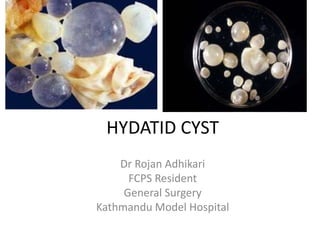
- 12. Cyst layers and contents
- 13. • Hydatid fluid is antigenic • This antigenicity is rarely of great clinical significance • Allergic reactions range from skin rash to a frank anaphylactic reaction • The antigenicity of hydatid fluid is the basis of serodiagnostics
- 14. Clinical presentation • The clinical features of hydatid liver disease depend on the site, size, stage of development, whether the cyst is alive or dead, and whether the cyst is infected or not. • Pain in the RUQ or epigastrium is the most common symptom, whereas hepatomegaly and a palpable mass are the most common signs.
- 15. Clinical Presentation SYMPTOMS Percentage Asymptomatic 75% Abdominal pain 20% Dyspepsia 13 % Fever and chills 8 % Jaundice 6%
- 16. Clinical Presentation SIGNS • Right upper quadrant mass •Right upper quadrant tenderness
- 17. Suppuration and Secondary Bacterial Infection • most frequent cause of infection is a cystobiliary communication • Clinically presents at pyogenic liver abscess • An infected hydatid cyst undergoes structural changes and the parasite dies
- 18. Pressure Effects • grow in the direction of the least resistance • Pressure effects appear sooner or later • symptoms result from direct pressure or distortion of neighboring structures or viscera. • An enlarging cyst – atrophy of surrounding hepatocytes – fibrosis – compensatory hypertrophy of the remaining liver parenchyma – replaces an entire liver lobe
- 19. • Serious consequence of cyst enlargement is cyst Rupture • Three types of cyst rupture have been addressed: – obscure – free – communicant rupture
- 20. Obscure (Internal) Rupture Injury or penetration of bile between pericyst and ectocyst Ruptue of ectocyst Protoscolesces occupies spaces Develops 100s of daughter cyst Unilocular multilocular Within yellow fluid of gelatin like amorphous mass inside pericyst
- 21. Free Rupture In free rupture, the hydatid contents disseminates throughout the peritoneal or pleural cavity
- 22. Intraperitoneal Rupture • Hydatid cyst grows in the direction of the least resistance • superficial portion of the pericyst is stretched, thinned out • cyst irregularly shaped, fibrous whitish structure protruding from normal liver parenchyma • Cysts reaching the anterior and inferior part of the liver continue to grow, protruding into the abdominalcavity •high intracystic pressure causes rupture of both univesicular and multivesicular cysts
- 23. Clinical presentations of intraperitoneal rupture (i) In acute symptomatic rupture, – peritoneal irritation and acute abdominal symptoms occur – The incidence is about 1% to 4%. (ii) In anaphylactic shock – rupture precipitates severe circulatory collapse, which may be fatal mask the abdominal manifestations (iii) In silent rupture, the patient presents with disseminated abdominal hydatidosis, unaware when the rupture occurred
- 24. • Intraperitoneal rupture is a life-threatening complication that results in secondary echinococcosis • •Multiple cysts develop throughout the peritoneal cavity causing – intestinal obstruction, – gross abdominal distention, – ascites, – and cachexia after years of the rupture. • This is the secondary, smaller life cycle for the parasite, occurring only in the intermediate host.
- 25. Intrathoracic Rupture • Elevated hemidiaphragm and a sterile pleural effusion can be the first signs of liver hydatid disease • Upward extension of a subdiaphragmatic cyst is usually asymptomatic, although it can cause dry cough, dyspnea, chest pain, and toxemia • The pleura and adherent basal lung segments often become inflamed and indurated • Frank intrapleural rupture with empyema (hydatopiothorax) is rare • pneumonitis or lung abscess
- 26. • The hydatid cyst may erode into a bronchiole and the contents can be evacuated • Rupture into bronchiole daughter cysts in the sputum • Ocassionally a bronchobiliary fistula will arise Expectoration of bile-tinged sputum • The incidence of diaphragmatic or transdiaphragmatic thoracic involvement by hydatid cysts in the dome of the liver is rare
- 27. Communicant Rupture Hydatid cysts can rupture into physiologic channels (e.g., biliary, blood vessels) or adjacent organs (e.g., digestive tract)
- 28. • In silent rupture, bile leaks from eroded small ducts into the cyst, causing – endogenic vesiculation – suppuration – eventually death of the parasite • Such cysts are filled with bile-stained fluid, although no visible bile duct communications can be seen. • Probably 80% to 90% of hydatid cyst bile duct ruptures are of the silent type.
- 29. • A triad of symptoms characterizes rupture into the bile ducts: I. biliary colic II. partial intermittent or complete ductal obstruction with cholangitis and jaundice III. germinative membranes in the feces.
- 30. Investigation • Casoni or intradermal test • Indirect hemagglutination test and enzyme-linked immunosorbent assay are the most widely used methods for detection of anti-Echinococcus antibodies (immunoglobulin G [IgG]). • These tests give false positive results in cases of schistosomiasis and nematode infestations that is why they are not specific for diagnosing hydatidosis.
- 31. Lab • Eosinophilia - 35% • Bilirubin >2 mg/Dl - 20% • WBC count <10,000/mm 3 - 10%
- 32. Investigation X-ray • Limited value • In endemic areas, elevation of the right hemidiaphragm in an otherwise healthy, asymptomatic patient is highly indicative of liver hydatidosis • Sometimes streaklike or round calcification of a senile hydatid cyst.
- 33. Ultrasound Imaging • comparatively cheap, noninvasive, enables interventional procedures
- 34. • Pathognomonic USG diagnostic features are I. unmistakable daughter cysts (rosettes) within the main cyst cavity II. detachment of the membrane of the cyst (double-contoured membrane) III. agglomeration of daughter cysts in the dependent portion of a hydatid cyst IV. calcification of the cyst wall
- 35. • Based on USG signs, Hassen Gharbi in 1981 classified liver hydatid cysts into five types I. pure fluid collection II. fluid collection with a split wall III. fluid collection with septa IV. heterogeneous appearance, and V. reflecting thick walls
- 38. CT • CT gives similar information to ultrasound, more specific information about the location and depth of the cyst within the liver. • Daughter cysts and exogenous cysts are also clearly visualized, and the volume of the cyst can be estimated. • CT is imperative for operative management, especially when a laparoscopic approach is utilized.
- 41. MRI & ERCP • MRI provides structural details of the hydatid cyst, but adds little more than ultrasound or CT, and is more expensive. • Endoscopic retrograde cholangio pancreatography (ERCP) may show communication between the cysts and bile ducts and can be used to drain the biliary tree before surgery.
- 42. Treatment • Medical, surgical, and percutaneous approaches may be part of the treatment. Small cysts (<4 cm) locateddeep in the parenchymaof the liver,if uncomplicated, canbe managed conservatively.
- 43. • Basic principles of treatment are (1)eradication of the parasite within the cyst, (2)protection of the host against spillage of scoleces, and (3)management of complications
- 44. Anti helmenthics • Medical therapy for echinococcosis is limited to the benzimidazoles (mebendazole and albendazole) • used alone is only 30% successful. • Albendazole is readily absorbed from the intestine and metabolized by the liver to an active form. Mebendazole is poorly absorbed and is inactivated by the liver. • 28-day course may be repeated, after 14 days without treatment to a total of 3 treatment cycles
- 45. PAIR technique (percutaneous aspiration, injection and re-aspiration) The most frequently utilized protoscolecidal agents 1. 15–20% saline, 2. 95% ethanol, 3. Mebendazole 4. 3% H2O2 5. Betadine 6. Silver nitrate 7. Formalin Combination is used Contraindicated in pregnancy, cyst communicating to billary tree and calcified cyst
- 46. PAIR technique Complication of PAIR • Spillage and anaphylaxis, • Recurrence • Mechanical damage to other tissue • Bilary fistula • Hemorrhage • Infection
- 48. Surgery • Surgery is still the treatment of choice for uncomplicated hydatid disease of the liver. • The objectives of surgical treatment are to: (1) inactivate the scoleces, (2) prevent spillage of cyst contents, (3) eliminate all viable elements of the cyst, and (4) manage the residual cavity of the cyst.
- 49. Surgery Indication • Large liver cysts with multiple daughter cysts • Superficially located single liver cysts that may rupture • Liver cyst with biliary tree communication or pressure effects on vital organs or structures • Infected cysts • Cysts in lungs, brain, kidneys, eyes, bones
- 50. Surgical Procedure • Early on, surgical management of hydatid cysts via cyst evacuation resulted in a high rate of peritoneal implantation. • This problem prompted the use of scolecidal agents for injection into the cyst and for use in the surrounding peritoneum. • The cyst is usually then aspirated through close suction
- 51. Surgical Procedures • The cyst is then unroofed which then can be followed by Conservative • Marsupialisation • Capittonage • Partial Pericystectomy This can be followed by omentoplasty
- 52. Surgical Procedure • Radical Pericystectomy: cyst and surrounding compressed liver tissue • Hepatic Resection: lobectomy or partial hepatectomy with entire cyst • Laparoscopic approach
- 53. 57 articles were selected for final analysis: one meta-analysis, 9 randomized clinical trials, 5 non-randomized comparative prospective studies, 7 non-comparative prospective studies, and 34 retrospective studies
- 54. Conclusion • Antihelminthic treatment alone is not the ideal treatment for liver hydatid cysts. • More studies in the literature support the effectiveness of radical treatment compared with conservative treatment. • Conservative surgery with omentoplasty is effective in preventing postoperative complications. • A laparoscopic approach is safe in some situations. • Percutaneous drainage with albendazole therapy is a safe and effective alternative treatment for hydatid cysts of the liver. • Radical surgery with pre- and post-operative administration of albendazole is the best treatment option for liver hydatid cysts due to low recurrence and complication rates.
- 59. References • Mastery of surgery • Sabiston text book of surgery • UpTodate online • Centre of Disease Control and Prevention • Parasitology “K D Chatterjee”
- 60. Thank you Wash your hands with soap and warm water after handling dogs, and before handling food.