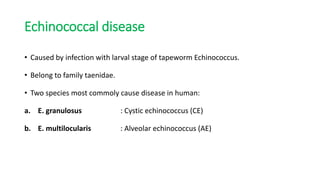
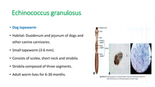
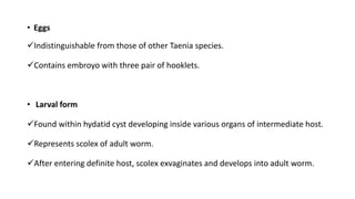
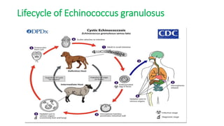
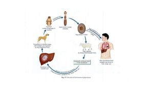
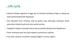
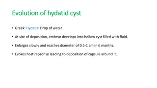
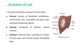
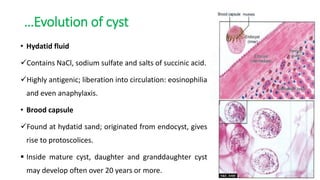
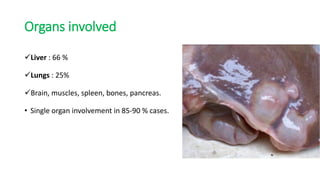
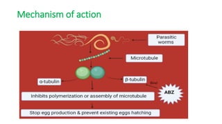
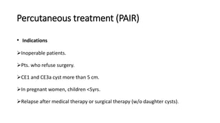
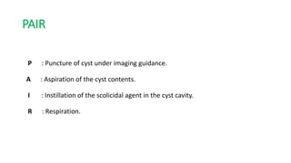
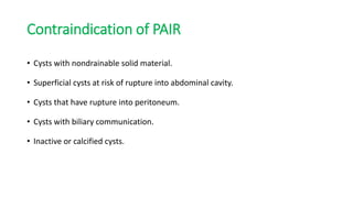
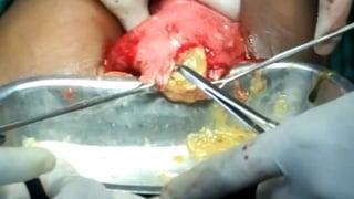
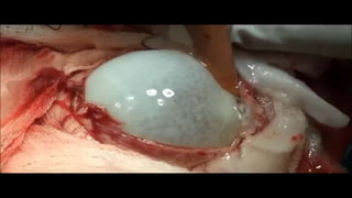
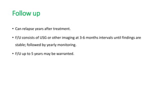
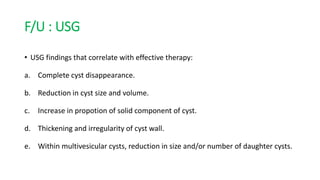
SlideShare a Scribd company logo
Hydatid cyst
Pranjal Rokaya
Resident General Surgery
KIST MCTH
29th December, 2022
Outline
Echinococcal disease
• Caused by infection with larval stage of tapeworm Echinococcus.
• Belong to family taenidae.
• Two species most commoly cause disease in human:
a. E. granulosus : Cystic echinococcus (CE)
b. E. multilocularis : Alveolar echinococcus (AE)
Echinococcus granulosus
• Dog tapeworm
• Habitat: Duodenum and jejunum of dogs and
other canine carnivores.
• Small tapeworm (3-6 mm).
• Consists of scolex, short neck and strobila.
• Strobila composed of three segments.
• Adult worm lives for 6-30 months.
• Eggs
Indistinguishable from those of other Taenia species.
Contains embroyo with three pair of hooklets.
• Larval form
Found within hydatid cyst developing inside various organs of intermediate host.
Represents scolex of adult worm.
After entering definite host, scolex exvaginates and develops into adult worm.
Lifecycle of Echinococcus granulosus
…Life cycle
• Infection follows ingestion of eggs due to intimate handling of dogs or eating raw
food contaminated with dog faeces.
• Ova liberated from chinitous wall by gastric juice liberating embroyos which
penetrate intestinal wall and enter portal venules.
• Trapped in hepatic sinusoids where they eventually develop into hydatid cyst.
• Some embroyos pass liver, get trapped in pulmonary capillary.
• Few enter systemic circulation and get lodged in various organs.
Evolution of hydatid cyst
• Greek: Hydatis: Drop of water.
• At site of deposition, embryo develops into hollow cyst filled with fluid.
• Enlarges slowly and reaches diameter of 0.5-1 cm in 6 months.
• Evokes host repsonse leading to deposition of capsule around it.
…Evolution of cyst
• Cyst wall secreted by embroyo has three layers:
a. Pericyst: Consists of fibroblastic proliferation,
mononuclear cells, eosinophils and giant cells;
eventually developing capsule.
b. Ectocyst: Composed of acellular, hyaline
material.
c. Endocyst: Germinal layer consisting of number
of nuclei; gives rise to brood capsule and hydatid
fluid.
…Evolution of cyst
• Hydatid fluid
Contains NaCl, sodium sulfate and salts of succinic acid.
Highly antigenic; liberation into circulation: eosinophilia
and even anaphylaxis.
• Brood capsule
Found at hydatid sand; originated from endocyst, gives
rise to protoscolices.
 Inside mature cyst, daughter and granddaughter cyst
may develop often over 20 years or more.
Organs involved
Liver : 66 %
Lungs : 25%
Brain, muscles, spleen, bones, pancreas.
• Single organ involvement in 85-90 % cases.
Clinical features
• Initial phase always asymptomatic.
• Many infections acquired in childhood but don’t cause symtoms until adulthood.
• 50% of detected cases in asymptomatic patients.
• Clinical feature depends upon site of cyst and their size.
Clinical features
a. Liver
Right lobe in 60-85% cases; symptoms unusual with cyst < 10 cm.
Hepatomegaly a/w RUQ pain, nausea and vomitting.
In 1/4th cases, cyst ruptures into biliary tree producing biliary colic, obstructive
jaundice, cholangitis and or pancreatitis.
Pressure effect: Cholestatis, portal hypertension, venous obstruction.
Rupture into peritoneum: peritonitis; transdiaphramatically: pulmonary hydatosis or
bronchial fistula.
b. Lungs
60% affects the right lung; 50-60% involve lower lobe.
• Cough (53-62%)
• Chest pain (49-91%)
• Dyspnea (10-70%)
• Hempotysis (12-21%)
Cyst rupture into pleural cavity: Pneumothorax, effusion or empyema.
Bacterial infection: Pulmonary abscess.
Other organs involvement
Heart: Mechanical rupture with widespread dissemination or tamponade.
CNS: Seizures, signs of raised ICP.
Kidneys: Hematuria , bilateral flank pain: glomerulonephritis leading to nephrotic
syndrome.
Bones: Asymptomatic until pathological fracture develops.
Complications
• Anaphylaxis
• Rupture of the cyst
• Obstructive jaundice
• Infection
• Calcification
• Liver failure
Investigations
Imaging Serology
Imaging
1. Ultrasonography
Sensitivity: 90-95%
Most common appearance: Anechoic, smooth, round cyst difficult to distinguish
from benign cyst.
In presence of liver cyst membrane, mixed echoes confused with abscess or
neoplasm.
Daughter cyst: Internal septations seen.
Classification based on USG
Gharbi classification
Type 1 Consists of pure fluid
Type 2 Fluid collection with split wall
Type 3 Contains daughter cysts (with
or without degenerated
material.
Type 4 Heteogenous echo pattern
Type 5 Calcified wall
WHO classification of hydatid cyst
WHO stage Description Stage
CE1 Unilocular, unechoic cystic lesion Active
CE2 Multiseptated cyst Active
CE3a Cyst with detatched membrane Transitional
CE3b Cyst with daughter cyst in solid matrix Transitional
CE4 Cyst with heterogenous hypo/hyperechoic
content; no daughter cysts
Inactive
CE5 Solid plus calcified wall Inactive
A non-calcified liver cyst with
with floating layers of germinal
membrane
Hydatid daughter cyst
Imaging
2. Computed tomography
Sensitivity: 95-100%
Best mode to determine number, size and anatomical
location of cysts.
Better for detection of extrahepatic cysts compared to
USG.
Used for monitoring lesions during therapy and detect
recurrence.
Imaging
3. MRI
No major advntage over CT for evaluation of abdominal or pulmonary cysts,
except in defining intra and extrahepatic venous system.
May delineate cyst capsule better than CT and better at diagnosing complications
(infection or biliary communication).
Usually not required, neither cost effective.
Serologic tests
Site of lesion Sensitivity of serologic tests
Liver a. IgG ELISA : 80-90 %
b. IgE ELISA. : 82-92%
c. Latex agglutination. : 65-75%
d. Hemagglutinin. : 80-90%
e. Immunoblot. : 80 %
Lung a. IgG ELISA : 60-85%
b. IgE ELISA. : 45-70%
c. Latex agglutination. : 50-70%
d. Hemagglutinaton. : 50-70%
e. Immunoblot. : 55-70%
Serologic tests
• Two antigens used in serologic tests: antigen 5 and antigen B.
• Utility of serology improved with combination of test or
sequential testing.
• No correlation between results and number or size of cyst.
• Negative test doesn’t rule out the disease.
• Less likely to be (+): cysts intact, calcified or noviable.
Interventional procedure
• In absence of (+) serology, percutaneous aspiration
or biopsy may be required for confirmation.
• Aspiration peformed under USG or CT guidance.
• ERCP may be required to evaluate biliary
involvement.
• May demonstrate hydatid membrane within
duodenum or impacted im pappila.
Differential diagnosis
a. Simple benign cyst
b. Hemangioma
c. Hepatocellular carcinoma
d. Abscess
e. Tuberculosis
Treatment
Antiparasitic
therapy
Percutaneous
aspiration and
instillation of
scolicidal
agent
Surgical
resection
Treatment based on WHO classification
Antiparasitic therapy
• Albendazole: Primary antiparasitic agent.
• Poorly absorbed; ideally ingested with fatty meal to increase bioavailability.
• Effective alone for cysts with single compartment and diameter <5 cm. (CE1,
CE3a).
• Drug t/t alone for multiple liver cyst <5 cm, cysts deep inside liver parenchyma
and intraperitoneal cyst.
• Definite therapy: one to three months
Adjunctive therapy
• Drug useful adjunct to surgery and percutaneous t/t.
• Perioperative drug therapy reduces recurrence by inactivatig protoscolices; aslo
softens cyst facilitating removal.
• Treatment initiated at least 4 days prior to surgery and continued at least one
month (albendazole) or three months (mebendazole) following surgery.
• Drug also required after spontaneous cyst rupture.
Mechanism of action
Percutaneous treatment (PAIR)
• Indications
Inoperable patients.
Pts. who refuse surgery.
CE1 and CE3a cyst more than 5 cm.
In pregnant women, children <5yrs.
Relapse after medical therapy or surgical therapy (w/o daughter cysts).
PAIR
P : Puncture of cyst under imaging guidance.
A : Aspiration of the cyst contents.
I : Instillation of the scolicidal agent in the cyst cavity.
R : Respiration.
Contraindication of PAIR
• Cysts with nondrainable solid material.
• Superficial cysts at risk of rupture into abdominal cavity.
• Cysts that have rupture into peritoneum.
• Cysts with biliary communication.
• Inactive or calcified cysts.
Modified catheterization techniques
• Large bore catheters and cutting devices used together to remove entire
endocyst and daughter cysts.
• Appropriate for:
 Cysts difficult to drain via PAIR.
 Stage CE2 and CE3b, which have many compartments, and commonly relapse
after PAIR.
Surgical treatment
• Indications
a. Complicated cysts
i. Ruptured cyst. ii. Cysts with biliary fistula
iii. Cysts compressing vital structure. iv. Cysts with secondary infection/hemorrhage
b. Cysts with many daughter vesicles not suitable for percutaneous t/t.
c. Cysts more than 10 cm.
d. Extrahepatic disease( lung, bone, brain, kidney, spleen).
Surgical treatment
• Goal is to evacuate the cyst and obliterating residual cavity.
• Effort made to avoid fluid spillage. (3-6 months albendazole)
• Different techniques:
Removal of intact cyst if feasible.
Cyst opened, sterilised, contents evacuated and pericyst removed.
 Laying open of cyst cavity w/o removing pericyst.
• Cyst excision followed by omentoplasty a/w lowest complication rates.
Surgical treatment
• Surgical field should be protected with pads soaked with scolicides.
• Scolicidal agents not used in case of biliary communication to prevent risk of
sclerosing cholangitis or pancreatitis.
No RCTs performed comparing laparoscopic approach vs open.
Laparoscopy may be a/w increased risk of spillage.
Exclusion criteria for laparoscopy: a. Deep intraparenchymal cysts
b. Posterior cysts close to venacava. c. Presence of >3 cysts with calcification.
Follow up
• Can relapse years after treatment.
• F/U consists of USG or other imaging at 3-6 months intervals until findings are
stable; followed by yearly monitoring.
• F/U up to 5 years may be warranted.
F/U : USG
• USG findings that correlate with effective therapy:
a. Complete cyst disappearance.
b. Reduction in cyst size and volume.
c. Increase in propotion of solid component of cyst.
d. Thickening and irregularity of cyst wall.
e. Within multivesicular cysts, reduction in size and/or number of daughter cysts.
F/U: Serology
• Serologic titres usually fall by 1 to 2 years following surgery and rise again in
setting of recurrence.
• Antibodies may remain elevated many years after succesful cyst removal.
Prevention
Ensuring pet dogs don’t eat animal carcass.
Periodic deworming of pet dogs.
Euthanization of infected dogs.
Washing hands after touching dogs; avoiding kissing pet dogs.
References
• Bailey and Love, 26th edition.
• Swartz texbook of surgery, 11th edition.
• Sabiston textbook of surgery, 21st edition.
Thank You.

More Related Content

What's hot

IMAGING IN ABDOMINAL TUBERCULOSIS
IMAGING IN ABDOMINAL TUBERCULOSISIMAGING IN ABDOMINAL TUBERCULOSIS
IMAGING IN ABDOMINAL TUBERCULOSIS
Navni Garg
 
Presentation1.pptx acute abdomen.
Presentation1.pptx acute abdomen.Presentation1.pptx acute abdomen.
Presentation1.pptx acute abdomen.Abdellah Nazeer
 
Git lymphomas
Git lymphomasGit lymphomas
Git lymphomasairwave12
 
Early gastric cancer
Early gastric cancerEarly gastric cancer
Early gastric cancer
dguin111
 
Acute and chronic mesenteric ischaemia(1)
Acute and chronic mesenteric ischaemia(1)Acute and chronic mesenteric ischaemia(1)
Acute and chronic mesenteric ischaemia(1)
Shambhavi Sharma
 
Cholecystitis & carcinoma of gallbladder
Cholecystitis & carcinoma of gallbladder Cholecystitis & carcinoma of gallbladder
Cholecystitis & carcinoma of gallbladder
Baiti Basheer
 
Carcinoma gall bladder
Carcinoma gall bladderCarcinoma gall bladder
Carcinoma gall bladder
Youttam Laudari
 
Hydatid cyst of liver by dr aakif
Hydatid cyst of liver by dr aakifHydatid cyst of liver by dr aakif
Hydatid cyst of liver by dr aakifdraakif
 
Biliary drainage
Biliary drainageBiliary drainage
Biliary drainage
Dr. Yash Kumar Achantani
 
Anomalies of biliary tree
Anomalies of biliary treeAnomalies of biliary tree
Anomalies of biliary tree
Shambhavi Sharma
 
Unilateral small kidney
Unilateral small kidneyUnilateral small kidney
Unilateral small kidney
maimusirdan
 
Presentation1.pptx, radiological imaging of divertiular disease and diverticu...
Presentation1.pptx, radiological imaging of divertiular disease and diverticu...Presentation1.pptx, radiological imaging of divertiular disease and diverticu...
Presentation1.pptx, radiological imaging of divertiular disease and diverticu...Abdellah Nazeer
 
imaging of benign hepatic masses
imaging of benign hepatic massesimaging of benign hepatic masses
imaging of benign hepatic masses
Navni Garg
 
Urinary bladder pathology radiology
Urinary bladder pathology radiologyUrinary bladder pathology radiology
Urinary bladder pathology radiology
Dr pradeep Kumar
 
Cystic artery anomalies
Cystic artery anomaliesCystic artery anomalies
Cystic artery anomalies
Jibran Mohsin
 
Pancreatic pseudocyst
Pancreatic pseudocystPancreatic pseudocyst
Pancreatic pseudocystdraakif
 
Cystic liver lesions - An ultrasound perspective
Cystic liver lesions - An ultrasound perspectiveCystic liver lesions - An ultrasound perspective
Cystic liver lesions - An ultrasound perspectiveSamir Haffar
 
Billiary tract
Billiary tractBilliary tract
Billiary tract
RMLIMS
 
Imaging in urology
Imaging in urologyImaging in urology
Imaging in urology
dr sajid Abbasi
 

What's hot (20)

IMAGING IN ABDOMINAL TUBERCULOSIS
IMAGING IN ABDOMINAL TUBERCULOSISIMAGING IN ABDOMINAL TUBERCULOSIS
IMAGING IN ABDOMINAL TUBERCULOSIS
 
Presentation1.pptx acute abdomen.
Presentation1.pptx acute abdomen.Presentation1.pptx acute abdomen.
Presentation1.pptx acute abdomen.
 
Git lymphomas
Git lymphomasGit lymphomas
Git lymphomas
 
Early gastric cancer
Early gastric cancerEarly gastric cancer
Early gastric cancer
 
Acute and chronic mesenteric ischaemia(1)
Acute and chronic mesenteric ischaemia(1)Acute and chronic mesenteric ischaemia(1)
Acute and chronic mesenteric ischaemia(1)
 
Cholecystitis & carcinoma of gallbladder
Cholecystitis & carcinoma of gallbladder Cholecystitis & carcinoma of gallbladder
Cholecystitis & carcinoma of gallbladder
 
Carcinoma gall bladder
Carcinoma gall bladderCarcinoma gall bladder
Carcinoma gall bladder
 
Hydatid cyst of liver by dr aakif
Hydatid cyst of liver by dr aakifHydatid cyst of liver by dr aakif
Hydatid cyst of liver by dr aakif
 
Biliary drainage
Biliary drainageBiliary drainage
Biliary drainage
 
Anomalies of biliary tree
Anomalies of biliary treeAnomalies of biliary tree
Anomalies of biliary tree
 
Unilateral small kidney
Unilateral small kidneyUnilateral small kidney
Unilateral small kidney
 
Large bowel obstruction
Large bowel obstructionLarge bowel obstruction
Large bowel obstruction
 
Presentation1.pptx, radiological imaging of divertiular disease and diverticu...
Presentation1.pptx, radiological imaging of divertiular disease and diverticu...Presentation1.pptx, radiological imaging of divertiular disease and diverticu...
Presentation1.pptx, radiological imaging of divertiular disease and diverticu...
 
imaging of benign hepatic masses
imaging of benign hepatic massesimaging of benign hepatic masses
imaging of benign hepatic masses
 
Urinary bladder pathology radiology
Urinary bladder pathology radiologyUrinary bladder pathology radiology
Urinary bladder pathology radiology
 
Cystic artery anomalies
Cystic artery anomaliesCystic artery anomalies
Cystic artery anomalies
 
Pancreatic pseudocyst
Pancreatic pseudocystPancreatic pseudocyst
Pancreatic pseudocyst
 
Cystic liver lesions - An ultrasound perspective
Cystic liver lesions - An ultrasound perspectiveCystic liver lesions - An ultrasound perspective
Cystic liver lesions - An ultrasound perspective
 
Billiary tract
Billiary tractBilliary tract
Billiary tract
 
Imaging in urology
Imaging in urologyImaging in urology
Imaging in urology
 

Similar to Hydatid Cyst

abc hdat.pdf
abc hdat.pdfabc hdat.pdf
abc hdat.pdf
aminf5388
 
Hydatid diseases
Hydatid diseasesHydatid diseases
Hydatid diseases
AIIMS, New Delhi, India
 
Dr Archit Gupta JR Surgery IGMC Shimla
Dr Archit Gupta JR Surgery IGMC ShimlaDr Archit Gupta JR Surgery IGMC Shimla
Dr Archit Gupta JR Surgery IGMC Shimla
Jagdish Gupta
 
Hydatid cyst
Hydatid cystHydatid cyst
Hydatid cyst
syed ubaid
 
Hydatid disease
Hydatid diseaseHydatid disease
Hydatid disease
Drbd Soni
 
Hydatidcystofliverby hegazy
Hydatidcystofliverby hegazyHydatidcystofliverby hegazy
Hydatidcystofliverby hegazy
mostafa hegazy
 
Hydatidcystofliverby hegazy
Hydatidcystofliverby hegazyHydatidcystofliverby hegazy
Hydatidcystofliverby hegazy
mostafa hegazy
 
Seminar on choledochal cyst
Seminar on choledochal cyst Seminar on choledochal cyst
Seminar on choledochal cyst
Biswajit Deka
 
Cystic disease of liver
Cystic disease of liverCystic disease of liver
Cystic disease of liver
Dr.Avijit Banerjee
 
Hydatid cyst disease of the liver الدكتور طارق المنيزل
Hydatid cyst disease of the liver  الدكتور طارق المنيزل Hydatid cyst disease of the liver  الدكتور طارق المنيزل
Hydatid cyst disease of the liver الدكتور طارق المنيزل
Tariq Al munaizel
 
Friday Presentation.pptx
Friday Presentation.pptxFriday Presentation.pptx
Friday Presentation.pptx
OMJHA20
 
Hydatid disease
Hydatid diseaseHydatid disease
Hydatid disease
BISHAL SAPKOTA
 
LIVER ABSCESS.pptx
LIVER ABSCESS.pptxLIVER ABSCESS.pptx
LIVER ABSCESS.pptx
SujanPandey11
 
Hydatid cyst
Hydatid cystHydatid cyst
Hydatid cyst
Youttam Laudari
 
Liver abcess
Liver abcessLiver abcess
Liver abcess
syed ubaid
 
Hydatid cyst.pptx
Hydatid cyst.pptxHydatid cyst.pptx
Hydatid cyst.pptx
IbrahimAboAlasaad
 
Hydatid cyst
Hydatid cystHydatid cyst
Hydatid cyst
Rojan Adhikari
 
Tb epididymitis, By Emad M.Qasem
Tb epididymitis, By Emad M.QasemTb epididymitis, By Emad M.Qasem
Tb epididymitis, By Emad M.Qasem
Emad Qasem
 
Liver Cysts and Abscesses
Liver Cysts and AbscessesLiver Cysts and Abscesses
Liver Cysts and Abscesses
Abdulkareem Kabir
 

Similar to Hydatid Cyst (20)

abc hdat.pdf
abc hdat.pdfabc hdat.pdf
abc hdat.pdf
 
Hydatid diseases
Hydatid diseasesHydatid diseases
Hydatid diseases
 
Dr Archit Gupta JR Surgery IGMC Shimla
Dr Archit Gupta JR Surgery IGMC ShimlaDr Archit Gupta JR Surgery IGMC Shimla
Dr Archit Gupta JR Surgery IGMC Shimla
 
Hydatid cyst
Hydatid cystHydatid cyst
Hydatid cyst
 
Hydatid disease
Hydatid diseaseHydatid disease
Hydatid disease
 
Hydatidcystofliverby hegazy
Hydatidcystofliverby hegazyHydatidcystofliverby hegazy
Hydatidcystofliverby hegazy
 
Hydatidcystofliverby hegazy
Hydatidcystofliverby hegazyHydatidcystofliverby hegazy
Hydatidcystofliverby hegazy
 
Seminar on choledochal cyst
Seminar on choledochal cyst Seminar on choledochal cyst
Seminar on choledochal cyst
 
Cystic disease of liver
Cystic disease of liverCystic disease of liver
Cystic disease of liver
 
Hydatid cyst disease of the liver الدكتور طارق المنيزل
Hydatid cyst disease of the liver  الدكتور طارق المنيزل Hydatid cyst disease of the liver  الدكتور طارق المنيزل
Hydatid cyst disease of the liver الدكتور طارق المنيزل
 
Friday Presentation.pptx
Friday Presentation.pptxFriday Presentation.pptx
Friday Presentation.pptx
 
Hydatid disease
Hydatid diseaseHydatid disease
Hydatid disease
 
Hydatid Cyst Of Liver
Hydatid Cyst Of LiverHydatid Cyst Of Liver
Hydatid Cyst Of Liver
 
LIVER ABSCESS.pptx
LIVER ABSCESS.pptxLIVER ABSCESS.pptx
LIVER ABSCESS.pptx
 
Hydatid cyst
Hydatid cystHydatid cyst
Hydatid cyst
 
Liver abcess
Liver abcessLiver abcess
Liver abcess
 
Hydatid cyst.pptx
Hydatid cyst.pptxHydatid cyst.pptx
Hydatid cyst.pptx
 
Hydatid cyst
Hydatid cystHydatid cyst
Hydatid cyst
 
Tb epididymitis, By Emad M.Qasem
Tb epididymitis, By Emad M.QasemTb epididymitis, By Emad M.Qasem
Tb epididymitis, By Emad M.Qasem
 
Liver Cysts and Abscesses
Liver Cysts and AbscessesLiver Cysts and Abscesses
Liver Cysts and Abscesses
 

More from KIST Surgery

surgical non specific infection
surgical non specific infectionsurgical non specific infection
surgical non specific infection
KIST Surgery
 
Splenic Injury.pptx
Splenic Injury.pptxSplenic Injury.pptx
Splenic Injury.pptx
KIST Surgery
 
Cleft lip & palate.ppt
Cleft lip & palate.pptCleft lip & palate.ppt
Cleft lip & palate.ppt
KIST Surgery
 
Fistula in ano
Fistula in anoFistula in ano
Fistula in ano
KIST Surgery
 
Pancreatic Cystic Neoplasm
Pancreatic Cystic NeoplasmPancreatic Cystic Neoplasm
Pancreatic Cystic Neoplasm
KIST Surgery
 
Journal club : Gallstone pancreatitis
Journal club : Gallstone pancreatitisJournal club : Gallstone pancreatitis
Journal club : Gallstone pancreatitis
KIST Surgery
 
Mesenteric Ischemia
Mesenteric Ischemia Mesenteric Ischemia
Mesenteric Ischemia
KIST Surgery
 
Raised intracranial pressure
Raised intracranial pressureRaised intracranial pressure
Raised intracranial pressure
KIST Surgery
 
Mesenteric Ischemia
Mesenteric IschemiaMesenteric Ischemia
Mesenteric Ischemia
KIST Surgery
 
PRE OPERATION PREPARATION
PRE OPERATION PREPARATIONPRE OPERATION PREPARATION
PRE OPERATION PREPARATION
KIST Surgery
 
MENINGIOMA
MENINGIOMAMENINGIOMA
MENINGIOMA
KIST Surgery
 
Optimization Of High Risk Surgical Patients
Optimization Of High Risk Surgical PatientsOptimization Of High Risk Surgical Patients
Optimization Of High Risk Surgical Patients
KIST Surgery
 
Intestinal ishaemia
Intestinal ishaemiaIntestinal ishaemia
Intestinal ishaemia
KIST Surgery
 
Intestinal Obstruction
Intestinal ObstructionIntestinal Obstruction
Intestinal Obstruction
KIST Surgery
 
Intravenous Fluids In Surgical Patients
Intravenous Fluids In Surgical PatientsIntravenous Fluids In Surgical Patients
Intravenous Fluids In Surgical Patients
KIST Surgery
 
Intracranial hematomas
Intracranial hematomasIntracranial hematomas
Intracranial hematomas
KIST Surgery
 
Journal club - Hernioplasty
Journal club - HernioplastyJournal club - Hernioplasty
Journal club - Hernioplasty
KIST Surgery
 
Journal club LCBDE+LC vs ERCP+LC
 Journal club LCBDE+LC vs ERCP+LC Journal club LCBDE+LC vs ERCP+LC
Journal club LCBDE+LC vs ERCP+LC
KIST Surgery
 
Breast disorders
Breast disordersBreast disorders
Breast disorders
KIST Surgery
 

More from KIST Surgery (20)

surgical non specific infection
surgical non specific infectionsurgical non specific infection
surgical non specific infection
 
Splenic Injury.pptx
Splenic Injury.pptxSplenic Injury.pptx
Splenic Injury.pptx
 
Cleft lip & palate.ppt
Cleft lip & palate.pptCleft lip & palate.ppt
Cleft lip & palate.ppt
 
Fistula in ano
Fistula in anoFistula in ano
Fistula in ano
 
Pancreatic Cystic Neoplasm
Pancreatic Cystic NeoplasmPancreatic Cystic Neoplasm
Pancreatic Cystic Neoplasm
 
Journal club : Gallstone pancreatitis
Journal club : Gallstone pancreatitisJournal club : Gallstone pancreatitis
Journal club : Gallstone pancreatitis
 
Mesenteric Ischemia
Mesenteric Ischemia Mesenteric Ischemia
Mesenteric Ischemia
 
Raised intracranial pressure
Raised intracranial pressureRaised intracranial pressure
Raised intracranial pressure
 
Mesenteric Ischemia
Mesenteric IschemiaMesenteric Ischemia
Mesenteric Ischemia
 
PRE OPERATION PREPARATION
PRE OPERATION PREPARATIONPRE OPERATION PREPARATION
PRE OPERATION PREPARATION
 
MENINGIOMA
MENINGIOMAMENINGIOMA
MENINGIOMA
 
Optimization Of High Risk Surgical Patients
Optimization Of High Risk Surgical PatientsOptimization Of High Risk Surgical Patients
Optimization Of High Risk Surgical Patients
 
Intestinal ishaemia
Intestinal ishaemiaIntestinal ishaemia
Intestinal ishaemia
 
Intestinal Obstruction
Intestinal ObstructionIntestinal Obstruction
Intestinal Obstruction
 
Intravenous Fluids In Surgical Patients
Intravenous Fluids In Surgical PatientsIntravenous Fluids In Surgical Patients
Intravenous Fluids In Surgical Patients
 
Intracranial hematomas
Intracranial hematomasIntracranial hematomas
Intracranial hematomas
 
Journal club - Hernioplasty
Journal club - HernioplastyJournal club - Hernioplasty
Journal club - Hernioplasty
 
Journal club LCBDE+LC vs ERCP+LC
 Journal club LCBDE+LC vs ERCP+LC Journal club LCBDE+LC vs ERCP+LC
Journal club LCBDE+LC vs ERCP+LC
 
Breast disorders
Breast disordersBreast disorders
Breast disorders
 
GIST
GISTGIST
GIST
 

Recently uploaded

ACUTE SCROTUM.....pdf. ACUTE SCROTAL CONDITIOND
ACUTE SCROTUM.....pdf. ACUTE SCROTAL CONDITIONDACUTE SCROTUM.....pdf. ACUTE SCROTAL CONDITIOND
ACUTE SCROTUM.....pdf. ACUTE SCROTAL CONDITIOND
DR SETH JOTHAM
 
Are There Any Natural Remedies To Treat Syphilis.pdf
Are There Any Natural Remedies To Treat Syphilis.pdfAre There Any Natural Remedies To Treat Syphilis.pdf
Are There Any Natural Remedies To Treat Syphilis.pdf
Little Cross Family Clinic
 
Ophthalmology Clinical Tests for OSCE exam
Ophthalmology Clinical Tests for OSCE examOphthalmology Clinical Tests for OSCE exam
Ophthalmology Clinical Tests for OSCE exam
KafrELShiekh University
 
Maxilla, Mandible & Hyoid Bone & Clinical Correlations by Dr. RIG.pptx
Maxilla, Mandible & Hyoid Bone & Clinical Correlations by Dr. RIG.pptxMaxilla, Mandible & Hyoid Bone & Clinical Correlations by Dr. RIG.pptx
Maxilla, Mandible & Hyoid Bone & Clinical Correlations by Dr. RIG.pptx
Dr. Rabia Inam Gandapore
 
How to Give Better Lectures: Some Tips for Doctors
How to Give Better Lectures: Some Tips for DoctorsHow to Give Better Lectures: Some Tips for Doctors
How to Give Better Lectures: Some Tips for Doctors
LanceCatedral
 
NVBDCP.pptx Nation vector borne disease control program
NVBDCP.pptx Nation vector borne disease control programNVBDCP.pptx Nation vector borne disease control program
NVBDCP.pptx Nation vector borne disease control program
Sapna Thakur
 
BRACHYTHERAPY OVERVIEW AND APPLICATORS
BRACHYTHERAPY OVERVIEW  AND  APPLICATORSBRACHYTHERAPY OVERVIEW  AND  APPLICATORS
BRACHYTHERAPY OVERVIEW AND APPLICATORS
Krishan Murari
 
24 Upakrama.pptx class ppt useful in all
24 Upakrama.pptx class ppt useful in all24 Upakrama.pptx class ppt useful in all
24 Upakrama.pptx class ppt useful in all
DrSathishMS1
 
ARTIFICIAL INTELLIGENCE IN HEALTHCARE.pdf
ARTIFICIAL INTELLIGENCE IN  HEALTHCARE.pdfARTIFICIAL INTELLIGENCE IN  HEALTHCARE.pdf
ARTIFICIAL INTELLIGENCE IN HEALTHCARE.pdf
Anujkumaranit
 
Tom Selleck Health: A Comprehensive Look at the Iconic Actor’s Wellness Journey
Tom Selleck Health: A Comprehensive Look at the Iconic Actor’s Wellness JourneyTom Selleck Health: A Comprehensive Look at the Iconic Actor’s Wellness Journey
Tom Selleck Health: A Comprehensive Look at the Iconic Actor’s Wellness Journey
greendigital
 
Charaka Samhita Sutra sthana Chapter 15 Upakalpaniyaadhyaya
Charaka Samhita Sutra sthana Chapter 15 UpakalpaniyaadhyayaCharaka Samhita Sutra sthana Chapter 15 Upakalpaniyaadhyaya
Charaka Samhita Sutra sthana Chapter 15 Upakalpaniyaadhyaya
Dr KHALID B.M
 
Ocular injury ppt Upendra pal optometrist upums saifai etawah
Ocular injury  ppt  Upendra pal  optometrist upums saifai etawahOcular injury  ppt  Upendra pal  optometrist upums saifai etawah
Ocular injury ppt Upendra pal optometrist upums saifai etawah
pal078100
 
Superficial & Deep Fascia of the NECK.pptx
Superficial & Deep Fascia of the NECK.pptxSuperficial & Deep Fascia of the NECK.pptx
Superficial & Deep Fascia of the NECK.pptx
Dr. Rabia Inam Gandapore
 
The Normal Electrocardiogram - Part I of II
The Normal Electrocardiogram - Part I of IIThe Normal Electrocardiogram - Part I of II
The Normal Electrocardiogram - Part I of II
MedicoseAcademics
 
POST OPERATIVE OLIGURIA and its management
POST OPERATIVE OLIGURIA and its managementPOST OPERATIVE OLIGURIA and its management
POST OPERATIVE OLIGURIA and its management
touseefaziz1
 
Lung Cancer: Artificial Intelligence, Synergetics, Complex System Analysis, S...
Lung Cancer: Artificial Intelligence, Synergetics, Complex System Analysis, S...Lung Cancer: Artificial Intelligence, Synergetics, Complex System Analysis, S...
Lung Cancer: Artificial Intelligence, Synergetics, Complex System Analysis, S...
Oleg Kshivets
 
Physiology of Chemical Sensation of smell.pdf
Physiology of Chemical Sensation of smell.pdfPhysiology of Chemical Sensation of smell.pdf
Physiology of Chemical Sensation of smell.pdf
MedicoseAcademics
 
BENIGN PROSTATIC HYPERPLASIA.BPH. BPHpdf
BENIGN PROSTATIC HYPERPLASIA.BPH. BPHpdfBENIGN PROSTATIC HYPERPLASIA.BPH. BPHpdf
BENIGN PROSTATIC HYPERPLASIA.BPH. BPHpdf
DR SETH JOTHAM
 
Cervical & Brachial Plexus By Dr. RIG.pptx
Cervical & Brachial Plexus By Dr. RIG.pptxCervical & Brachial Plexus By Dr. RIG.pptx
Cervical & Brachial Plexus By Dr. RIG.pptx
Dr. Rabia Inam Gandapore
 
Prix Galien International 2024 Forum Program
Prix Galien International 2024 Forum ProgramPrix Galien International 2024 Forum Program
Prix Galien International 2024 Forum Program
Levi Shapiro
 

Recently uploaded (20)

ACUTE SCROTUM.....pdf. ACUTE SCROTAL CONDITIOND
ACUTE SCROTUM.....pdf. ACUTE SCROTAL CONDITIONDACUTE SCROTUM.....pdf. ACUTE SCROTAL CONDITIOND
ACUTE SCROTUM.....pdf. ACUTE SCROTAL CONDITIOND
 
Are There Any Natural Remedies To Treat Syphilis.pdf
Are There Any Natural Remedies To Treat Syphilis.pdfAre There Any Natural Remedies To Treat Syphilis.pdf
Are There Any Natural Remedies To Treat Syphilis.pdf
 
Ophthalmology Clinical Tests for OSCE exam
Ophthalmology Clinical Tests for OSCE examOphthalmology Clinical Tests for OSCE exam
Ophthalmology Clinical Tests for OSCE exam
 
Maxilla, Mandible & Hyoid Bone & Clinical Correlations by Dr. RIG.pptx
Maxilla, Mandible & Hyoid Bone & Clinical Correlations by Dr. RIG.pptxMaxilla, Mandible & Hyoid Bone & Clinical Correlations by Dr. RIG.pptx
Maxilla, Mandible & Hyoid Bone & Clinical Correlations by Dr. RIG.pptx
 
How to Give Better Lectures: Some Tips for Doctors
How to Give Better Lectures: Some Tips for DoctorsHow to Give Better Lectures: Some Tips for Doctors
How to Give Better Lectures: Some Tips for Doctors
 
NVBDCP.pptx Nation vector borne disease control program
NVBDCP.pptx Nation vector borne disease control programNVBDCP.pptx Nation vector borne disease control program
NVBDCP.pptx Nation vector borne disease control program
 
BRACHYTHERAPY OVERVIEW AND APPLICATORS
BRACHYTHERAPY OVERVIEW  AND  APPLICATORSBRACHYTHERAPY OVERVIEW  AND  APPLICATORS
BRACHYTHERAPY OVERVIEW AND APPLICATORS
 
24 Upakrama.pptx class ppt useful in all
24 Upakrama.pptx class ppt useful in all24 Upakrama.pptx class ppt useful in all
24 Upakrama.pptx class ppt useful in all
 
ARTIFICIAL INTELLIGENCE IN HEALTHCARE.pdf
ARTIFICIAL INTELLIGENCE IN  HEALTHCARE.pdfARTIFICIAL INTELLIGENCE IN  HEALTHCARE.pdf
ARTIFICIAL INTELLIGENCE IN HEALTHCARE.pdf
 
Tom Selleck Health: A Comprehensive Look at the Iconic Actor’s Wellness Journey
Tom Selleck Health: A Comprehensive Look at the Iconic Actor’s Wellness JourneyTom Selleck Health: A Comprehensive Look at the Iconic Actor’s Wellness Journey
Tom Selleck Health: A Comprehensive Look at the Iconic Actor’s Wellness Journey
 
Charaka Samhita Sutra sthana Chapter 15 Upakalpaniyaadhyaya
Charaka Samhita Sutra sthana Chapter 15 UpakalpaniyaadhyayaCharaka Samhita Sutra sthana Chapter 15 Upakalpaniyaadhyaya
Charaka Samhita Sutra sthana Chapter 15 Upakalpaniyaadhyaya
 
Ocular injury ppt Upendra pal optometrist upums saifai etawah
Ocular injury  ppt  Upendra pal  optometrist upums saifai etawahOcular injury  ppt  Upendra pal  optometrist upums saifai etawah
Ocular injury ppt Upendra pal optometrist upums saifai etawah
 
Superficial & Deep Fascia of the NECK.pptx
Superficial & Deep Fascia of the NECK.pptxSuperficial & Deep Fascia of the NECK.pptx
Superficial & Deep Fascia of the NECK.pptx
 
The Normal Electrocardiogram - Part I of II
The Normal Electrocardiogram - Part I of IIThe Normal Electrocardiogram - Part I of II
The Normal Electrocardiogram - Part I of II
 
POST OPERATIVE OLIGURIA and its management
POST OPERATIVE OLIGURIA and its managementPOST OPERATIVE OLIGURIA and its management
POST OPERATIVE OLIGURIA and its management
 
Lung Cancer: Artificial Intelligence, Synergetics, Complex System Analysis, S...
Lung Cancer: Artificial Intelligence, Synergetics, Complex System Analysis, S...Lung Cancer: Artificial Intelligence, Synergetics, Complex System Analysis, S...
Lung Cancer: Artificial Intelligence, Synergetics, Complex System Analysis, S...
 
Physiology of Chemical Sensation of smell.pdf
Physiology of Chemical Sensation of smell.pdfPhysiology of Chemical Sensation of smell.pdf
Physiology of Chemical Sensation of smell.pdf
 
BENIGN PROSTATIC HYPERPLASIA.BPH. BPHpdf
BENIGN PROSTATIC HYPERPLASIA.BPH. BPHpdfBENIGN PROSTATIC HYPERPLASIA.BPH. BPHpdf
BENIGN PROSTATIC HYPERPLASIA.BPH. BPHpdf
 
Cervical & Brachial Plexus By Dr. RIG.pptx
Cervical & Brachial Plexus By Dr. RIG.pptxCervical & Brachial Plexus By Dr. RIG.pptx
Cervical & Brachial Plexus By Dr. RIG.pptx
 
Prix Galien International 2024 Forum Program
Prix Galien International 2024 Forum ProgramPrix Galien International 2024 Forum Program
Prix Galien International 2024 Forum Program
 

Hydatid Cyst

  • 1. Hydatid cyst Pranjal Rokaya Resident General Surgery KIST MCTH 29th December, 2022
  • 3. Echinococcal disease • Caused by infection with larval stage of tapeworm Echinococcus. • Belong to family taenidae. • Two species most commoly cause disease in human: a. E. granulosus : Cystic echinococcus (CE) b. E. multilocularis : Alveolar echinococcus (AE)
  • 4. Echinococcus granulosus • Dog tapeworm • Habitat: Duodenum and jejunum of dogs and other canine carnivores. • Small tapeworm (3-6 mm). • Consists of scolex, short neck and strobila. • Strobila composed of three segments. • Adult worm lives for 6-30 months.
  • 5. • Eggs Indistinguishable from those of other Taenia species. Contains embroyo with three pair of hooklets. • Larval form Found within hydatid cyst developing inside various organs of intermediate host. Represents scolex of adult worm. After entering definite host, scolex exvaginates and develops into adult worm.
  • 7.
  • 8. …Life cycle • Infection follows ingestion of eggs due to intimate handling of dogs or eating raw food contaminated with dog faeces. • Ova liberated from chinitous wall by gastric juice liberating embroyos which penetrate intestinal wall and enter portal venules. • Trapped in hepatic sinusoids where they eventually develop into hydatid cyst. • Some embroyos pass liver, get trapped in pulmonary capillary. • Few enter systemic circulation and get lodged in various organs.
  • 9. Evolution of hydatid cyst • Greek: Hydatis: Drop of water. • At site of deposition, embryo develops into hollow cyst filled with fluid. • Enlarges slowly and reaches diameter of 0.5-1 cm in 6 months. • Evokes host repsonse leading to deposition of capsule around it.
  • 10. …Evolution of cyst • Cyst wall secreted by embroyo has three layers: a. Pericyst: Consists of fibroblastic proliferation, mononuclear cells, eosinophils and giant cells; eventually developing capsule. b. Ectocyst: Composed of acellular, hyaline material. c. Endocyst: Germinal layer consisting of number of nuclei; gives rise to brood capsule and hydatid fluid.
  • 11. …Evolution of cyst • Hydatid fluid Contains NaCl, sodium sulfate and salts of succinic acid. Highly antigenic; liberation into circulation: eosinophilia and even anaphylaxis. • Brood capsule Found at hydatid sand; originated from endocyst, gives rise to protoscolices.  Inside mature cyst, daughter and granddaughter cyst may develop often over 20 years or more.
  • 12. Organs involved Liver : 66 % Lungs : 25% Brain, muscles, spleen, bones, pancreas. • Single organ involvement in 85-90 % cases.
  • 13. Clinical features • Initial phase always asymptomatic. • Many infections acquired in childhood but don’t cause symtoms until adulthood. • 50% of detected cases in asymptomatic patients. • Clinical feature depends upon site of cyst and their size.
  • 14. Clinical features a. Liver Right lobe in 60-85% cases; symptoms unusual with cyst < 10 cm. Hepatomegaly a/w RUQ pain, nausea and vomitting. In 1/4th cases, cyst ruptures into biliary tree producing biliary colic, obstructive jaundice, cholangitis and or pancreatitis. Pressure effect: Cholestatis, portal hypertension, venous obstruction. Rupture into peritoneum: peritonitis; transdiaphramatically: pulmonary hydatosis or bronchial fistula.
  • 15. b. Lungs 60% affects the right lung; 50-60% involve lower lobe. • Cough (53-62%) • Chest pain (49-91%) • Dyspnea (10-70%) • Hempotysis (12-21%) Cyst rupture into pleural cavity: Pneumothorax, effusion or empyema. Bacterial infection: Pulmonary abscess.
  • 16. Other organs involvement Heart: Mechanical rupture with widespread dissemination or tamponade. CNS: Seizures, signs of raised ICP. Kidneys: Hematuria , bilateral flank pain: glomerulonephritis leading to nephrotic syndrome. Bones: Asymptomatic until pathological fracture develops.
  • 17. Complications • Anaphylaxis • Rupture of the cyst • Obstructive jaundice • Infection • Calcification • Liver failure
  • 19. Imaging 1. Ultrasonography Sensitivity: 90-95% Most common appearance: Anechoic, smooth, round cyst difficult to distinguish from benign cyst. In presence of liver cyst membrane, mixed echoes confused with abscess or neoplasm. Daughter cyst: Internal septations seen.
  • 20. Classification based on USG Gharbi classification Type 1 Consists of pure fluid Type 2 Fluid collection with split wall Type 3 Contains daughter cysts (with or without degenerated material. Type 4 Heteogenous echo pattern Type 5 Calcified wall
  • 21. WHO classification of hydatid cyst WHO stage Description Stage CE1 Unilocular, unechoic cystic lesion Active CE2 Multiseptated cyst Active CE3a Cyst with detatched membrane Transitional CE3b Cyst with daughter cyst in solid matrix Transitional CE4 Cyst with heterogenous hypo/hyperechoic content; no daughter cysts Inactive CE5 Solid plus calcified wall Inactive
  • 22.
  • 23. A non-calcified liver cyst with with floating layers of germinal membrane Hydatid daughter cyst
  • 24. Imaging 2. Computed tomography Sensitivity: 95-100% Best mode to determine number, size and anatomical location of cysts. Better for detection of extrahepatic cysts compared to USG. Used for monitoring lesions during therapy and detect recurrence.
  • 25. Imaging 3. MRI No major advntage over CT for evaluation of abdominal or pulmonary cysts, except in defining intra and extrahepatic venous system. May delineate cyst capsule better than CT and better at diagnosing complications (infection or biliary communication). Usually not required, neither cost effective.
  • 26. Serologic tests Site of lesion Sensitivity of serologic tests Liver a. IgG ELISA : 80-90 % b. IgE ELISA. : 82-92% c. Latex agglutination. : 65-75% d. Hemagglutinin. : 80-90% e. Immunoblot. : 80 % Lung a. IgG ELISA : 60-85% b. IgE ELISA. : 45-70% c. Latex agglutination. : 50-70% d. Hemagglutinaton. : 50-70% e. Immunoblot. : 55-70%
  • 27. Serologic tests • Two antigens used in serologic tests: antigen 5 and antigen B. • Utility of serology improved with combination of test or sequential testing. • No correlation between results and number or size of cyst. • Negative test doesn’t rule out the disease. • Less likely to be (+): cysts intact, calcified or noviable.
  • 28. Interventional procedure • In absence of (+) serology, percutaneous aspiration or biopsy may be required for confirmation. • Aspiration peformed under USG or CT guidance. • ERCP may be required to evaluate biliary involvement. • May demonstrate hydatid membrane within duodenum or impacted im pappila.
  • 29. Differential diagnosis a. Simple benign cyst b. Hemangioma c. Hepatocellular carcinoma d. Abscess e. Tuberculosis
  • 31. Treatment based on WHO classification
  • 32. Antiparasitic therapy • Albendazole: Primary antiparasitic agent. • Poorly absorbed; ideally ingested with fatty meal to increase bioavailability. • Effective alone for cysts with single compartment and diameter <5 cm. (CE1, CE3a). • Drug t/t alone for multiple liver cyst <5 cm, cysts deep inside liver parenchyma and intraperitoneal cyst. • Definite therapy: one to three months
  • 33. Adjunctive therapy • Drug useful adjunct to surgery and percutaneous t/t. • Perioperative drug therapy reduces recurrence by inactivatig protoscolices; aslo softens cyst facilitating removal. • Treatment initiated at least 4 days prior to surgery and continued at least one month (albendazole) or three months (mebendazole) following surgery. • Drug also required after spontaneous cyst rupture.
  • 35. Percutaneous treatment (PAIR) • Indications Inoperable patients. Pts. who refuse surgery. CE1 and CE3a cyst more than 5 cm. In pregnant women, children <5yrs. Relapse after medical therapy or surgical therapy (w/o daughter cysts).
  • 36. PAIR P : Puncture of cyst under imaging guidance. A : Aspiration of the cyst contents. I : Instillation of the scolicidal agent in the cyst cavity. R : Respiration.
  • 37. Contraindication of PAIR • Cysts with nondrainable solid material. • Superficial cysts at risk of rupture into abdominal cavity. • Cysts that have rupture into peritoneum. • Cysts with biliary communication. • Inactive or calcified cysts.
  • 38. Modified catheterization techniques • Large bore catheters and cutting devices used together to remove entire endocyst and daughter cysts. • Appropriate for:  Cysts difficult to drain via PAIR.  Stage CE2 and CE3b, which have many compartments, and commonly relapse after PAIR.
  • 39. Surgical treatment • Indications a. Complicated cysts i. Ruptured cyst. ii. Cysts with biliary fistula iii. Cysts compressing vital structure. iv. Cysts with secondary infection/hemorrhage b. Cysts with many daughter vesicles not suitable for percutaneous t/t. c. Cysts more than 10 cm. d. Extrahepatic disease( lung, bone, brain, kidney, spleen).
  • 40. Surgical treatment • Goal is to evacuate the cyst and obliterating residual cavity. • Effort made to avoid fluid spillage. (3-6 months albendazole) • Different techniques: Removal of intact cyst if feasible. Cyst opened, sterilised, contents evacuated and pericyst removed.  Laying open of cyst cavity w/o removing pericyst. • Cyst excision followed by omentoplasty a/w lowest complication rates.
  • 41. Surgical treatment • Surgical field should be protected with pads soaked with scolicides. • Scolicidal agents not used in case of biliary communication to prevent risk of sclerosing cholangitis or pancreatitis. No RCTs performed comparing laparoscopic approach vs open. Laparoscopy may be a/w increased risk of spillage. Exclusion criteria for laparoscopy: a. Deep intraparenchymal cysts b. Posterior cysts close to venacava. c. Presence of >3 cysts with calcification.
  • 42.
  • 43.
  • 44. Follow up • Can relapse years after treatment. • F/U consists of USG or other imaging at 3-6 months intervals until findings are stable; followed by yearly monitoring. • F/U up to 5 years may be warranted.
  • 45. F/U : USG • USG findings that correlate with effective therapy: a. Complete cyst disappearance. b. Reduction in cyst size and volume. c. Increase in propotion of solid component of cyst. d. Thickening and irregularity of cyst wall. e. Within multivesicular cysts, reduction in size and/or number of daughter cysts.
  • 46. F/U: Serology • Serologic titres usually fall by 1 to 2 years following surgery and rise again in setting of recurrence. • Antibodies may remain elevated many years after succesful cyst removal.
  • 47. Prevention Ensuring pet dogs don’t eat animal carcass. Periodic deworming of pet dogs. Euthanization of infected dogs. Washing hands after touching dogs; avoiding kissing pet dogs.
  • 48. References • Bailey and Love, 26th edition. • Swartz texbook of surgery, 11th edition. • Sabiston textbook of surgery, 21st edition.