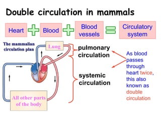
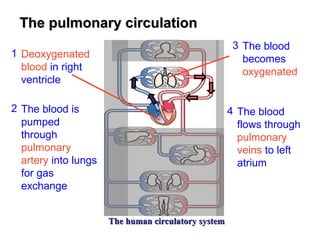
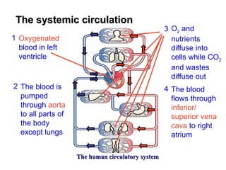
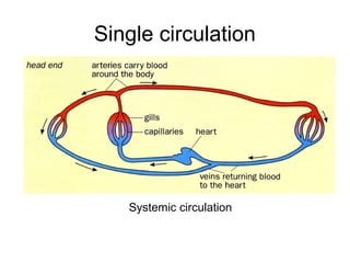
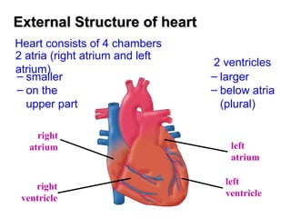
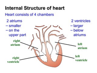
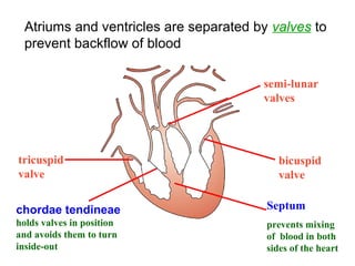
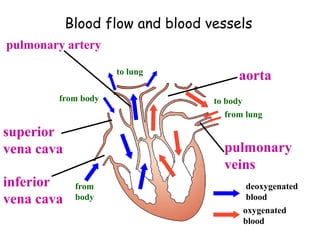
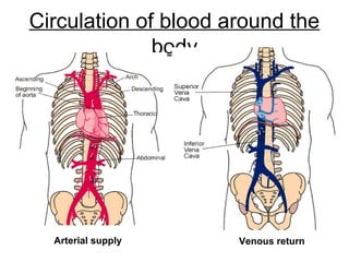
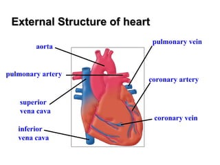
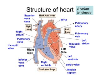
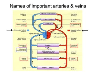
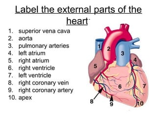
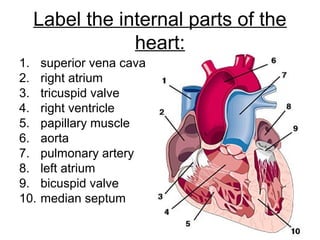
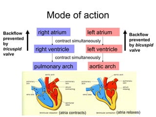
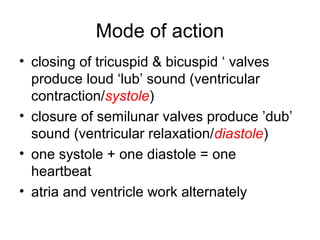
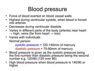
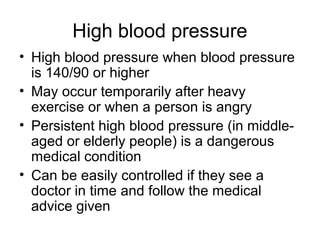
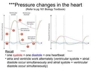
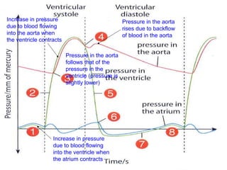
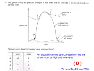
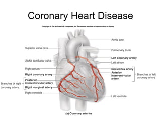
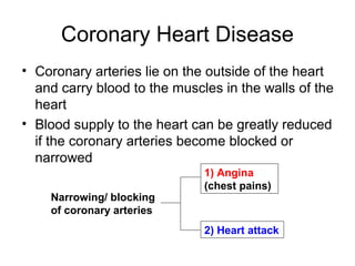
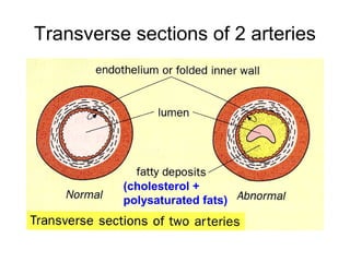
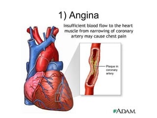
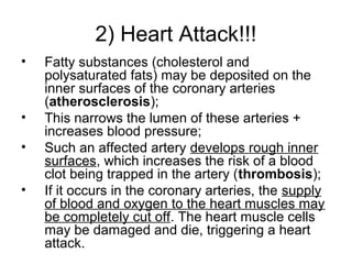
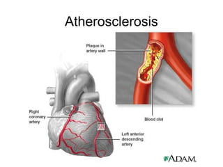
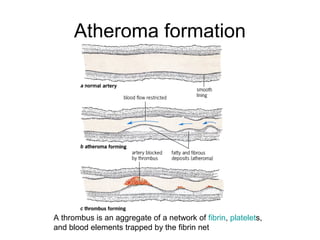
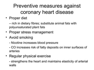
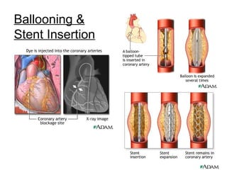
3) Heart
Lesson Objectives:
• Double & single circulation
• Structure of heart
• Mode of action of heart
Lung
All other parts
of the body
The mammalianThe mammalian
circulation plancirculation plan
Double circulation in mammals
Heart Blood
Blood
vessels
Circulatory
system
pulmonary
circulation
systemic
circulation
As blood
passes
through
heart twice,
this also
known as
double
circulation
The human circulatory systemThe human circulatory system
The pulmonary circulationThe pulmonary circulation
2 The blood is
pumped
through
pulmonary
artery into lungs
for gas
exchange
3 The blood
becomes
oxygenated
4 The blood
flows through
pulmonary
veins to left
atrium
1 Deoxygenated
blood in right
ventricle
The human circulatory systemThe human circulatory system
The systemic circulationThe systemic circulation
1 Oxygenated
blood in left
ventricle
2 The blood is
pumped
through aorta
to all parts of
the body
except lungs
3 O2 and
nutrients
diffuse into
cells while CO2
and wastes
diffuse out
4 The blood
flows through
inferior/
superior vena
cava to right
atrium
Single circulation
Systemic circulation
Heart
Click me!
External Structure of heartExternal Structure of heart
Heart consists of 4 chambers
2 atria (right atrium and left
atrium)
– smaller
– on the
upper part
2 ventricles
– larger
– below atria
(plural)
left
atrium
right
atrium
right
ventricle
left
ventricle
Internal Structure of heartInternal Structure of heart
Heart consists of 4 chambers
2 atriums
– smaller
– on the
upper part
2 ventricles
– larger
– below
atriums
left
atrium
right
atrium
right
ventricle
left
ventricle
Atriums and ventricles are separated by valves to
prevent backflow of blood
bicuspid
valve
Septum
prevents mixing
of blood in both
sides of the heart
chordae tendineae
holds valves in position
and avoids them to turn
inside-out
tricuspid
valve
semi-lunar
valves
Blood flow and blood vessels
aorta
pulmonary artery
superior
vena cava
inferior
vena cava
pulmonary
veins
from body
from
body
to lung
from lung
deoxygenated
blood
oxygenated
blood
to body
Circulation of blood around the
body
Arterial supply Venous return
External Structure of heartExternal Structure of heart
aorta
pulmonary artery
superior
vena cava
inferior
vena cava
pulmonary vein
coronary artery
coronary vein
Structure of heart
bicuspid
valve
tricuspid
valve
Inferior
vena
cava
Superior
vena
cava
Pulmonary
artery
Pulmonary
vein
Right
atrium
Right
ventricle
Left
atrium
Left
ventricle
aortic valve
Median
septum
aorta
chordae
tendineae
Pulmonary
valve
Activity:
Label the following diagram of the
heart in the worksheet provided
Names of important arteries & veins
quiz…
Label the external parts of the
heart:
1. superior vena cava
2. aorta
3. pulmonary arteries
4. left atrium
5. right atrium
6. right ventricle
7. left ventricle
8. right coronary vein
9. right coronary artery
10. apex
Label the internal parts of the
heart:
1. superior vena cava
2. right atrium
3. tricuspid valve
4. right ventricle
5. papillary muscle
6. aorta
7. pulmonary artery
8. left atrium
9. bicuspid valve
10. median septum
Mode of action of heart
Mode of action
right atrium left atrium
right ventricle
contract simultaneously
left ventricle
pulmonary arch
contract simultaneously
aortic arch
Backflow
prevented
by
tricuspid
valve
Backflow
prevented
by bicuspid
valve
(atria contracts) (atria relaxes)
Mode of action
• closing of tricuspid & bicuspid ‘ valves
produce loud ‘lub’ sound (ventricular
contraction/systole)
• closure of semilunar valves produce ’dub’
sound (ventricular relaxation/diastole)
• one systole + one diastole = one
heartbeat
• atria and ventricle work alternately
Blood pressure
Blood pressure
• Force of blood exerted on blood vessel walls
• Highest during ventricular systole, when blood is forced
into arteries
• Decreases during ventricular diastole
• Varies in different parts of the body (arteries near heart
→ high; veins (far from heart → low)
• Varies with individuals
Normal person:
systolic pressure = 120-140mm of mercury
diastolic pressure = 75-90mm of mercury
• Blood pressure is given as the systolic pressure being
the first number than diastolic pressure being the second
number e.g. 120/80 (120 over 80)
• High blood pressure when blood pressure is 140/90 or
higher
High blood pressure
• High blood pressure when blood pressure
is 140/90 or higher
• May occur temporarily after heavy
exercise or when a person is angry
• Persistent high blood pressure (in middle-
aged or elderly people) is a dangerous
medical condition
• Can be easily controlled if they see a
doctor in time and follow the medical
advice given
***Pressure changes in the heart
(Refer to pg 161 Biology Textbook)
Recall
• one systole + one diastole = one heartbeat
• atria and ventricle work alternately (ventricular systole + atrial
diastole occur simultaneously and atrial systole + ventricular
diastole occur simultaneously)
Increase in pressure
due to blood flowing
into the ventricle when
the atrium contracts
Increase in pressure
due to blood flowing
into the aorta when
the ventricle contracts
Pressure in the aorta
follows that of the
pressure in the
ventricle (pressure is
slightly lower)
Pressure in the aorta
rises due to backflow
of blood in the aorta
‘O’ Level Bio P1 Nov 2008
( D )
For bicuspid valve to open, pressure in the left
atrium must be high and vice versa
‘O’ Level Bio P1 Nov 2006
( B )
Atrio-ventricular valve = bicuspid or tricupid
valves
Semi-lunar valve = valve in aorta
Between W and X, ventricular pressure is
increasing hence atrio ventricular valves
would be closed and semi lunar valve
would open as blood is pushed through the
aorta
‘O’ Level Bio P1 Nov 2006
( C )
0.6 s = one heartbeat
60/0.6 = 100 heartbeats per min
Pulse
Pulse (produced after every ventricular contraction)
ventricular contraction
aortic arch
blood is pumped into
arteries
blood is pumped into
arteries dilate
increased pressure causes
walls of arteries recoil
Blood forced along in series of waves
[Imagine a fireman’s hose]
Experiment to demonstrate the
presence of valves in veins
Demonstrating the presence of
valves
Procedure:
• Bandage the upper arm (valves in veins appear as small
swellings) [see above]
• Place 2 fingers at point Y
• Using one finger push blood to point X
• Blood flowed back to from X to b and no further
Heart diseases
Coronary Heart Disease
• Coronary arteries lie on the outside of the heart
and carry blood to the muscles in the walls of the
heart
• Blood supply to the heart can be greatly reduced
if the coronary arteries become blocked or
narrowed
Narrowing/ blocking
of coronary arteries
1) Angina
(chest pains)
2) Heart attack
Transverse sections of 2 arteries
(cholesterol +
polysaturated fats)
1) Angina
2) Heart Attack!!!
• Fatty substances (cholesterol and
polysaturated fats) may be deposited on the
inner surfaces of the coronary arteries
(atherosclerosis);
• This narrows the lumen of these arteries +
increases blood pressure;
• Such an affected artery develops rough inner
surfaces, which increases the risk of a blood
clot being trapped in the artery (thrombosis);
• If it occurs in the coronary arteries, the supply
of blood and oxygen to the heart muscles may
be completely cut off. The heart muscle cells
may be damaged and die, triggering a heart
attack.
Atherosclerosis
Atheroma formation
A thrombus is an aggregate of a network of fibrin, platelets,
and blood elements trapped by the fibrin net
Factors that increase the risk of
atherosclerosis and coronary heart
disease
1. A diet rich in cholesterol and
saturated animal fats
2. Emotional stress
3. Smoking
Preventive measures against
coronary heart disease
• Proper diet
– rich in dietary fibres; substitute animal fats with
polyunsaturated plant fats
• Proper stress management
• Avoid smoking
- Nicotine increases blood pressure
- CO increases risk of fatty deposits on inner surfaces of
arteries
• Regular physical exercise
- strengthens the heart and maintains elasticity of arterial
walls
Ballooning &
Stent Insertion

Chapter 8 Transport in Humans Lesson 3 - Structure and function of the human heart

  • 1.
  • 2.
    Lesson Objectives: • Double& single circulation • Structure of heart • Mode of action of heart
  • 3.
    Lung All other parts ofthe body The mammalianThe mammalian circulation plancirculation plan Double circulation in mammals Heart Blood Blood vessels Circulatory system pulmonary circulation systemic circulation As blood passes through heart twice, this also known as double circulation
  • 4.
    The human circulatorysystemThe human circulatory system The pulmonary circulationThe pulmonary circulation 2 The blood is pumped through pulmonary artery into lungs for gas exchange 3 The blood becomes oxygenated 4 The blood flows through pulmonary veins to left atrium 1 Deoxygenated blood in right ventricle
  • 5.
    The human circulatorysystemThe human circulatory system The systemic circulationThe systemic circulation 1 Oxygenated blood in left ventricle 2 The blood is pumped through aorta to all parts of the body except lungs 3 O2 and nutrients diffuse into cells while CO2 and wastes diffuse out 4 The blood flows through inferior/ superior vena cava to right atrium
  • 6.
  • 7.
  • 8.
    External Structure ofheartExternal Structure of heart Heart consists of 4 chambers 2 atria (right atrium and left atrium) – smaller – on the upper part 2 ventricles – larger – below atria (plural) left atrium right atrium right ventricle left ventricle
  • 9.
    Internal Structure ofheartInternal Structure of heart Heart consists of 4 chambers 2 atriums – smaller – on the upper part 2 ventricles – larger – below atriums left atrium right atrium right ventricle left ventricle
  • 10.
    Atriums and ventriclesare separated by valves to prevent backflow of blood bicuspid valve Septum prevents mixing of blood in both sides of the heart chordae tendineae holds valves in position and avoids them to turn inside-out tricuspid valve semi-lunar valves
  • 11.
    Blood flow andblood vessels aorta pulmonary artery superior vena cava inferior vena cava pulmonary veins from body from body to lung from lung deoxygenated blood oxygenated blood to body
  • 12.
    Circulation of bloodaround the body Arterial supply Venous return
  • 13.
    External Structure ofheartExternal Structure of heart aorta pulmonary artery superior vena cava inferior vena cava pulmonary vein coronary artery coronary vein
  • 14.
  • 15.
    Activity: Label the followingdiagram of the heart in the worksheet provided
  • 17.
    Names of importantarteries & veins
  • 18.
  • 19.
    Label the externalparts of the heart: 1. superior vena cava 2. aorta 3. pulmonary arteries 4. left atrium 5. right atrium 6. right ventricle 7. left ventricle 8. right coronary vein 9. right coronary artery 10. apex
  • 20.
    Label the internalparts of the heart: 1. superior vena cava 2. right atrium 3. tricuspid valve 4. right ventricle 5. papillary muscle 6. aorta 7. pulmonary artery 8. left atrium 9. bicuspid valve 10. median septum
  • 21.
  • 22.
    Mode of action rightatrium left atrium right ventricle contract simultaneously left ventricle pulmonary arch contract simultaneously aortic arch Backflow prevented by tricuspid valve Backflow prevented by bicuspid valve (atria contracts) (atria relaxes)
  • 23.
    Mode of action •closing of tricuspid & bicuspid ‘ valves produce loud ‘lub’ sound (ventricular contraction/systole) • closure of semilunar valves produce ’dub’ sound (ventricular relaxation/diastole) • one systole + one diastole = one heartbeat • atria and ventricle work alternately
  • 24.
  • 25.
    Blood pressure • Forceof blood exerted on blood vessel walls • Highest during ventricular systole, when blood is forced into arteries • Decreases during ventricular diastole • Varies in different parts of the body (arteries near heart → high; veins (far from heart → low) • Varies with individuals Normal person: systolic pressure = 120-140mm of mercury diastolic pressure = 75-90mm of mercury • Blood pressure is given as the systolic pressure being the first number than diastolic pressure being the second number e.g. 120/80 (120 over 80) • High blood pressure when blood pressure is 140/90 or higher
  • 26.
    High blood pressure •High blood pressure when blood pressure is 140/90 or higher • May occur temporarily after heavy exercise or when a person is angry • Persistent high blood pressure (in middle- aged or elderly people) is a dangerous medical condition • Can be easily controlled if they see a doctor in time and follow the medical advice given
  • 27.
    ***Pressure changes inthe heart (Refer to pg 161 Biology Textbook) Recall • one systole + one diastole = one heartbeat • atria and ventricle work alternately (ventricular systole + atrial diastole occur simultaneously and atrial systole + ventricular diastole occur simultaneously)
  • 28.
    Increase in pressure dueto blood flowing into the ventricle when the atrium contracts Increase in pressure due to blood flowing into the aorta when the ventricle contracts Pressure in the aorta follows that of the pressure in the ventricle (pressure is slightly lower) Pressure in the aorta rises due to backflow of blood in the aorta
  • 29.
    ‘O’ Level BioP1 Nov 2008 ( D ) For bicuspid valve to open, pressure in the left atrium must be high and vice versa
  • 30.
    ‘O’ Level BioP1 Nov 2006 ( B ) Atrio-ventricular valve = bicuspid or tricupid valves Semi-lunar valve = valve in aorta Between W and X, ventricular pressure is increasing hence atrio ventricular valves would be closed and semi lunar valve would open as blood is pushed through the aorta
  • 31.
    ‘O’ Level BioP1 Nov 2006 ( C ) 0.6 s = one heartbeat 60/0.6 = 100 heartbeats per min
  • 32.
  • 33.
    Pulse (produced afterevery ventricular contraction) ventricular contraction aortic arch blood is pumped into arteries blood is pumped into arteries dilate increased pressure causes walls of arteries recoil Blood forced along in series of waves [Imagine a fireman’s hose]
  • 34.
    Experiment to demonstratethe presence of valves in veins
  • 35.
    Demonstrating the presenceof valves Procedure: • Bandage the upper arm (valves in veins appear as small swellings) [see above] • Place 2 fingers at point Y • Using one finger push blood to point X • Blood flowed back to from X to b and no further
  • 36.
  • 37.
  • 38.
    Coronary Heart Disease •Coronary arteries lie on the outside of the heart and carry blood to the muscles in the walls of the heart • Blood supply to the heart can be greatly reduced if the coronary arteries become blocked or narrowed Narrowing/ blocking of coronary arteries 1) Angina (chest pains) 2) Heart attack
  • 39.
    Transverse sections of2 arteries (cholesterol + polysaturated fats)
  • 40.
  • 41.
    2) Heart Attack!!! •Fatty substances (cholesterol and polysaturated fats) may be deposited on the inner surfaces of the coronary arteries (atherosclerosis); • This narrows the lumen of these arteries + increases blood pressure; • Such an affected artery develops rough inner surfaces, which increases the risk of a blood clot being trapped in the artery (thrombosis); • If it occurs in the coronary arteries, the supply of blood and oxygen to the heart muscles may be completely cut off. The heart muscle cells may be damaged and die, triggering a heart attack.
  • 42.
  • 43.
    Atheroma formation A thrombusis an aggregate of a network of fibrin, platelets, and blood elements trapped by the fibrin net
  • 44.
    Factors that increasethe risk of atherosclerosis and coronary heart disease 1. A diet rich in cholesterol and saturated animal fats 2. Emotional stress 3. Smoking
  • 45.
    Preventive measures against coronaryheart disease • Proper diet – rich in dietary fibres; substitute animal fats with polyunsaturated plant fats • Proper stress management • Avoid smoking - Nicotine increases blood pressure - CO increases risk of fatty deposits on inner surfaces of arteries • Regular physical exercise - strengthens the heart and maintains elasticity of arterial walls
  • 47.