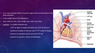
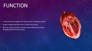
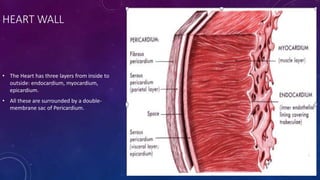
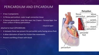
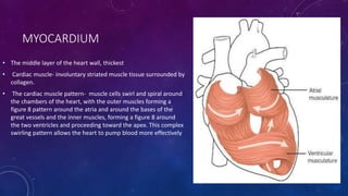
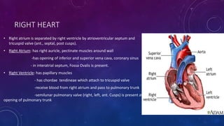
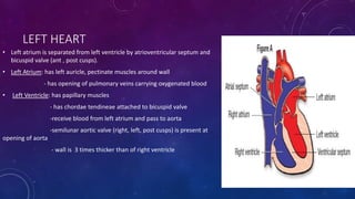
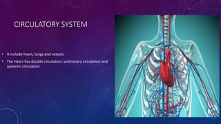
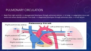
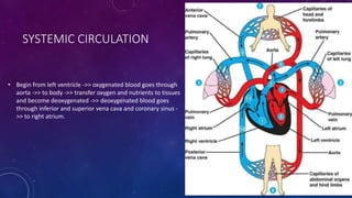
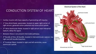
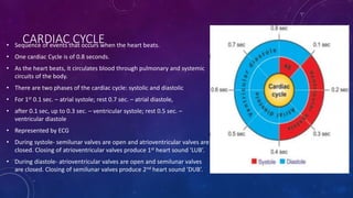
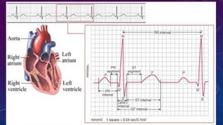
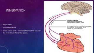
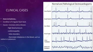
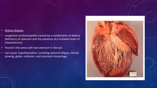
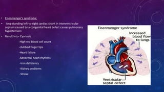
SlideShare a Scribd company logo
THE HEART
THE HEART
• It is a cone-shaped hollow muscular organ which pumps blood to the
body and back.
• It has weight about 250-300 grams.
• Size is that of a fist: 12cm length, 8cm wide, 6cm thick
• Location: -in middle mediastinum.
-1/3rd to right of midsternal plane and 2/3rd to left of it.
-posterior to body of sternum and 2nd-6th costal cartilages.
-anterior to vertebral column at level of T5-T8
-superior to superior surface of diaphragm.
• Base -quadrilateral directed upward, backward and posteriorly
-made of –left atrium
--parts of right atrium
• Apex –directed forward, downward and to left
-made of –parts of left ventricle
• 4 Surfaces: sternocostal/anterior surface- consist of right ventricle, some right atrium, some left ventricle.
diaphragmatic/inferior surface- consist of left ventricle, some right ventricle.
left pulmonary surface- consist of left atrium and left ventricle.
right pulmonary surface- consist of right atrium.
• 4 Margins: right margin
left margin
inferior/ acute margin- between anterior and diaphragmatic surfaces
obtuse margin- between anterior and left pulmonary surfaces
• 4 Grooves: coronary sulci- divide atria from ventricles
interatrial groove- divide left and right atria
interventricular grooves - anterior
- posterior
FUNCTION
• Pumps blood throughout the body via the circulatory system.
• Supply oxygenated blood with nutrients to tissues.
• Remove carbon dioxide and other wastes therefore receive
deoxygenated blood from body.
HEART WALL
• The Heart has three layers from inside to
outside: endocardium, myocardium,
epicardium.
• All these are surrounded by a double-
membrane sac of Pericardium.
PERICARDIUM AND EPICARDIUM
• It has 2 components:
• 1) Fibrous pericardium- outer tough connective tissue
• 2) Serous pericardium- inner thin layer, has 2 layers – Parietal layer- line
inner surface of fibrous pericardium
--Visceral layer-
attached to heart {EPICARDIUM}
• In between these two present the pericardial cavity having serous fluid.
• It allow lubrication of heart for friction-free movements.
• Prevent overfilling of heart with blood.
MYOCARDIUM
• The middle layer of the heart wall, thickest
• Cardiac muscle- involuntary striated muscle tissue surrounded by
collagen.
• The cardiac muscle pattern- muscle cells swirl and spiral around
the chambers of the heart, with the outer muscles forming a
figure 8 pattern around the atria and around the bases of the
great vessels and the inner muscles, forming a figure 8 around
the two ventricles and proceeding toward the apex. This complex
swirling pattern allows the heart to pump blood more effectively
ENDOCARDIUM
• The innermost layer.
• Lining of simple squamous epithelium.
• Covers heart chambers and valves and is continuous with the
endothelium of the veins and arteries of the heart.
• Secrete Endothelin- regulate contraction of the myocardium.
• Conduction system is present here.
CHAMBERS
• Heart has 4 chambers divided by the grooves, septum and valves.
• Right Heart: right atrium and right ventricle
• Left Heart: left atrium and left ventricle
• There are interatrial septum, interventricular septum, and
atrioventricular septum.
• There are bicuspid (mitral) valve, tricuspid (bazian) valve, aortic valve and
pulmonary valve.
RIGHT HEART
• Right atrium is separated by right ventricle by atrioventricular septum and
tricuspid valve (ant., septal, post cusps).
• Right Atrium: has right auricle, pectinate muscles around wall
-has opening of inferior and superior vena cava, coronary sinus
- in interatrial septum, Fossa Ovalis is present.
• Right Ventricle: has papillary muscles
- has chordae tendineae which attach to tricuspid valve
-receive blood from right atrium and pass to pulmonary trunk
-semilunar pulmonary valve (right, left, ant. Cusps) is present at
opening of pulmonary trunk
LEFT HEART
• Left atrium is separated from left ventricle by atrioventricular septum and
bicuspid valve (ant , post cusps).
• Left Atrium: has left auricle, pectinate muscles around wall
- has opening of pulmonary veins carrying oxygenated blood
• Left Ventricle: has papillary muscles
- has chordae tendineae attached to bicuspid valve
-receive blood from left atrium and pass to aorta
-semilunar aortic valve (right, left, post cusps) is present at
opening of aorta
- wall is 3 times thicker than of right ventricle
CIRCULATORY SYSTEM
• It include heart, lungs and vessels.
• The Heart has double circulation: pulmonary circulation and
systemic circulation
PULMONARY CIRCULATION
• Begin from right ventricle ->> deoxygenated blood goes through pulmonary arteries ->> to lungs ->> oxygenation occur and
waste and carbon dioxide exhaled from body ->> oxygenated blood goes through pulmonary veins ->> to left atrium.
SYSTEMIC CIRCULATION
• Begin from left ventricle ->> oxygenated blood goes through
aorta ->> to body ->> transfer oxygen and nutrients to tissues
and become deoxygenated ->> deoxygenated blood goes
through inferior and superior vena cava and coronary sinus -
>> to right atrium.
CONDUCTION SYSTEM OF HEART
• Cardiac muscle cells have capacity of generating self-impulse.
• 1st Sino-Atrial Node: pacemaker, located at upper right corner of
right atrium, generate action potential and cause atrial systole.
• 2nd Atrio-Ventricular Node: located in right atrium near interatrial
septum, delay the signal.
• Between these 2 are present internodal pathways.
• 3rd Bundle of Hiss: AV bundle, located in interventricular septum, has
right and left bundle branches.
• 4th Purkinje Fibers: located in right and left ventricles, cause
ventricular systole.
CARDIAC CYCLE• Sequence of events that occurs when the heart beats.
• One cardiac Cycle is of 0.8 seconds.
• As the heart beats, it circulates blood through pulmonary and systemic
circuits of the body.
• There are two phases of the cardiac cycle: systolic and diastolic
• For 1st 0.1 sec. – atrial systole; rest 0.7 sec. – atrial diastole,
• after 0.1 sec, up to 0.3 sec. – ventricular systole; rest 0.5 sec. –
ventricular diastole
• Represented by ECG
• During systole- semilunar valves are open and atrioventricular valves are
closed. Closing of atrioventricular valves produce 1st heart sound ‘LUB’.
• During diastole- atrioventricular valves are open and semilunar valves
are closed. Closing of semilunar valves produce 2nd heart sound ‘DUB’.
VASCULATURE OF HEART
• Arteries:
• 1) Right coronary artery- supply right atrium and
posterior surface of both ventricles.
• Branches- marginal artery
- posterior interventricular artery
• 2) Left coronary artery- supply left atrium and ventricle.
• Branches- circumflex artery
- anterior interventricular artery
• Veins:
• Coronary sinus- receive blood from – great cardiac vein
- anterior cardiac vein
- middle cardiac vein
- small cardiac vein
INNERVATION
• Vagus nerve
• Sympathetic trunk
• These nerves form a network of nerves that lies over
the heart called the cardiac plexus.
CLINICAL CASES
• Heart Arrhythmia :
• Condition of irregular heart beat.
• Causes- Coronary artery disease.
-High blood pressure.
-cardiomyopathy
-Valve disorders.
-Electrolyte imbalances in the blood, such as
sodium or potassium.
• Cardiogenic Shock:
• condition in which heart suddenly can't pump enough blood to meet body's needs.
• Causes- damage to heart's right ventricle, which sends blood to lungs to receive oxygen.
- myocardial infarction
-pulmonary embolism
-pericardial tamponade
-sudden valvular regurgitation
-inability of heart muscle to work properly
-ventricular tachycardia.
• Keshan disease:
• congestive cardiomyopathy caused by a combination of dietary
deficiency of selenium and the presence of a mutated strain of
Coxsackievirus.
• Found in the areas with low selenium in the soil.
• Can cause- hypothyroidism, including extreme fatigue, mental
slowing, goiter, cretinism, and recurrent miscarriage
• Eisenmenger's syndrome:
• long-standing left-to-right cardiac shunt in interventricular
septum caused by a congenital heart defect causes pulmonary
hypertension
• Result into- Cyanosis
-High red blood cell count
-clubbed finger tips
-Heart failure
-Abnormal heart rhythms
-Iron deficiency
-Kidney problems
-Stroke
THANK YOU
-AKSHMALA SHARMA

More Related Content

What's hot

Heart anatomy
Heart anatomyHeart anatomy
Heart anatomy
Asha damodar
 
Heart Anatomy & Basic Function
Heart Anatomy & Basic FunctionHeart Anatomy & Basic Function
Heart Anatomy & Basic Function
Vivek Raman
 
Structure of human heart
Structure of human heartStructure of human heart
Structure of human heart
Suprabha Panda
 
Heart
HeartHeart
The Human Heart
The Human HeartThe Human Heart
The Human Heart
Marona Ysabel Julabar
 
Anatomy and physiology of heart
Anatomy and physiology of heartAnatomy and physiology of heart
Anatomy and physiology of heart
Amrutha nayaka
 
structure of heart
structure of heartstructure of heart
structure of heart
saranyanaturalscience
 
Anatomy of Cardiac System
Anatomy of Cardiac SystemAnatomy of Cardiac System
Anatomy of Cardiac System
DR .PALLAVI PATHANIA
 
Anatomy and physiology of the heart
Anatomy and physiology of the heartAnatomy and physiology of the heart
Anatomy and physiology of the heart
CharlesSsekawu
 
Location & structure of the heart
Location & structure  of the heartLocation & structure  of the heart
Location & structure of the heart
Vyramuthu Atputhan
 
3. internal structure of the heart
3. internal structure of the heart3. internal structure of the heart
3. internal structure of the heart
Dr. Mohammad Mahmoud
 
Anatomy of the heart
Anatomy of the heartAnatomy of the heart
Anatomy of the heart
Zeyad Magdy
 
Heart
HeartHeart
Embrology and Anatomy of Cardiovascular System
Embrology and Anatomy of Cardiovascular SystemEmbrology and Anatomy of Cardiovascular System
Embrology and Anatomy of Cardiovascular System
The Medical Post
 
Anatomy of heart
Anatomy of heartAnatomy of heart
Copy of cardio vascular system
Copy of cardio vascular systemCopy of cardio vascular system
Copy of cardio vascular system
mo7ammed9ale7
 
Anatomy of the Cardiovascular system
Anatomy of the Cardiovascular systemAnatomy of the Cardiovascular system
Anatomy of the Cardiovascular system
romainsperera
 
Presentation on anatomy of heart ,coronary circulation ,mi and ecg interpreta...
Presentation on anatomy of heart ,coronary circulation ,mi and ecg interpreta...Presentation on anatomy of heart ,coronary circulation ,mi and ecg interpreta...
Presentation on anatomy of heart ,coronary circulation ,mi and ecg interpreta...
febin rony
 
Cardiovascular system ppt
Cardiovascular system pptCardiovascular system ppt

What's hot (19)

Heart anatomy
Heart anatomyHeart anatomy
Heart anatomy
 
Heart Anatomy & Basic Function
Heart Anatomy & Basic FunctionHeart Anatomy & Basic Function
Heart Anatomy & Basic Function
 
Structure of human heart
Structure of human heartStructure of human heart
Structure of human heart
 
Heart
HeartHeart
Heart
 
The Human Heart
The Human HeartThe Human Heart
The Human Heart
 
Anatomy and physiology of heart
Anatomy and physiology of heartAnatomy and physiology of heart
Anatomy and physiology of heart
 
structure of heart
structure of heartstructure of heart
structure of heart
 
Anatomy of Cardiac System
Anatomy of Cardiac SystemAnatomy of Cardiac System
Anatomy of Cardiac System
 
Anatomy and physiology of the heart
Anatomy and physiology of the heartAnatomy and physiology of the heart
Anatomy and physiology of the heart
 
Location & structure of the heart
Location & structure  of the heartLocation & structure  of the heart
Location & structure of the heart
 
3. internal structure of the heart
3. internal structure of the heart3. internal structure of the heart
3. internal structure of the heart
 
Anatomy of the heart
Anatomy of the heartAnatomy of the heart
Anatomy of the heart
 
Heart
HeartHeart
Heart
 
Embrology and Anatomy of Cardiovascular System
Embrology and Anatomy of Cardiovascular SystemEmbrology and Anatomy of Cardiovascular System
Embrology and Anatomy of Cardiovascular System
 
Anatomy of heart
Anatomy of heartAnatomy of heart
Anatomy of heart
 
Copy of cardio vascular system
Copy of cardio vascular systemCopy of cardio vascular system
Copy of cardio vascular system
 
Anatomy of the Cardiovascular system
Anatomy of the Cardiovascular systemAnatomy of the Cardiovascular system
Anatomy of the Cardiovascular system
 
Presentation on anatomy of heart ,coronary circulation ,mi and ecg interpreta...
Presentation on anatomy of heart ,coronary circulation ,mi and ecg interpreta...Presentation on anatomy of heart ,coronary circulation ,mi and ecg interpreta...
Presentation on anatomy of heart ,coronary circulation ,mi and ecg interpreta...
 
Cardiovascular system ppt
Cardiovascular system pptCardiovascular system ppt
Cardiovascular system ppt
 

Similar to The heart

Cardiovascular ststem
Cardiovascular ststemCardiovascular ststem
Cardiovascular ststem
suchismita sethi
 
Anatomy of the heart lecture.pptx
Anatomy of the heart lecture.pptxAnatomy of the heart lecture.pptx
Anatomy of the heart lecture.pptx
SayaliPatil790915
 
Anatomy of Human Heart along with internal and external features.
Anatomy of Human Heart along with internal and external features.Anatomy of Human Heart along with internal and external features.
Anatomy of Human Heart along with internal and external features.
ssuser7bf0a2
 
The heart
The heartThe heart
CARDIOVASCULAR SYSTEM - ANATOMY & PHYSIOLOGY
CARDIOVASCULAR SYSTEM - ANATOMY & PHYSIOLOGYCARDIOVASCULAR SYSTEM - ANATOMY & PHYSIOLOGY
CARDIOVASCULAR SYSTEM - ANATOMY & PHYSIOLOGY
Kameshwaran Sugavanam
 
2 Cardio Vascular System.pptx
2 Cardio Vascular System.pptx2 Cardio Vascular System.pptx
2 Cardio Vascular System.pptx
RamarGurusamy2
 
UNIT 4-WPS Office.pptx
UNIT 4-WPS Office.pptxUNIT 4-WPS Office.pptx
UNIT 4-WPS Office.pptx
DNYANESHWARBIRADAR1
 
Human heart
Human heartHuman heart
Human heart
Hussein Al-tameemi
 
A&P -The Cardiovascular System
A&P -The Cardiovascular SystemA&P -The Cardiovascular System
A&P -The Cardiovascular System
Jeffrey Giles RN BSN MBA
 
CARDIO VASCULAR SYSTEM THE HEART
CARDIO VASCULAR SYSTEM THE HEARTCARDIO VASCULAR SYSTEM THE HEART
CARDIO VASCULAR SYSTEM THE HEART
PARUL UNIVERSITY
 
Final heart
Final heart Final heart
Final heart
Sohan Patel
 
7. cardiovascular system
7. cardiovascular system7. cardiovascular system
7. cardiovascular system
Yogeshwary Bhongade
 
CVS ANATOMY- The Heart.pptx
CVS ANATOMY- The Heart.pptxCVS ANATOMY- The Heart.pptx
CVS ANATOMY- The Heart.pptx
Shaly Jose
 
Anatomy of Heart- Internal structures pt
Anatomy of Heart- Internal structures ptAnatomy of Heart- Internal structures pt
Anatomy of Heart- Internal structures pt
Saili Gaude
 
Cardiovascular physiology for university students
Cardiovascular physiology for university studentsCardiovascular physiology for university students
Cardiovascular physiology for university students
ItsOnyii
 
The Circulatory System.pptx
The Circulatory System.pptxThe Circulatory System.pptx
The Circulatory System.pptx
NehaPandey199
 
Human cardiovascular system
Human cardiovascular systemHuman cardiovascular system
Human cardiovascular system
Sigfrid Mustapha
 
The Heart
The HeartThe Heart
The Heart
Bruce Coulter
 
anatomy and physiology of heart..ppt....
anatomy and physiology of heart..ppt....anatomy and physiology of heart..ppt....
anatomy and physiology of heart..ppt....
DeepikaLingam2
 
Cardiovascular system
Cardiovascular systemCardiovascular system
Cardiovascular system
mariaidrees3
 

Similar to The heart (20)

Cardiovascular ststem
Cardiovascular ststemCardiovascular ststem
Cardiovascular ststem
 
Anatomy of the heart lecture.pptx
Anatomy of the heart lecture.pptxAnatomy of the heart lecture.pptx
Anatomy of the heart lecture.pptx
 
Anatomy of Human Heart along with internal and external features.
Anatomy of Human Heart along with internal and external features.Anatomy of Human Heart along with internal and external features.
Anatomy of Human Heart along with internal and external features.
 
The heart
The heartThe heart
The heart
 
CARDIOVASCULAR SYSTEM - ANATOMY & PHYSIOLOGY
CARDIOVASCULAR SYSTEM - ANATOMY & PHYSIOLOGYCARDIOVASCULAR SYSTEM - ANATOMY & PHYSIOLOGY
CARDIOVASCULAR SYSTEM - ANATOMY & PHYSIOLOGY
 
2 Cardio Vascular System.pptx
2 Cardio Vascular System.pptx2 Cardio Vascular System.pptx
2 Cardio Vascular System.pptx
 
UNIT 4-WPS Office.pptx
UNIT 4-WPS Office.pptxUNIT 4-WPS Office.pptx
UNIT 4-WPS Office.pptx
 
Human heart
Human heartHuman heart
Human heart
 
A&P -The Cardiovascular System
A&P -The Cardiovascular SystemA&P -The Cardiovascular System
A&P -The Cardiovascular System
 
CARDIO VASCULAR SYSTEM THE HEART
CARDIO VASCULAR SYSTEM THE HEARTCARDIO VASCULAR SYSTEM THE HEART
CARDIO VASCULAR SYSTEM THE HEART
 
Final heart
Final heart Final heart
Final heart
 
7. cardiovascular system
7. cardiovascular system7. cardiovascular system
7. cardiovascular system
 
CVS ANATOMY- The Heart.pptx
CVS ANATOMY- The Heart.pptxCVS ANATOMY- The Heart.pptx
CVS ANATOMY- The Heart.pptx
 
Anatomy of Heart- Internal structures pt
Anatomy of Heart- Internal structures ptAnatomy of Heart- Internal structures pt
Anatomy of Heart- Internal structures pt
 
Cardiovascular physiology for university students
Cardiovascular physiology for university studentsCardiovascular physiology for university students
Cardiovascular physiology for university students
 
The Circulatory System.pptx
The Circulatory System.pptxThe Circulatory System.pptx
The Circulatory System.pptx
 
Human cardiovascular system
Human cardiovascular systemHuman cardiovascular system
Human cardiovascular system
 
The Heart
The HeartThe Heart
The Heart
 
anatomy and physiology of heart..ppt....
anatomy and physiology of heart..ppt....anatomy and physiology of heart..ppt....
anatomy and physiology of heart..ppt....
 
Cardiovascular system
Cardiovascular systemCardiovascular system
Cardiovascular system
 

More from Akshmala Sharma

PCOS (polycystic ovarian syndrome)
PCOS (polycystic ovarian syndrome)PCOS (polycystic ovarian syndrome)
PCOS (polycystic ovarian syndrome)
Akshmala Sharma
 
Hiv
HivHiv
Heart failure...
Heart failure...Heart failure...
Heart failure...
Akshmala Sharma
 
Orthomyxoviruses...
Orthomyxoviruses...Orthomyxoviruses...
Orthomyxoviruses...
Akshmala Sharma
 
General adaptation syndrome
General adaptation syndromeGeneral adaptation syndrome
General adaptation syndrome
Akshmala Sharma
 
Filariasis
FilariasisFilariasis
Filariasis
Akshmala Sharma
 
Anaphylaxis
AnaphylaxisAnaphylaxis
Anaphylaxis
Akshmala Sharma
 
Staging of cancer
Staging of cancerStaging of cancer
Staging of cancer
Akshmala Sharma
 

More from Akshmala Sharma (8)

PCOS (polycystic ovarian syndrome)
PCOS (polycystic ovarian syndrome)PCOS (polycystic ovarian syndrome)
PCOS (polycystic ovarian syndrome)
 
Hiv
HivHiv
Hiv
 
Heart failure...
Heart failure...Heart failure...
Heart failure...
 
Orthomyxoviruses...
Orthomyxoviruses...Orthomyxoviruses...
Orthomyxoviruses...
 
General adaptation syndrome
General adaptation syndromeGeneral adaptation syndrome
General adaptation syndrome
 
Filariasis
FilariasisFilariasis
Filariasis
 
Anaphylaxis
AnaphylaxisAnaphylaxis
Anaphylaxis
 
Staging of cancer
Staging of cancerStaging of cancer
Staging of cancer
 

Recently uploaded

3D Hybrid PIC simulation of the plasma expansion (ISSS-14)
3D Hybrid PIC simulation of the plasma expansion (ISSS-14)3D Hybrid PIC simulation of the plasma expansion (ISSS-14)
3D Hybrid PIC simulation of the plasma expansion (ISSS-14)
David Osipyan
 
Thornton ESPP slides UK WW Network 4_6_24.pdf
Thornton ESPP slides UK WW Network 4_6_24.pdfThornton ESPP slides UK WW Network 4_6_24.pdf
Thornton ESPP slides UK WW Network 4_6_24.pdf
European Sustainable Phosphorus Platform
 
Bob Reedy - Nitrate in Texas Groundwater.pdf
Bob Reedy - Nitrate in Texas Groundwater.pdfBob Reedy - Nitrate in Texas Groundwater.pdf
Bob Reedy - Nitrate in Texas Groundwater.pdf
Texas Alliance of Groundwater Districts
 
Topic: SICKLE CELL DISEASE IN CHILDREN-3.pdf
Topic: SICKLE CELL DISEASE IN CHILDREN-3.pdfTopic: SICKLE CELL DISEASE IN CHILDREN-3.pdf
Topic: SICKLE CELL DISEASE IN CHILDREN-3.pdf
TinyAnderson
 
NuGOweek 2024 Ghent programme overview flyer
NuGOweek 2024 Ghent programme overview flyerNuGOweek 2024 Ghent programme overview flyer
NuGOweek 2024 Ghent programme overview flyer
pablovgd
 
ESR spectroscopy in liquid food and beverages.pptx
ESR spectroscopy in liquid food and beverages.pptxESR spectroscopy in liquid food and beverages.pptx
ESR spectroscopy in liquid food and beverages.pptx
PRIYANKA PATEL
 
原版制作(carleton毕业证书)卡尔顿大学毕业证硕士文凭原版一模一样
原版制作(carleton毕业证书)卡尔顿大学毕业证硕士文凭原版一模一样原版制作(carleton毕业证书)卡尔顿大学毕业证硕士文凭原版一模一样
原版制作(carleton毕业证书)卡尔顿大学毕业证硕士文凭原版一模一样
yqqaatn0
 
SAR of Medicinal Chemistry 1st by dk.pdf
SAR of Medicinal Chemistry 1st by dk.pdfSAR of Medicinal Chemistry 1st by dk.pdf
SAR of Medicinal Chemistry 1st by dk.pdf
KrushnaDarade1
 
The use of Nauplii and metanauplii artemia in aquaculture (brine shrimp).pptx
The use of Nauplii and metanauplii artemia in aquaculture (brine shrimp).pptxThe use of Nauplii and metanauplii artemia in aquaculture (brine shrimp).pptx
The use of Nauplii and metanauplii artemia in aquaculture (brine shrimp).pptx
MAGOTI ERNEST
 
Nucleophilic Addition of carbonyl compounds.pptx
Nucleophilic Addition of carbonyl  compounds.pptxNucleophilic Addition of carbonyl  compounds.pptx
Nucleophilic Addition of carbonyl compounds.pptx
SSR02
 
bordetella pertussis.................................ppt
bordetella pertussis.................................pptbordetella pertussis.................................ppt
bordetella pertussis.................................ppt
kejapriya1
 
Equivariant neural networks and representation theory
Equivariant neural networks and representation theoryEquivariant neural networks and representation theory
Equivariant neural networks and representation theory
Daniel Tubbenhauer
 
ANAMOLOUS SECONDARY GROWTH IN DICOT ROOTS.pptx
ANAMOLOUS SECONDARY GROWTH IN DICOT ROOTS.pptxANAMOLOUS SECONDARY GROWTH IN DICOT ROOTS.pptx
ANAMOLOUS SECONDARY GROWTH IN DICOT ROOTS.pptx
RASHMI M G
 
Cytokines and their role in immune regulation.pptx
Cytokines and their role in immune regulation.pptxCytokines and their role in immune regulation.pptx
Cytokines and their role in immune regulation.pptx
Hitesh Sikarwar
 
Oedema_types_causes_pathophysiology.pptx
Oedema_types_causes_pathophysiology.pptxOedema_types_causes_pathophysiology.pptx
Oedema_types_causes_pathophysiology.pptx
muralinath2
 
Micronuclei test.M.sc.zoology.fisheries.
Micronuclei test.M.sc.zoology.fisheries.Micronuclei test.M.sc.zoology.fisheries.
Micronuclei test.M.sc.zoology.fisheries.
Aditi Bajpai
 
Medical Orthopedic PowerPoint Templates.pptx
Medical Orthopedic PowerPoint Templates.pptxMedical Orthopedic PowerPoint Templates.pptx
Medical Orthopedic PowerPoint Templates.pptx
terusbelajar5
 
The binding of cosmological structures by massless topological defects
The binding of cosmological structures by massless topological defectsThe binding of cosmological structures by massless topological defects
The binding of cosmological structures by massless topological defects
Sérgio Sacani
 
Deep Software Variability and Frictionless Reproducibility
Deep Software Variability and Frictionless ReproducibilityDeep Software Variability and Frictionless Reproducibility
Deep Software Variability and Frictionless Reproducibility
University of Rennes, INSA Rennes, Inria/IRISA, CNRS
 
Remote Sensing and Computational, Evolutionary, Supercomputing, and Intellige...
Remote Sensing and Computational, Evolutionary, Supercomputing, and Intellige...Remote Sensing and Computational, Evolutionary, Supercomputing, and Intellige...
Remote Sensing and Computational, Evolutionary, Supercomputing, and Intellige...
University of Maribor
 

Recently uploaded (20)

3D Hybrid PIC simulation of the plasma expansion (ISSS-14)
3D Hybrid PIC simulation of the plasma expansion (ISSS-14)3D Hybrid PIC simulation of the plasma expansion (ISSS-14)
3D Hybrid PIC simulation of the plasma expansion (ISSS-14)
 
Thornton ESPP slides UK WW Network 4_6_24.pdf
Thornton ESPP slides UK WW Network 4_6_24.pdfThornton ESPP slides UK WW Network 4_6_24.pdf
Thornton ESPP slides UK WW Network 4_6_24.pdf
 
Bob Reedy - Nitrate in Texas Groundwater.pdf
Bob Reedy - Nitrate in Texas Groundwater.pdfBob Reedy - Nitrate in Texas Groundwater.pdf
Bob Reedy - Nitrate in Texas Groundwater.pdf
 
Topic: SICKLE CELL DISEASE IN CHILDREN-3.pdf
Topic: SICKLE CELL DISEASE IN CHILDREN-3.pdfTopic: SICKLE CELL DISEASE IN CHILDREN-3.pdf
Topic: SICKLE CELL DISEASE IN CHILDREN-3.pdf
 
NuGOweek 2024 Ghent programme overview flyer
NuGOweek 2024 Ghent programme overview flyerNuGOweek 2024 Ghent programme overview flyer
NuGOweek 2024 Ghent programme overview flyer
 
ESR spectroscopy in liquid food and beverages.pptx
ESR spectroscopy in liquid food and beverages.pptxESR spectroscopy in liquid food and beverages.pptx
ESR spectroscopy in liquid food and beverages.pptx
 
原版制作(carleton毕业证书)卡尔顿大学毕业证硕士文凭原版一模一样
原版制作(carleton毕业证书)卡尔顿大学毕业证硕士文凭原版一模一样原版制作(carleton毕业证书)卡尔顿大学毕业证硕士文凭原版一模一样
原版制作(carleton毕业证书)卡尔顿大学毕业证硕士文凭原版一模一样
 
SAR of Medicinal Chemistry 1st by dk.pdf
SAR of Medicinal Chemistry 1st by dk.pdfSAR of Medicinal Chemistry 1st by dk.pdf
SAR of Medicinal Chemistry 1st by dk.pdf
 
The use of Nauplii and metanauplii artemia in aquaculture (brine shrimp).pptx
The use of Nauplii and metanauplii artemia in aquaculture (brine shrimp).pptxThe use of Nauplii and metanauplii artemia in aquaculture (brine shrimp).pptx
The use of Nauplii and metanauplii artemia in aquaculture (brine shrimp).pptx
 
Nucleophilic Addition of carbonyl compounds.pptx
Nucleophilic Addition of carbonyl  compounds.pptxNucleophilic Addition of carbonyl  compounds.pptx
Nucleophilic Addition of carbonyl compounds.pptx
 
bordetella pertussis.................................ppt
bordetella pertussis.................................pptbordetella pertussis.................................ppt
bordetella pertussis.................................ppt
 
Equivariant neural networks and representation theory
Equivariant neural networks and representation theoryEquivariant neural networks and representation theory
Equivariant neural networks and representation theory
 
ANAMOLOUS SECONDARY GROWTH IN DICOT ROOTS.pptx
ANAMOLOUS SECONDARY GROWTH IN DICOT ROOTS.pptxANAMOLOUS SECONDARY GROWTH IN DICOT ROOTS.pptx
ANAMOLOUS SECONDARY GROWTH IN DICOT ROOTS.pptx
 
Cytokines and their role in immune regulation.pptx
Cytokines and their role in immune regulation.pptxCytokines and their role in immune regulation.pptx
Cytokines and their role in immune regulation.pptx
 
Oedema_types_causes_pathophysiology.pptx
Oedema_types_causes_pathophysiology.pptxOedema_types_causes_pathophysiology.pptx
Oedema_types_causes_pathophysiology.pptx
 
Micronuclei test.M.sc.zoology.fisheries.
Micronuclei test.M.sc.zoology.fisheries.Micronuclei test.M.sc.zoology.fisheries.
Micronuclei test.M.sc.zoology.fisheries.
 
Medical Orthopedic PowerPoint Templates.pptx
Medical Orthopedic PowerPoint Templates.pptxMedical Orthopedic PowerPoint Templates.pptx
Medical Orthopedic PowerPoint Templates.pptx
 
The binding of cosmological structures by massless topological defects
The binding of cosmological structures by massless topological defectsThe binding of cosmological structures by massless topological defects
The binding of cosmological structures by massless topological defects
 
Deep Software Variability and Frictionless Reproducibility
Deep Software Variability and Frictionless ReproducibilityDeep Software Variability and Frictionless Reproducibility
Deep Software Variability and Frictionless Reproducibility
 
Remote Sensing and Computational, Evolutionary, Supercomputing, and Intellige...
Remote Sensing and Computational, Evolutionary, Supercomputing, and Intellige...Remote Sensing and Computational, Evolutionary, Supercomputing, and Intellige...
Remote Sensing and Computational, Evolutionary, Supercomputing, and Intellige...
 

The heart

  • 2. • It is a cone-shaped hollow muscular organ which pumps blood to the body and back. • It has weight about 250-300 grams. • Size is that of a fist: 12cm length, 8cm wide, 6cm thick • Location: -in middle mediastinum. -1/3rd to right of midsternal plane and 2/3rd to left of it. -posterior to body of sternum and 2nd-6th costal cartilages. -anterior to vertebral column at level of T5-T8 -superior to superior surface of diaphragm.
  • 3. • Base -quadrilateral directed upward, backward and posteriorly -made of –left atrium --parts of right atrium • Apex –directed forward, downward and to left -made of –parts of left ventricle
  • 4. • 4 Surfaces: sternocostal/anterior surface- consist of right ventricle, some right atrium, some left ventricle. diaphragmatic/inferior surface- consist of left ventricle, some right ventricle. left pulmonary surface- consist of left atrium and left ventricle. right pulmonary surface- consist of right atrium. • 4 Margins: right margin left margin inferior/ acute margin- between anterior and diaphragmatic surfaces obtuse margin- between anterior and left pulmonary surfaces • 4 Grooves: coronary sulci- divide atria from ventricles interatrial groove- divide left and right atria interventricular grooves - anterior - posterior
  • 5. FUNCTION • Pumps blood throughout the body via the circulatory system. • Supply oxygenated blood with nutrients to tissues. • Remove carbon dioxide and other wastes therefore receive deoxygenated blood from body.
  • 6. HEART WALL • The Heart has three layers from inside to outside: endocardium, myocardium, epicardium. • All these are surrounded by a double- membrane sac of Pericardium.
  • 7. PERICARDIUM AND EPICARDIUM • It has 2 components: • 1) Fibrous pericardium- outer tough connective tissue • 2) Serous pericardium- inner thin layer, has 2 layers – Parietal layer- line inner surface of fibrous pericardium --Visceral layer- attached to heart {EPICARDIUM} • In between these two present the pericardial cavity having serous fluid. • It allow lubrication of heart for friction-free movements. • Prevent overfilling of heart with blood.
  • 8. MYOCARDIUM • The middle layer of the heart wall, thickest • Cardiac muscle- involuntary striated muscle tissue surrounded by collagen. • The cardiac muscle pattern- muscle cells swirl and spiral around the chambers of the heart, with the outer muscles forming a figure 8 pattern around the atria and around the bases of the great vessels and the inner muscles, forming a figure 8 around the two ventricles and proceeding toward the apex. This complex swirling pattern allows the heart to pump blood more effectively
  • 9. ENDOCARDIUM • The innermost layer. • Lining of simple squamous epithelium. • Covers heart chambers and valves and is continuous with the endothelium of the veins and arteries of the heart. • Secrete Endothelin- regulate contraction of the myocardium. • Conduction system is present here.
  • 10. CHAMBERS • Heart has 4 chambers divided by the grooves, septum and valves. • Right Heart: right atrium and right ventricle • Left Heart: left atrium and left ventricle • There are interatrial septum, interventricular septum, and atrioventricular septum. • There are bicuspid (mitral) valve, tricuspid (bazian) valve, aortic valve and pulmonary valve.
  • 11. RIGHT HEART • Right atrium is separated by right ventricle by atrioventricular septum and tricuspid valve (ant., septal, post cusps). • Right Atrium: has right auricle, pectinate muscles around wall -has opening of inferior and superior vena cava, coronary sinus - in interatrial septum, Fossa Ovalis is present. • Right Ventricle: has papillary muscles - has chordae tendineae which attach to tricuspid valve -receive blood from right atrium and pass to pulmonary trunk -semilunar pulmonary valve (right, left, ant. Cusps) is present at opening of pulmonary trunk
  • 12. LEFT HEART • Left atrium is separated from left ventricle by atrioventricular septum and bicuspid valve (ant , post cusps). • Left Atrium: has left auricle, pectinate muscles around wall - has opening of pulmonary veins carrying oxygenated blood • Left Ventricle: has papillary muscles - has chordae tendineae attached to bicuspid valve -receive blood from left atrium and pass to aorta -semilunar aortic valve (right, left, post cusps) is present at opening of aorta - wall is 3 times thicker than of right ventricle
  • 13.
  • 14. CIRCULATORY SYSTEM • It include heart, lungs and vessels. • The Heart has double circulation: pulmonary circulation and systemic circulation
  • 15. PULMONARY CIRCULATION • Begin from right ventricle ->> deoxygenated blood goes through pulmonary arteries ->> to lungs ->> oxygenation occur and waste and carbon dioxide exhaled from body ->> oxygenated blood goes through pulmonary veins ->> to left atrium.
  • 16. SYSTEMIC CIRCULATION • Begin from left ventricle ->> oxygenated blood goes through aorta ->> to body ->> transfer oxygen and nutrients to tissues and become deoxygenated ->> deoxygenated blood goes through inferior and superior vena cava and coronary sinus - >> to right atrium.
  • 17. CONDUCTION SYSTEM OF HEART • Cardiac muscle cells have capacity of generating self-impulse. • 1st Sino-Atrial Node: pacemaker, located at upper right corner of right atrium, generate action potential and cause atrial systole. • 2nd Atrio-Ventricular Node: located in right atrium near interatrial septum, delay the signal. • Between these 2 are present internodal pathways. • 3rd Bundle of Hiss: AV bundle, located in interventricular septum, has right and left bundle branches. • 4th Purkinje Fibers: located in right and left ventricles, cause ventricular systole.
  • 18. CARDIAC CYCLE• Sequence of events that occurs when the heart beats. • One cardiac Cycle is of 0.8 seconds. • As the heart beats, it circulates blood through pulmonary and systemic circuits of the body. • There are two phases of the cardiac cycle: systolic and diastolic • For 1st 0.1 sec. – atrial systole; rest 0.7 sec. – atrial diastole, • after 0.1 sec, up to 0.3 sec. – ventricular systole; rest 0.5 sec. – ventricular diastole • Represented by ECG • During systole- semilunar valves are open and atrioventricular valves are closed. Closing of atrioventricular valves produce 1st heart sound ‘LUB’. • During diastole- atrioventricular valves are open and semilunar valves are closed. Closing of semilunar valves produce 2nd heart sound ‘DUB’.
  • 19.
  • 20. VASCULATURE OF HEART • Arteries: • 1) Right coronary artery- supply right atrium and posterior surface of both ventricles. • Branches- marginal artery - posterior interventricular artery • 2) Left coronary artery- supply left atrium and ventricle. • Branches- circumflex artery - anterior interventricular artery
  • 21. • Veins: • Coronary sinus- receive blood from – great cardiac vein - anterior cardiac vein - middle cardiac vein - small cardiac vein
  • 22. INNERVATION • Vagus nerve • Sympathetic trunk • These nerves form a network of nerves that lies over the heart called the cardiac plexus.
  • 23. CLINICAL CASES • Heart Arrhythmia : • Condition of irregular heart beat. • Causes- Coronary artery disease. -High blood pressure. -cardiomyopathy -Valve disorders. -Electrolyte imbalances in the blood, such as sodium or potassium.
  • 24. • Cardiogenic Shock: • condition in which heart suddenly can't pump enough blood to meet body's needs. • Causes- damage to heart's right ventricle, which sends blood to lungs to receive oxygen. - myocardial infarction -pulmonary embolism -pericardial tamponade -sudden valvular regurgitation -inability of heart muscle to work properly -ventricular tachycardia.
  • 25. • Keshan disease: • congestive cardiomyopathy caused by a combination of dietary deficiency of selenium and the presence of a mutated strain of Coxsackievirus. • Found in the areas with low selenium in the soil. • Can cause- hypothyroidism, including extreme fatigue, mental slowing, goiter, cretinism, and recurrent miscarriage
  • 26. • Eisenmenger's syndrome: • long-standing left-to-right cardiac shunt in interventricular septum caused by a congenital heart defect causes pulmonary hypertension • Result into- Cyanosis -High red blood cell count -clubbed finger tips -Heart failure -Abnormal heart rhythms -Iron deficiency -Kidney problems -Stroke