Downloaded 797 times
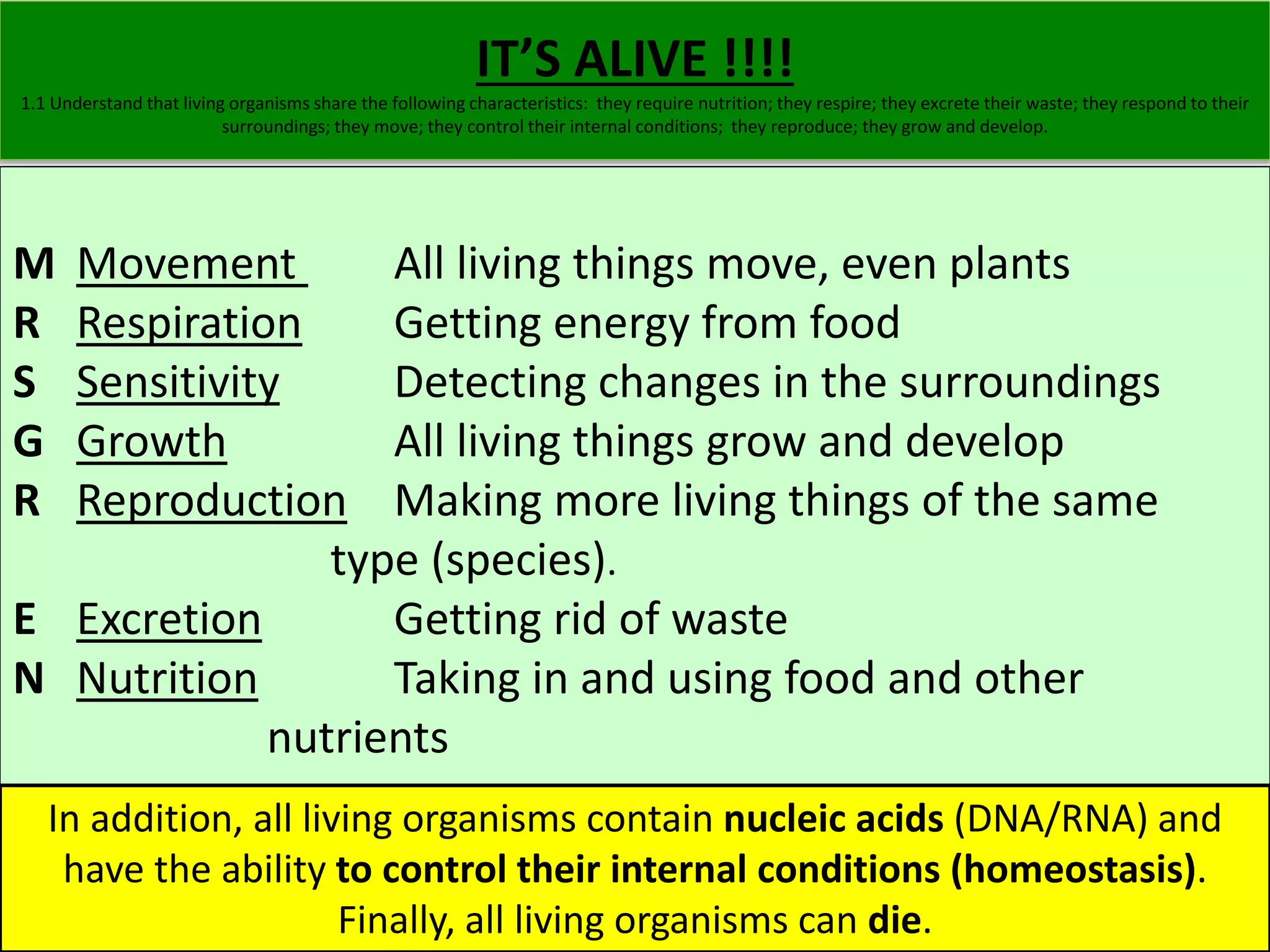
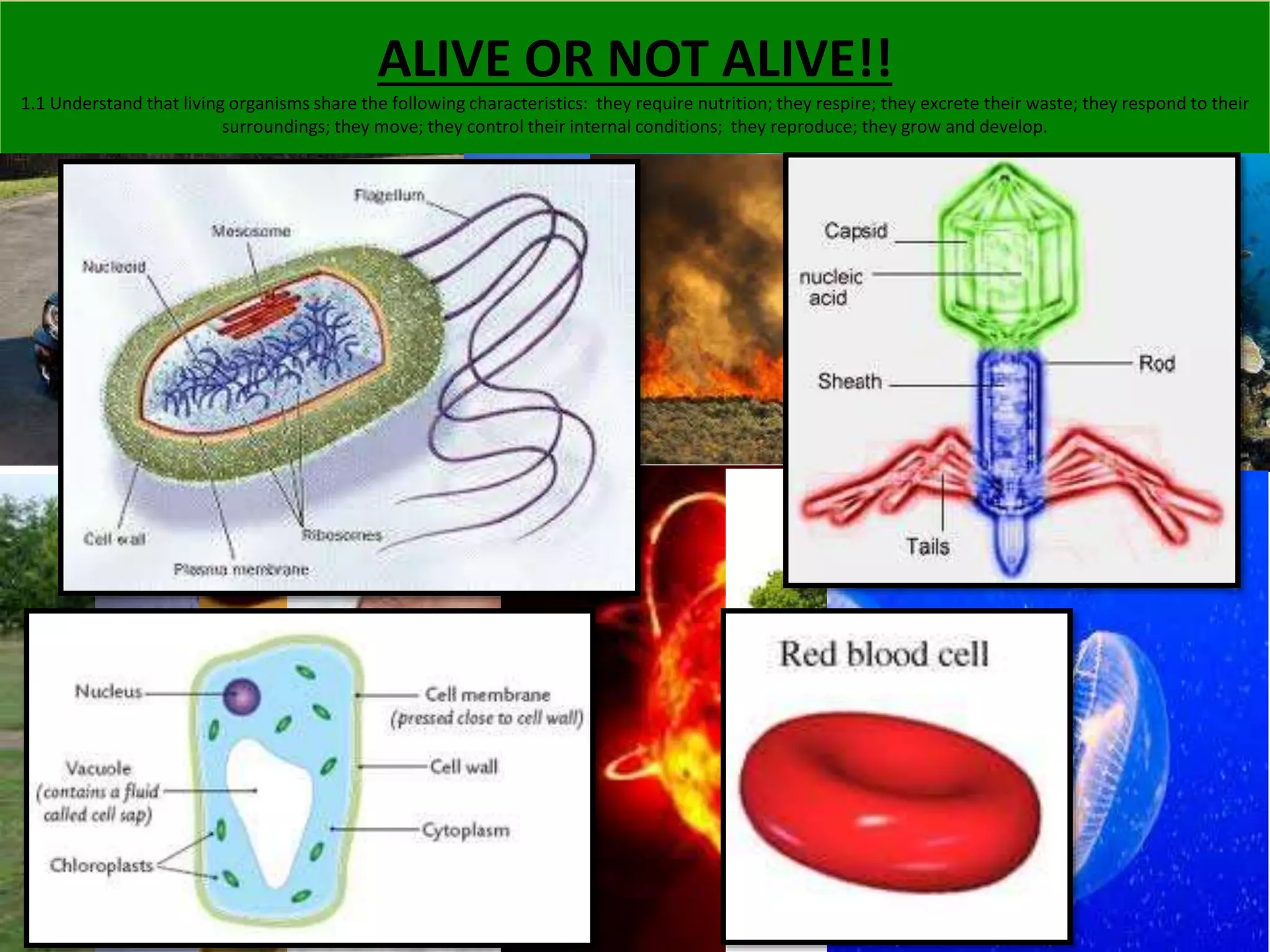
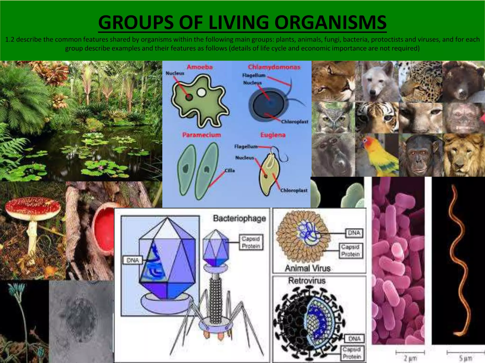
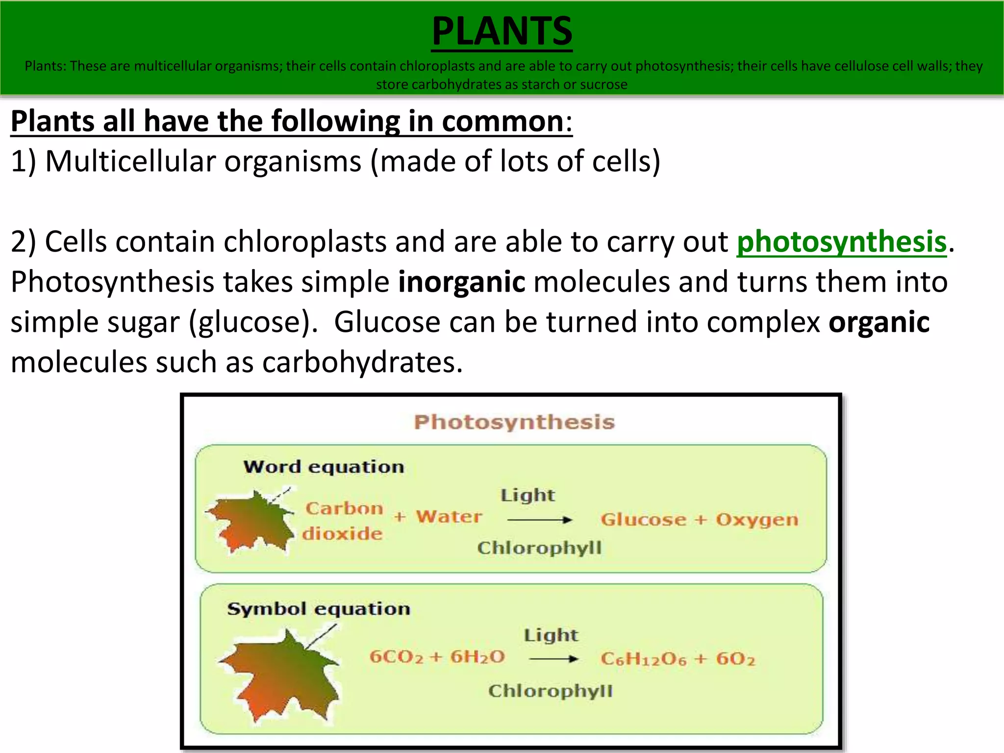


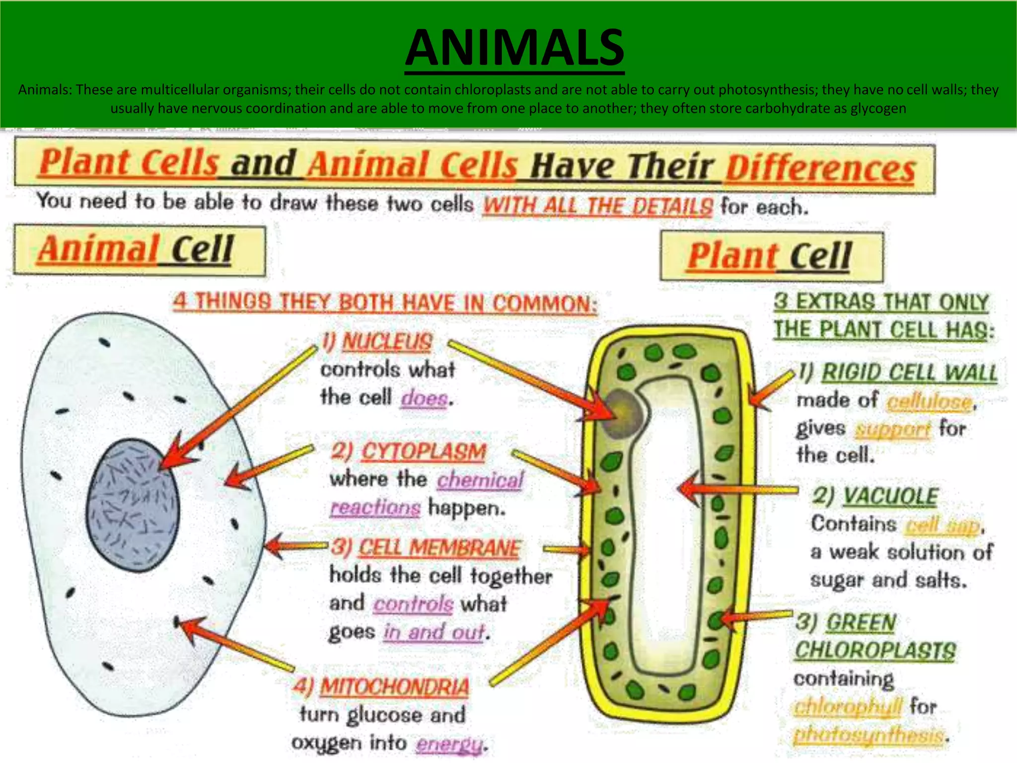


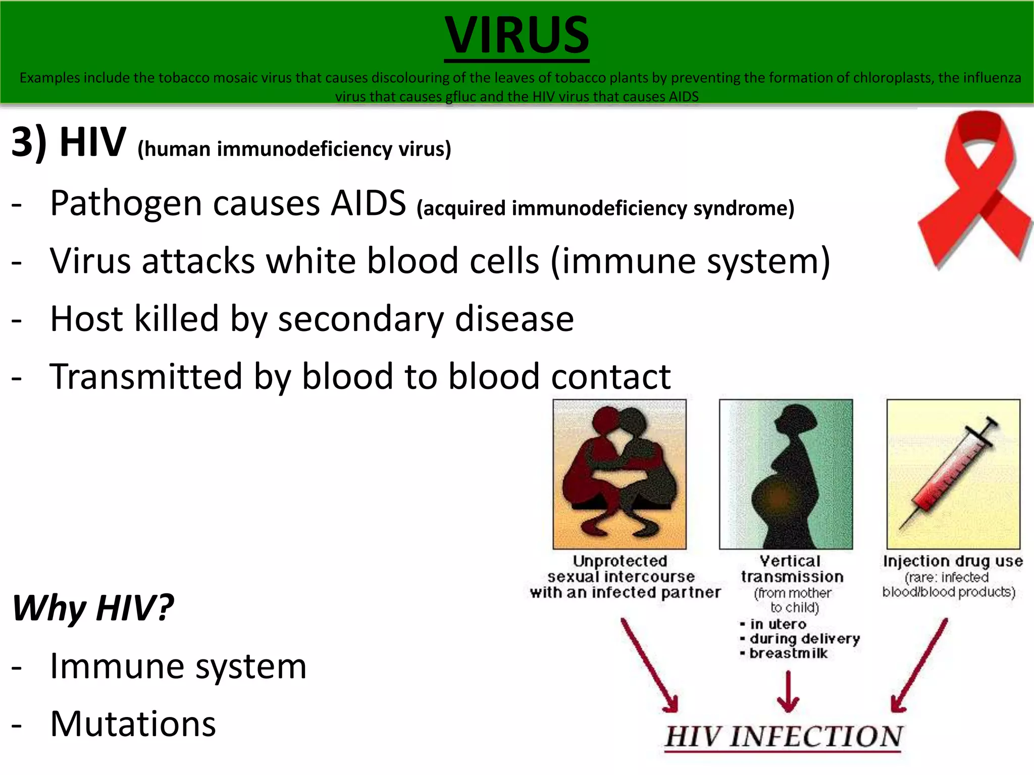
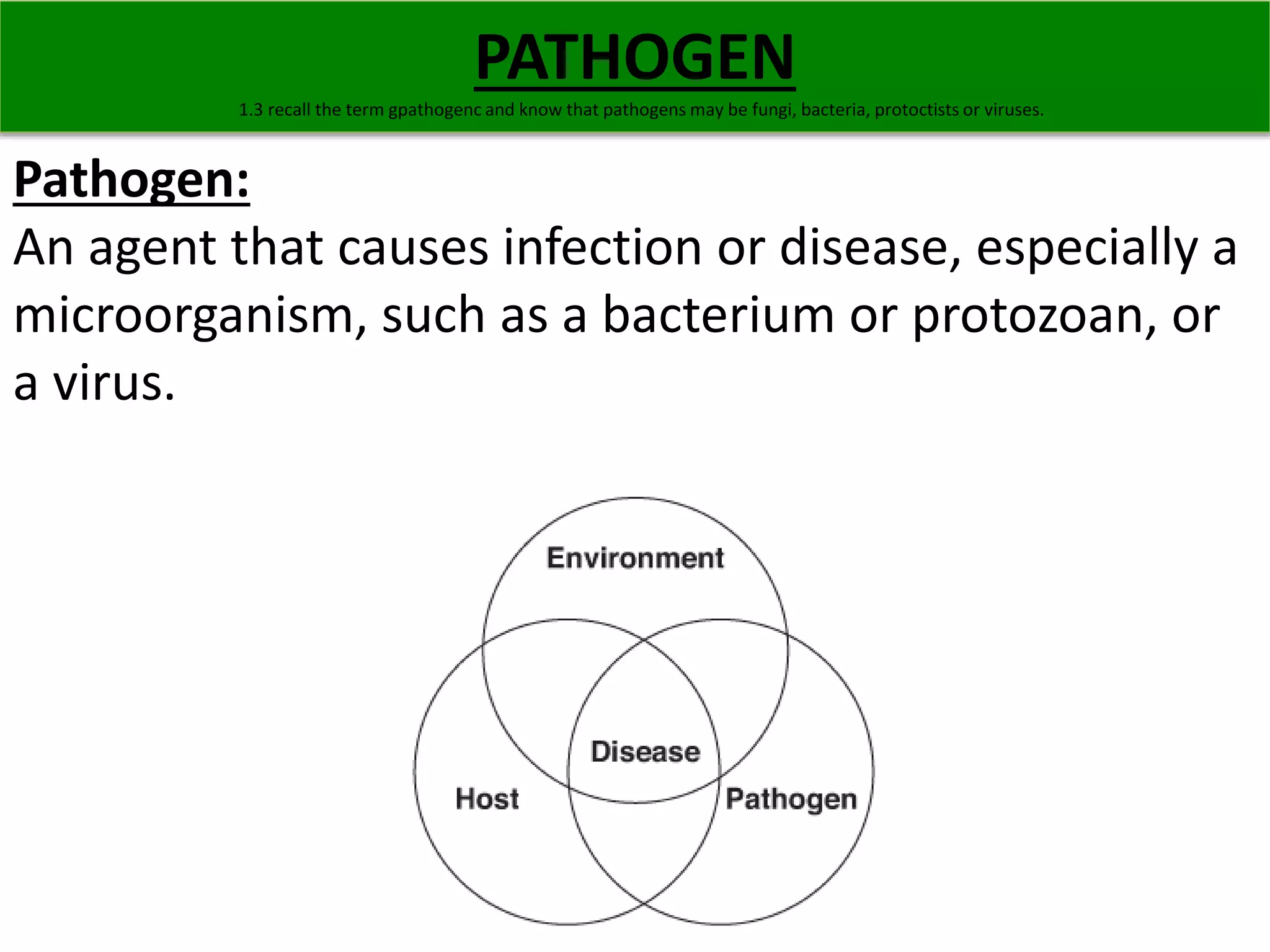
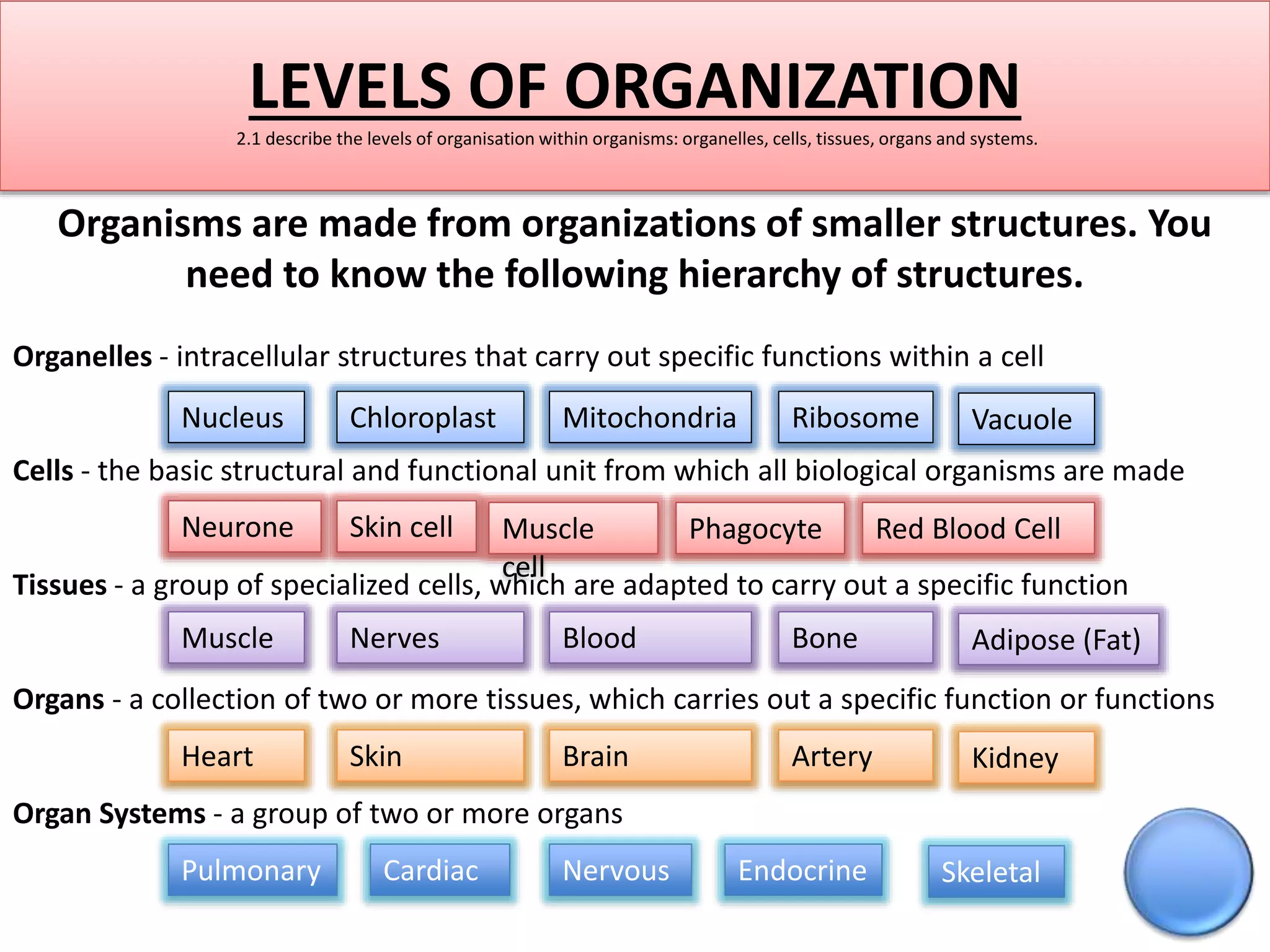
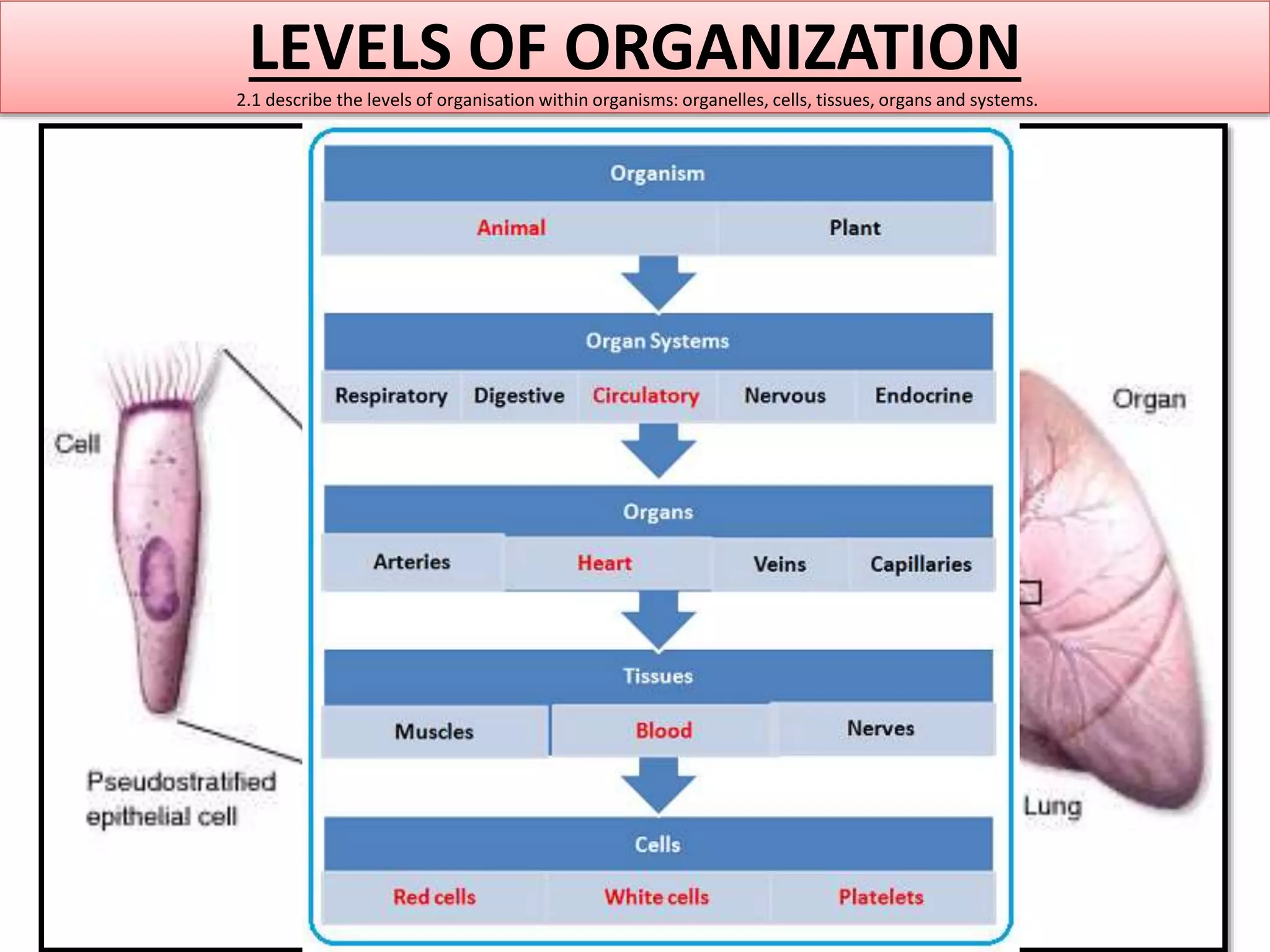
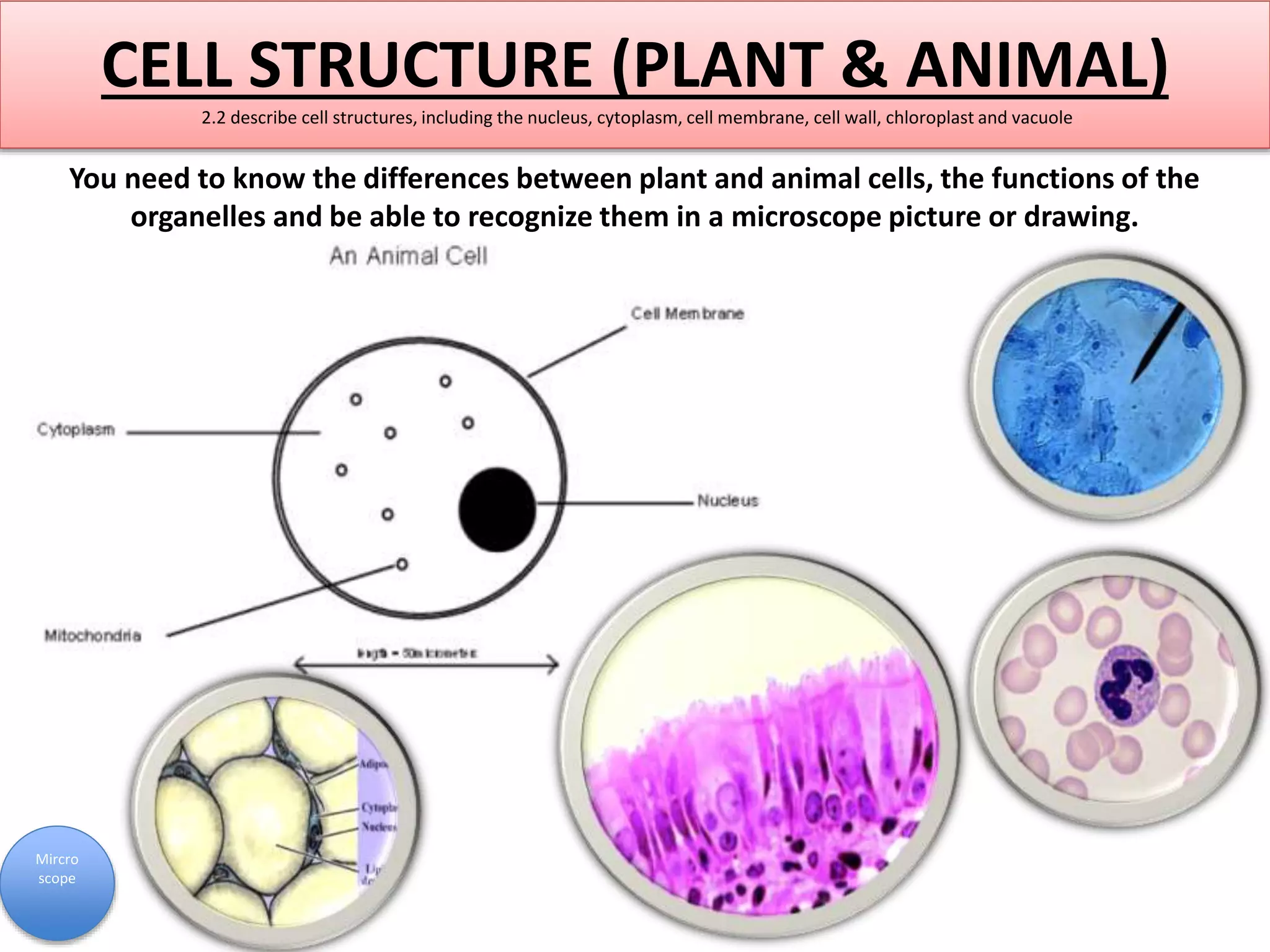
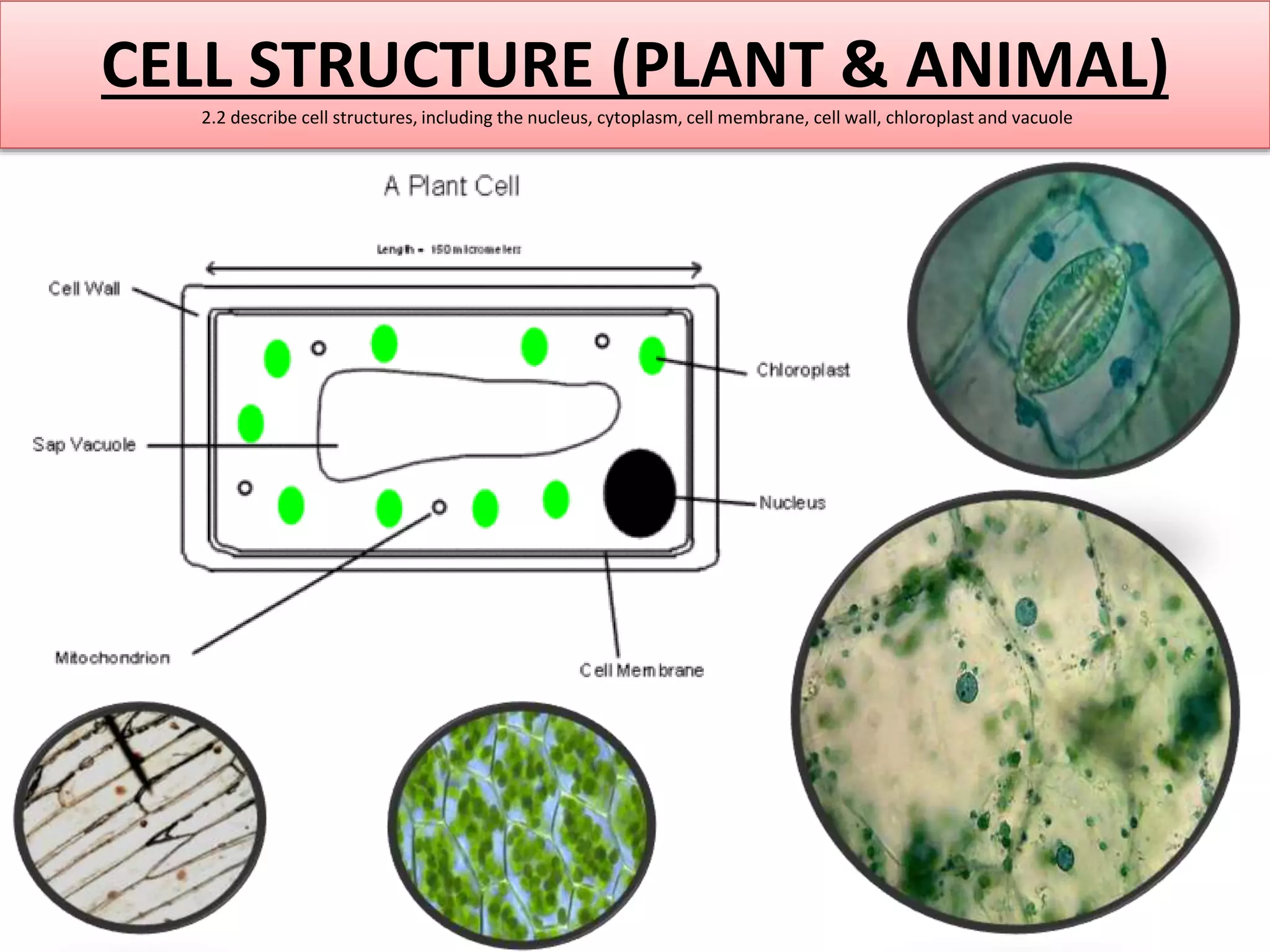
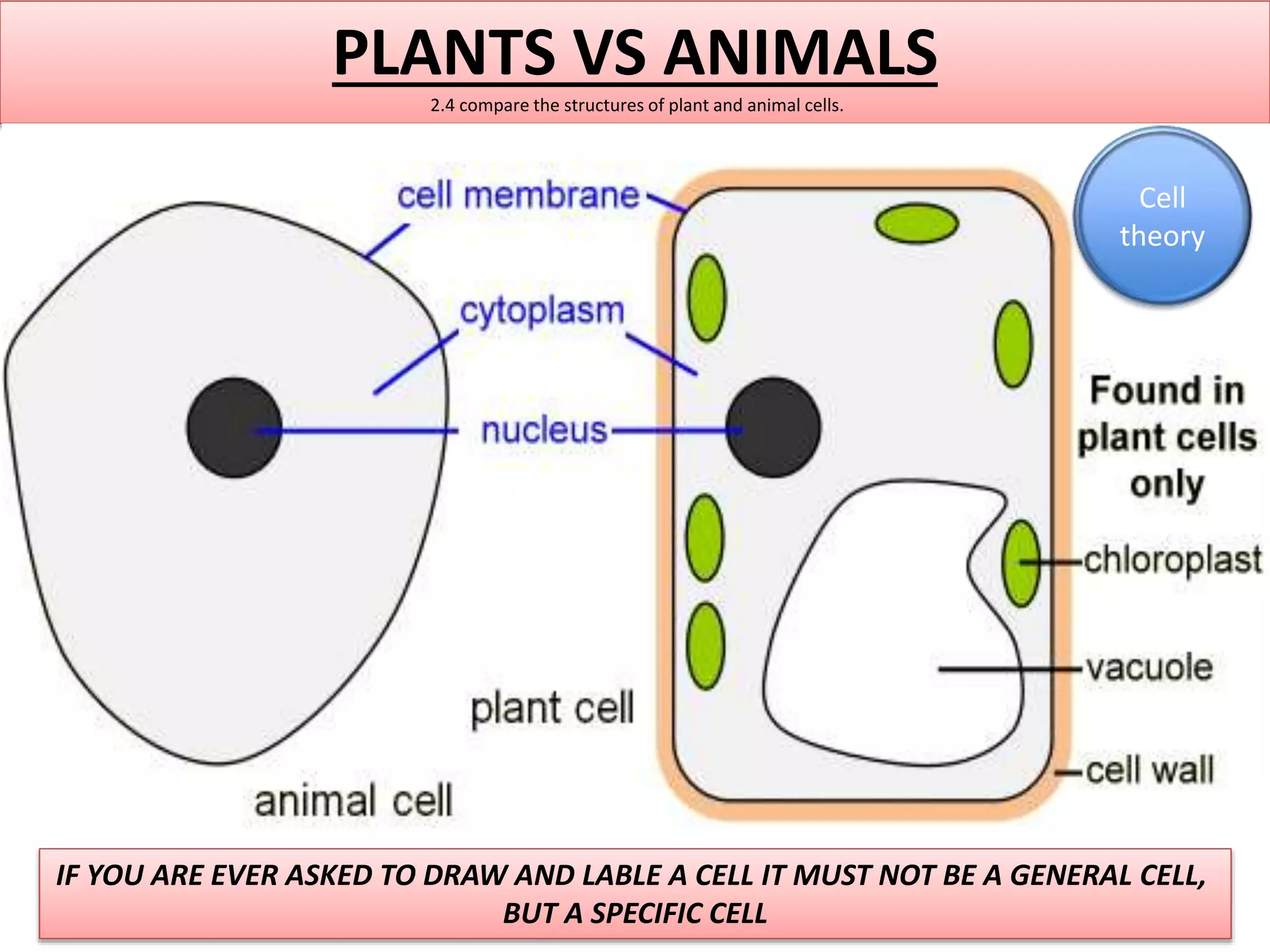
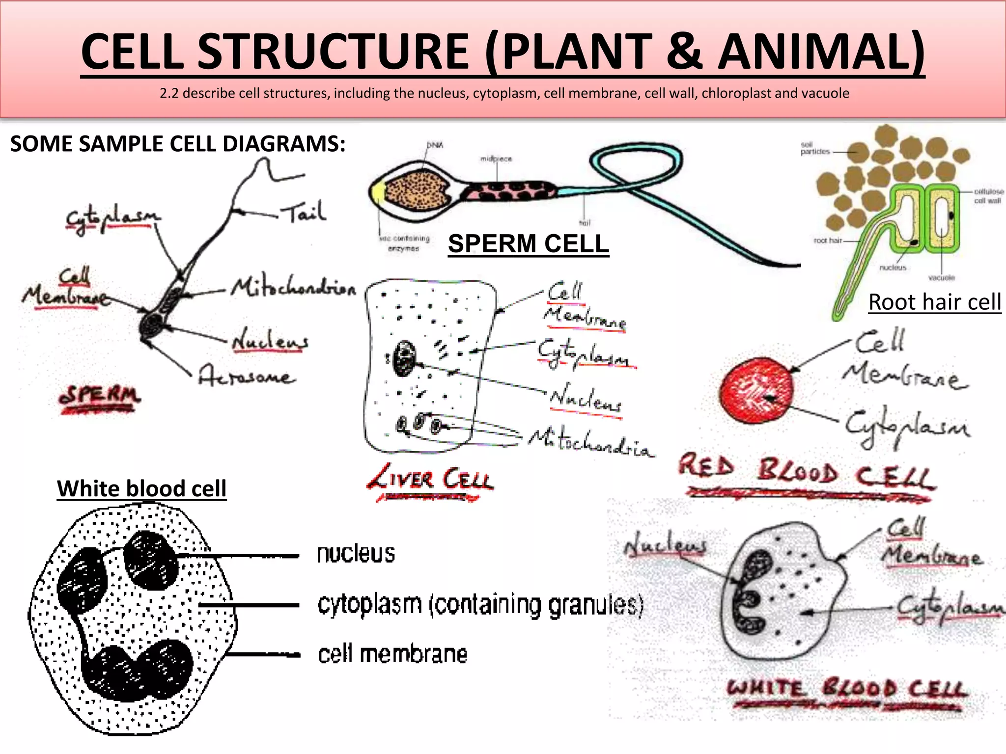


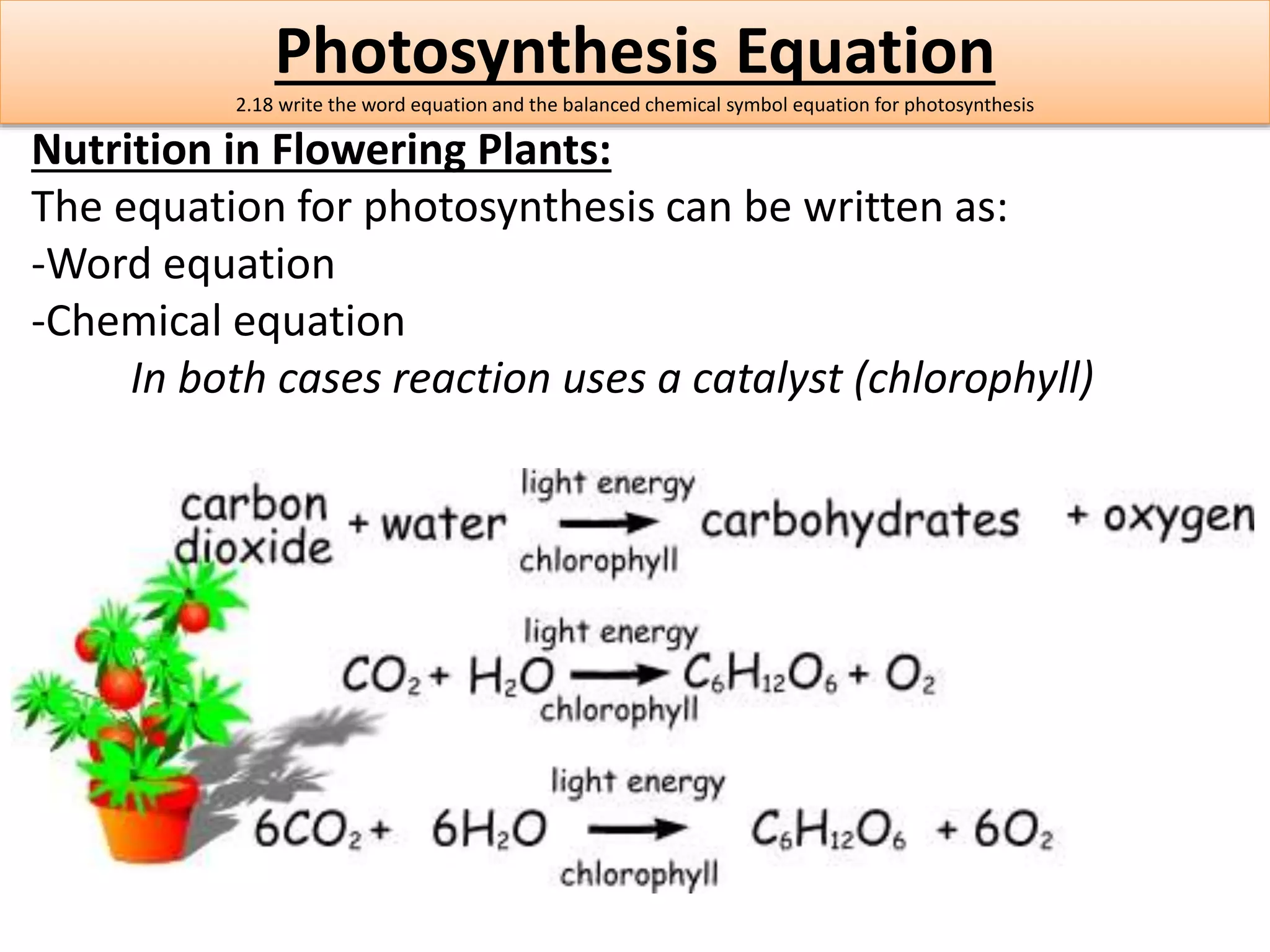

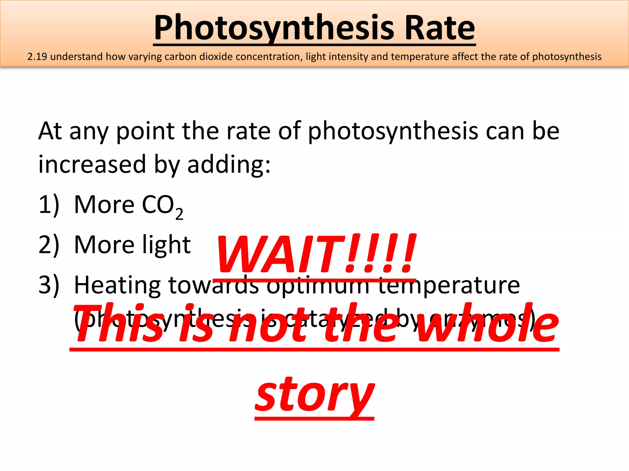
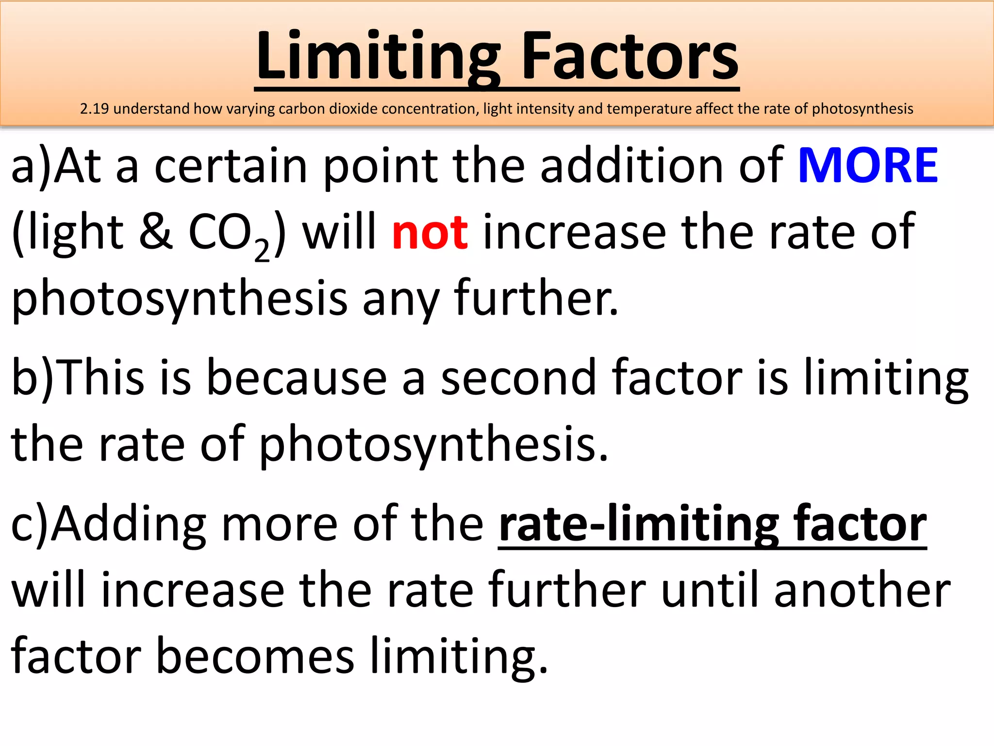
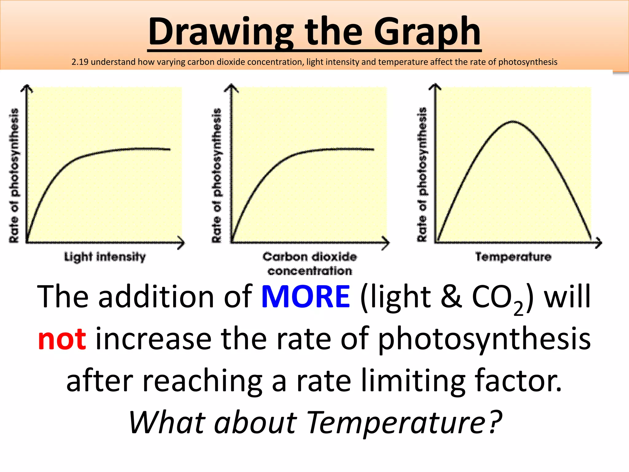
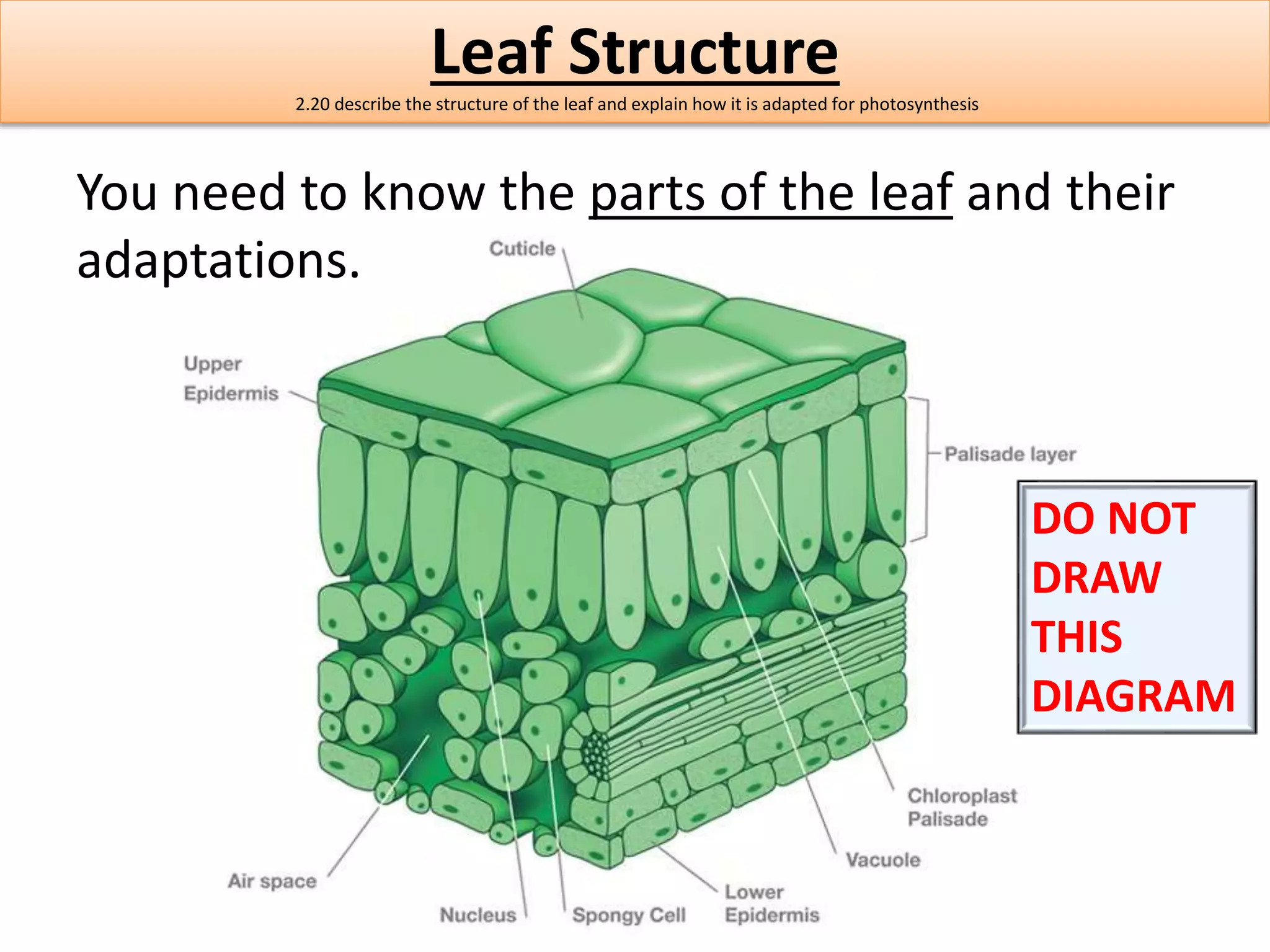
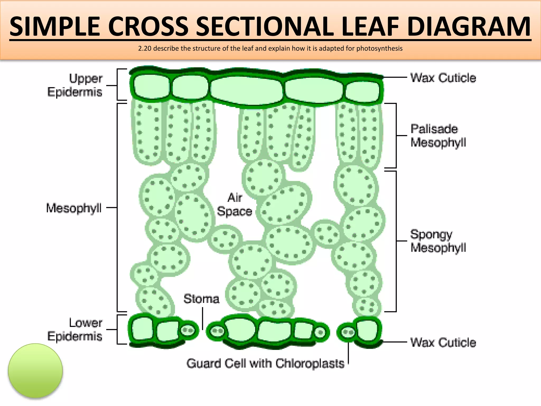
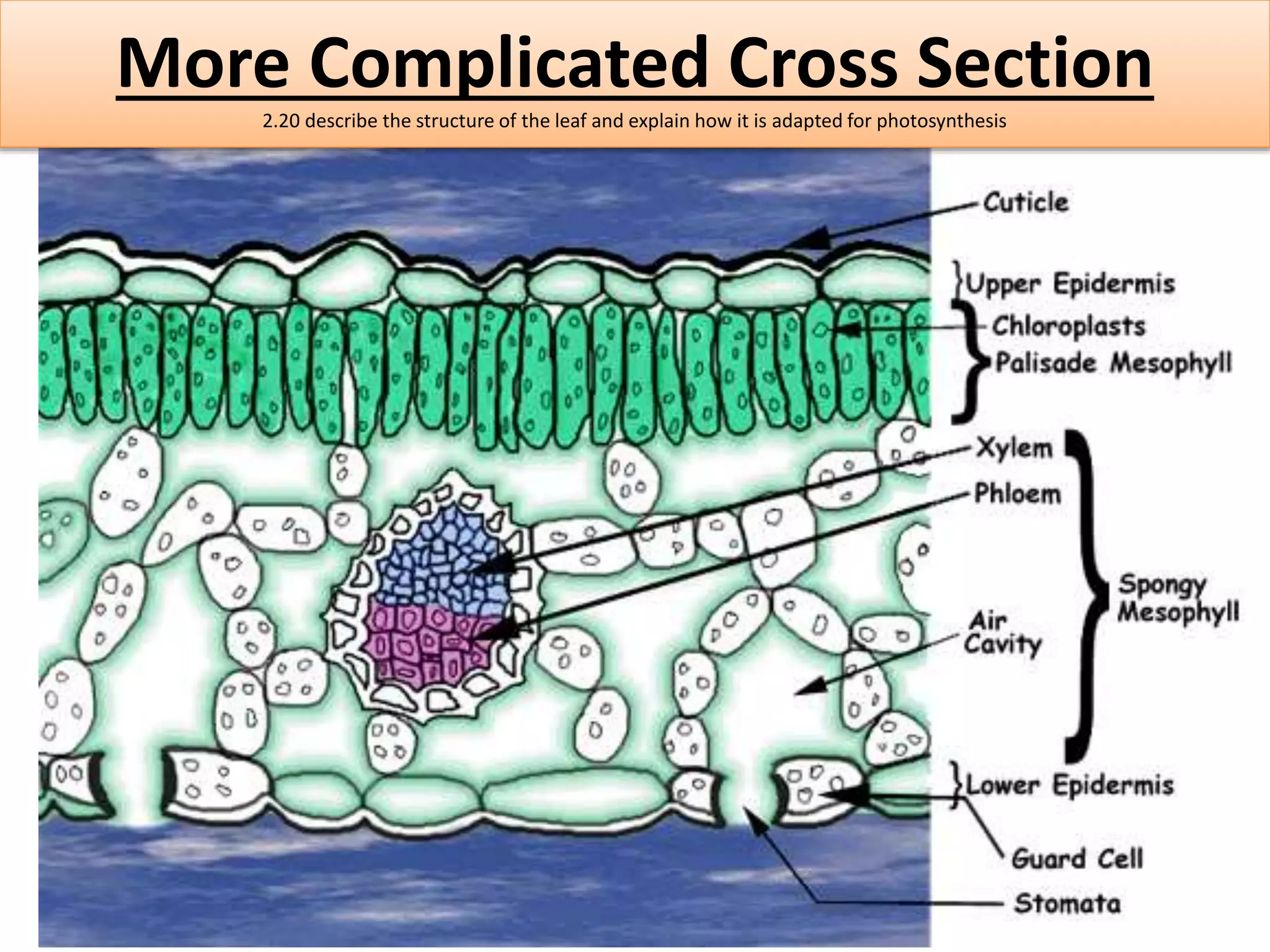
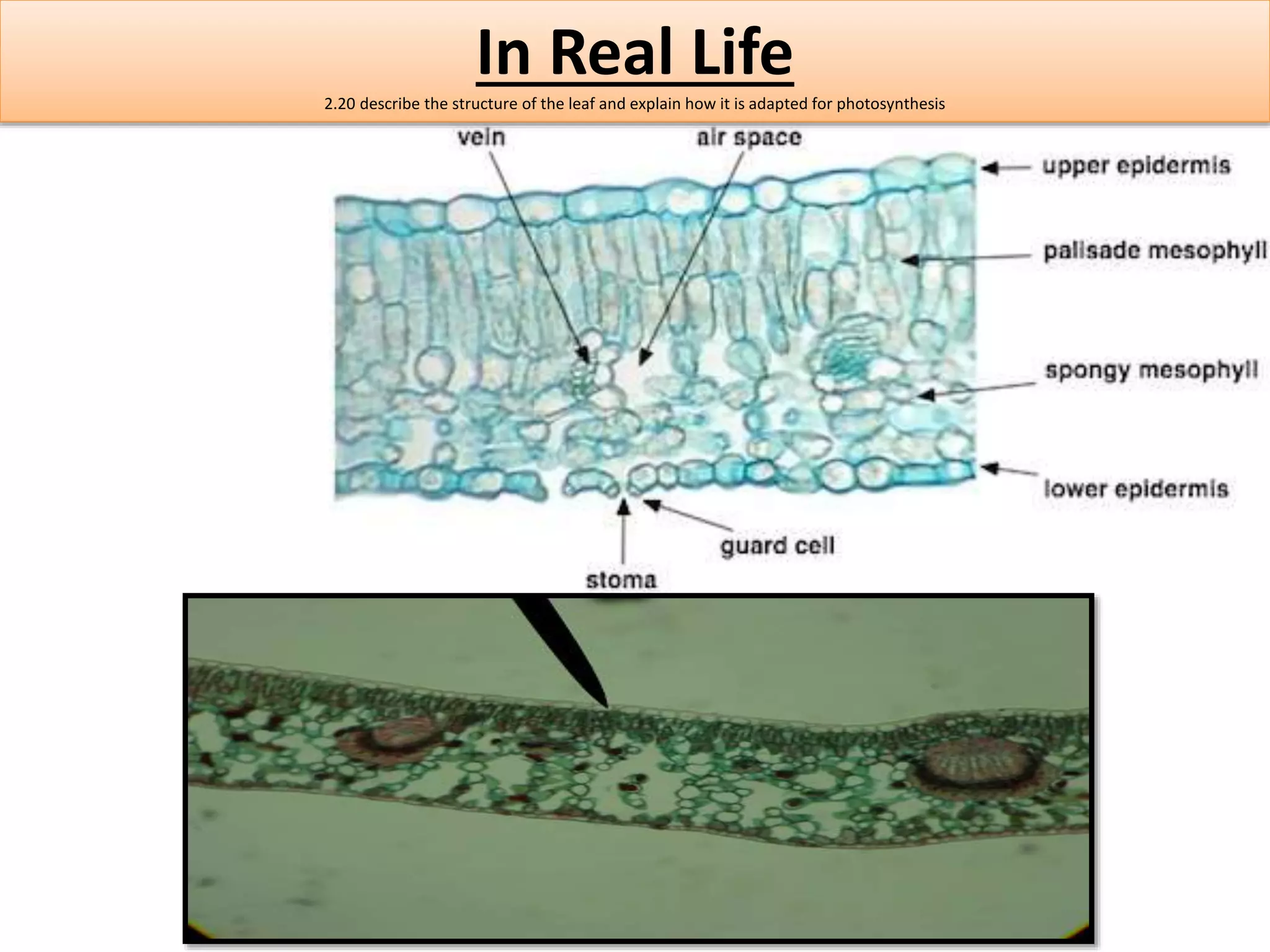


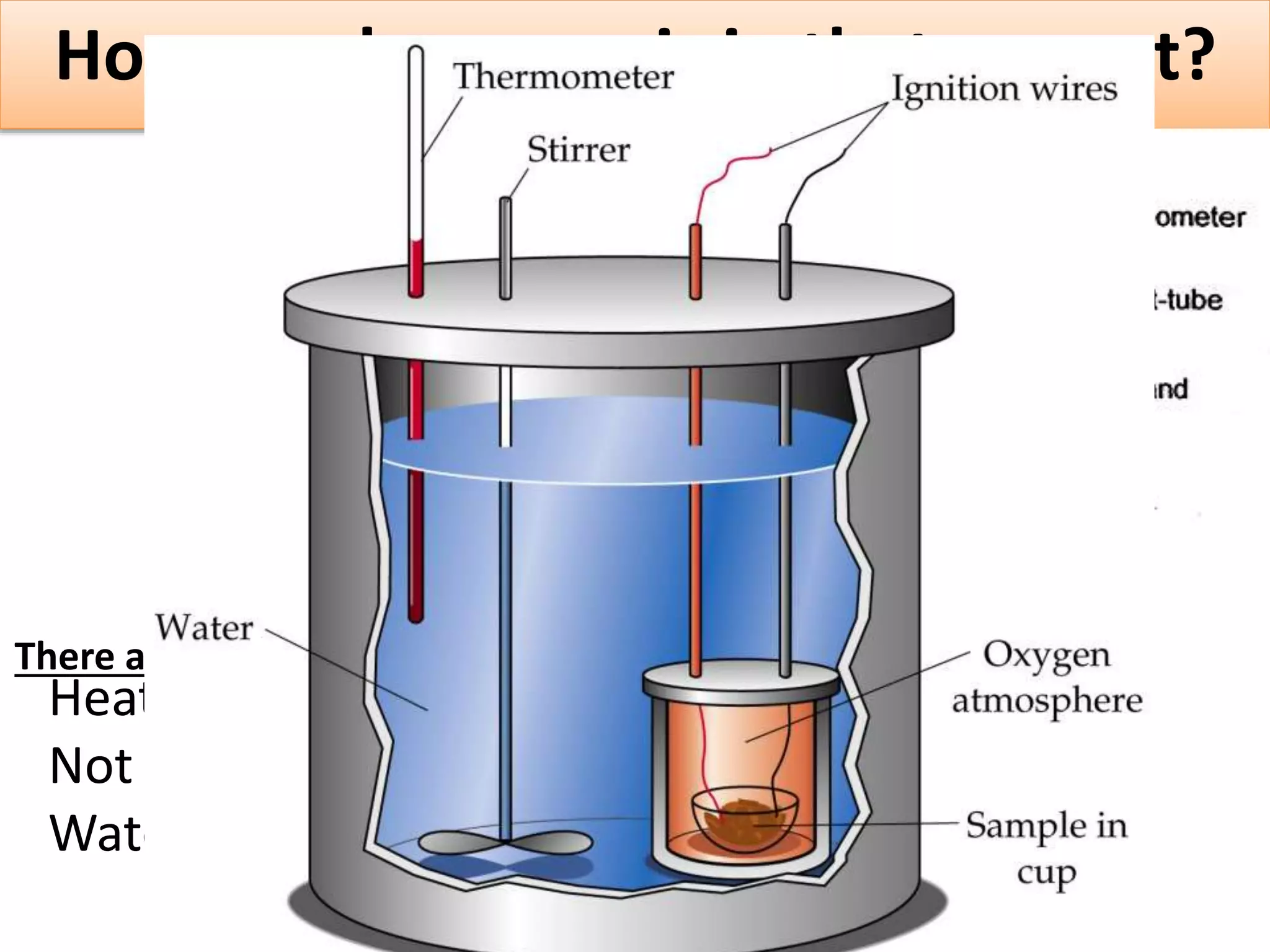


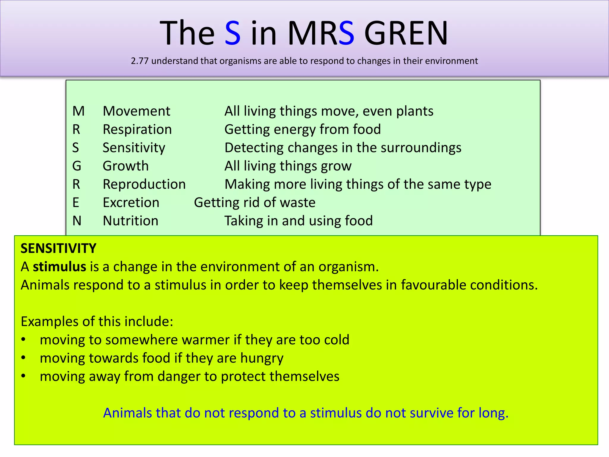


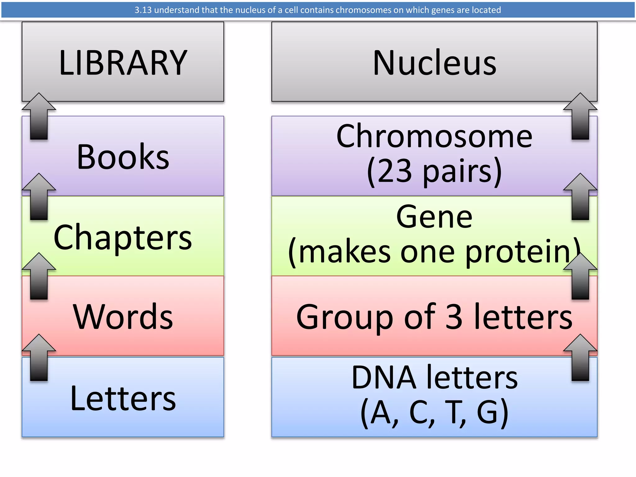
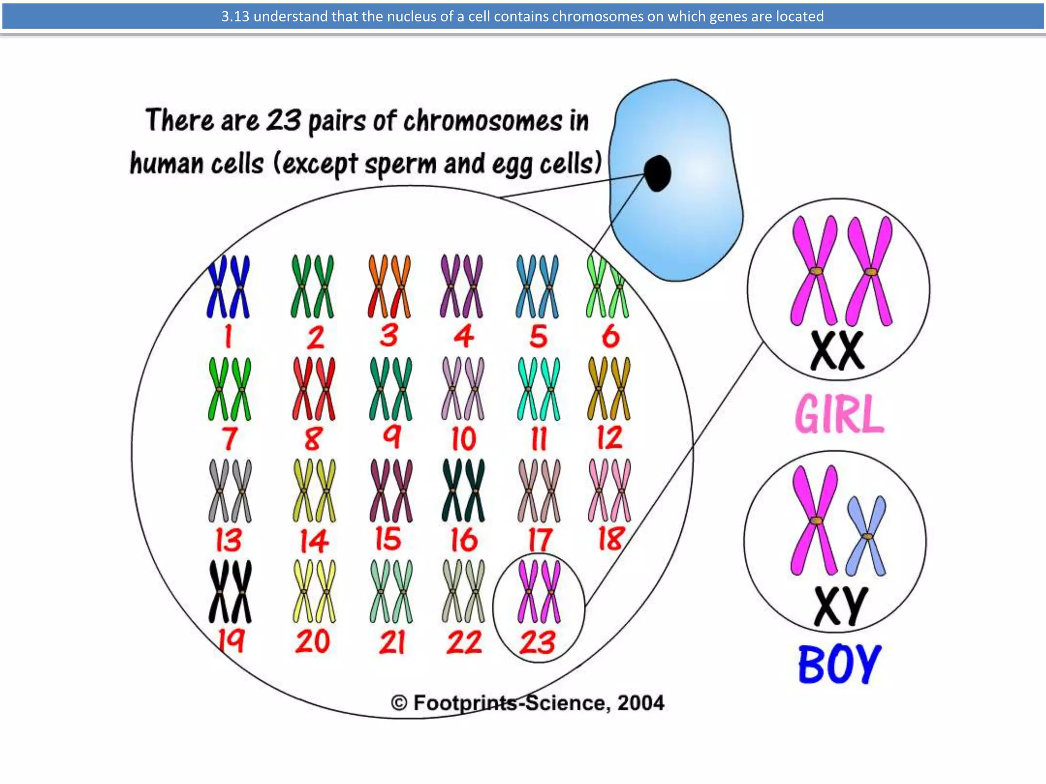
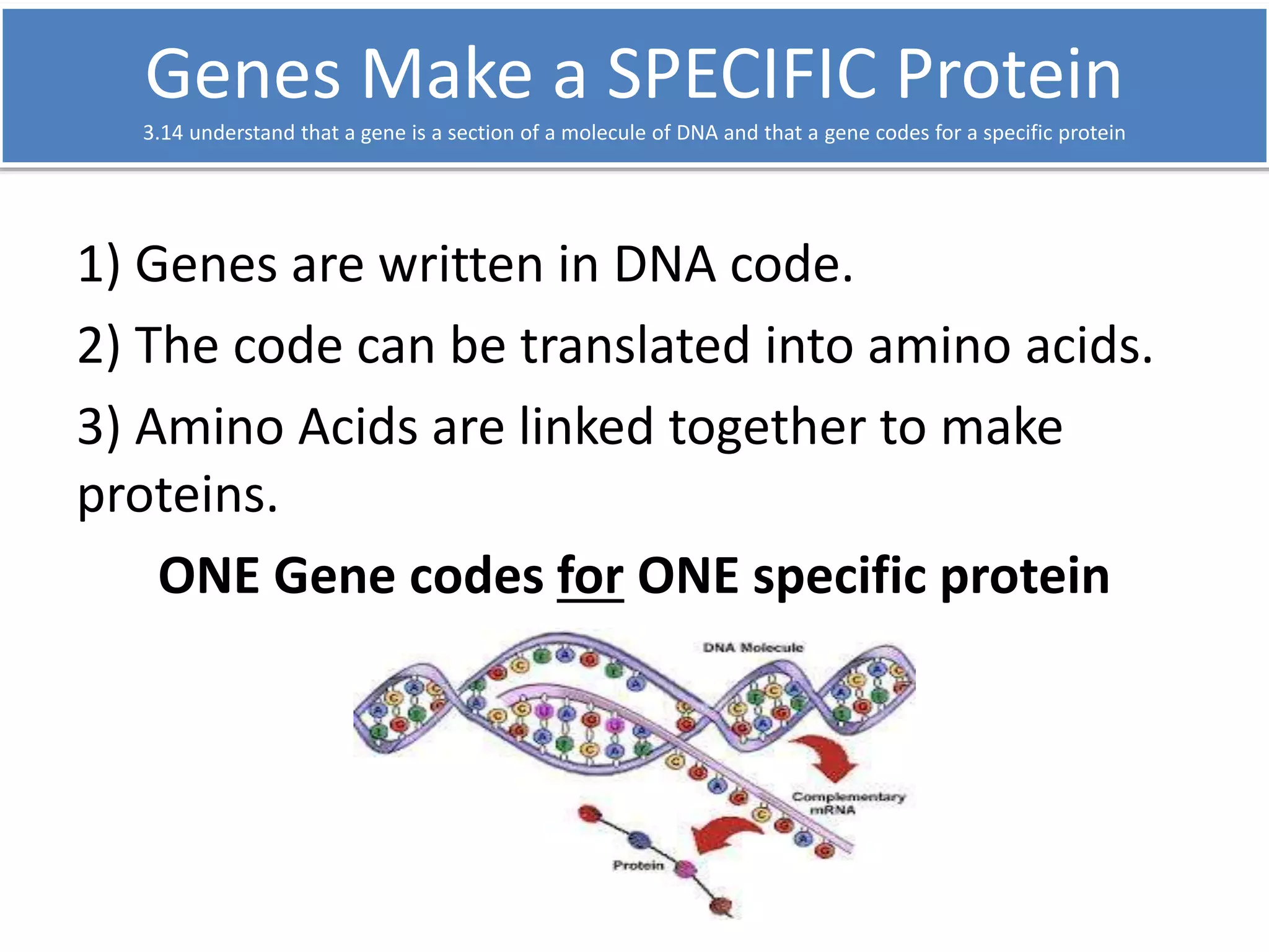
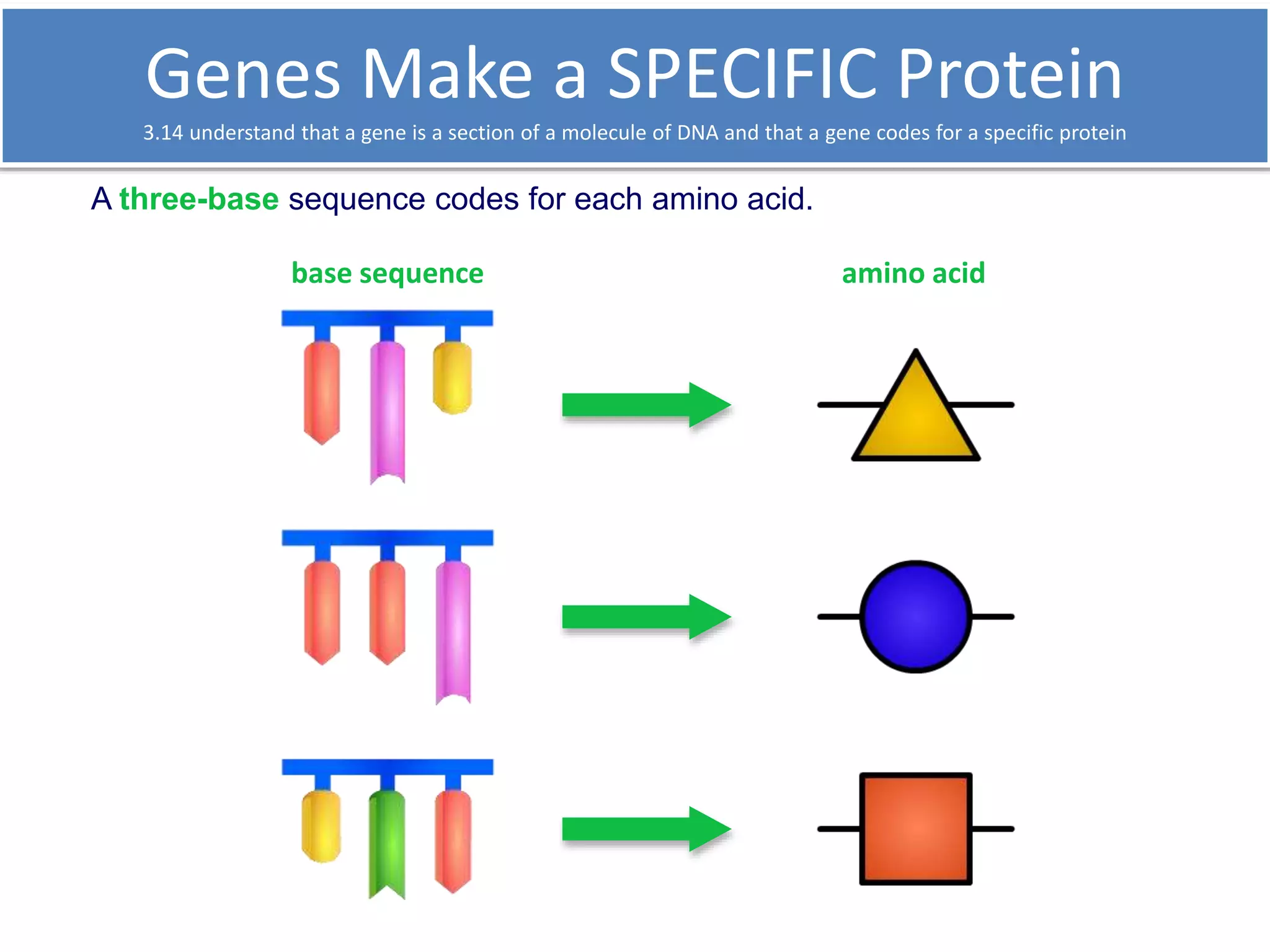


The document outlines the fundamental characteristics that define living organisms, including nutrition, respiration, and reproduction. It categorizes the main groups of living organisms—plants, animals, fungi, bacteria, protoctists, and viruses—detailing their unique features and examples. Additionally, it discusses the hierarchical organization within organisms, emphasizing the structure and function of cells and their components.


