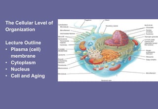
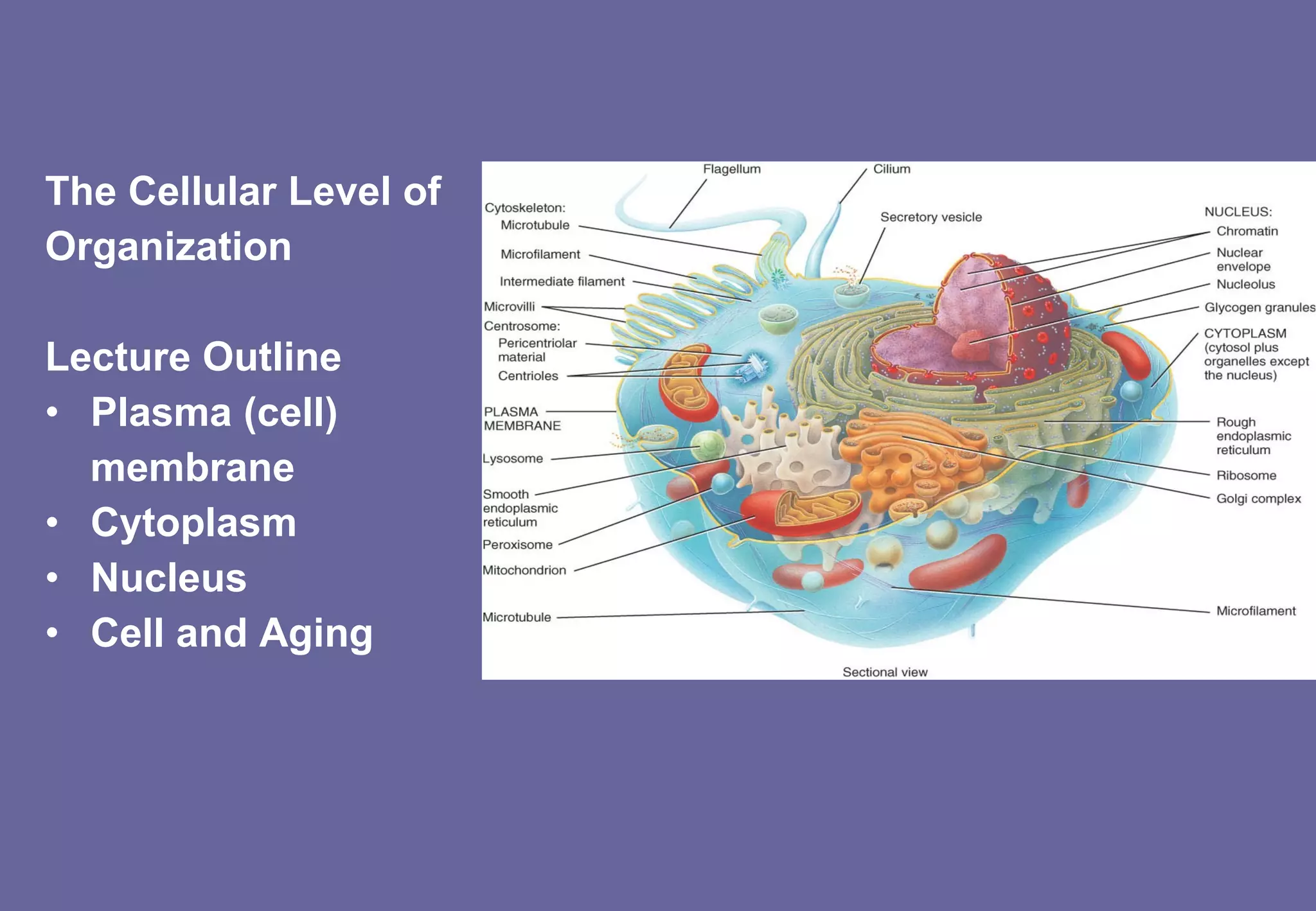
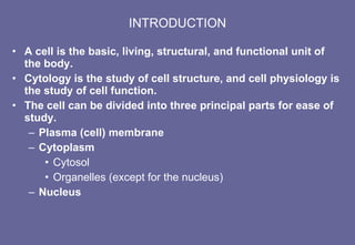
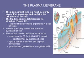
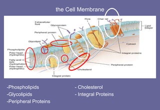
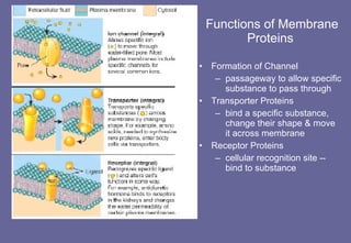
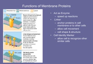
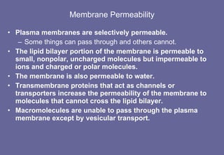
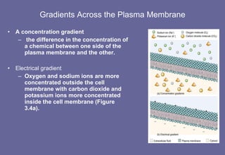
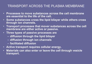
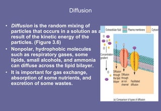
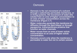
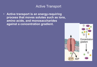
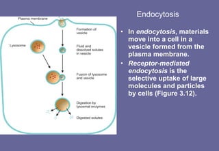
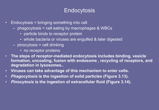
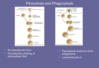
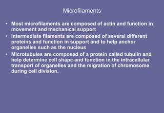
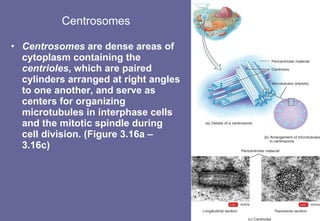
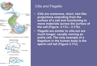
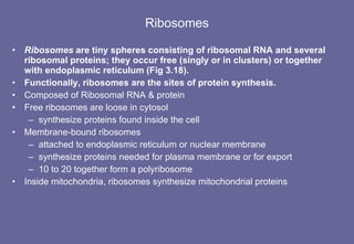
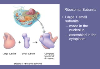
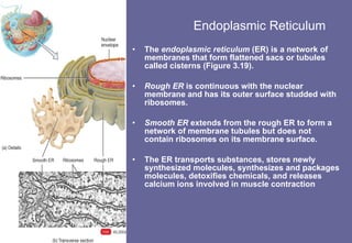
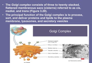
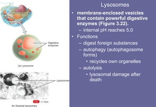
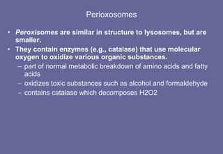
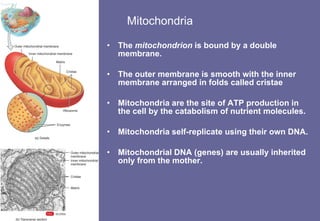
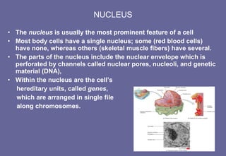
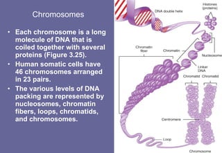
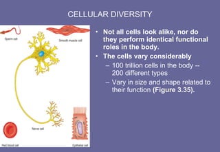
The Cellular Level of  Organization Lecture Outline Plasma (cell) membrane Cytoplasm Nucleus Cell and Aging
INTRODUCTION A cell is the basic, living, structural, and functional unit of the body. Cytology is the study of cell structure, and cell physiology is the study of cell function. The cell can be divided into three principal parts for ease of study. Plasma (cell) membrane Cytoplasm Cytosol Organelles (except for the nucleus) Nucleus
THE PLASMA MEMBRANE The plasma membrane is a flexible, sturdy barrier that surrounds and contains the cytoplasm of the cell. The fluid mosaic model describes its structure (Figure 3.2). The membrane consists of proteins in a sea of lipids. Flexible but sturdy barrier that surround cytoplasm of cell Fluid mosaic model describes its structure membrane is  50 %  lipid & 50 %  protein  held together by hydrogen bonds lipid is barrier to entry or exit of polar substances proteins are “gatekeepers” -- regulate traffic
the Cell Membrane -Phospholipids  - Cholesterol -Glycolipids  - Integral Proteins -Peripheral Proteins
Functions of Membrane Proteins   Formation of Channel passageway to allow specific substance to pass through Transporter Proteins bind a specific substance, change their shape & move it across membrane Receptor Proteins cellular recognition site -- bind to substance
Functions of Membrane Proteins Act as Enzyme speed up reactions Linker anchor proteins in cell membrane or to other cells allow cell movement cell shape & structure  Cell Identity Marker allow cell to recognize other similar cells
Membrane Permeability Plasma membranes are selectively permeable. Some things can pass through and others cannot. The lipid bilayer portion of the membrane is permeable to small, nonpolar, uncharged molecules but impermeable to ions and charged or polar molecules.  The membrane is also permeable to water. Transmembrane proteins that act as channels or transporters increase the permeability of the membrane to molecules that cannot cross the lipid bilayer. Macromolecules are unable to pass through the plasma membrane except by vesicular transport.
Gradients Across the Plasma Membrane A concentration gradient the difference in the concentration of a chemical between one side of the plasma membrane and the other.  Electrical gradient Oxygen and sodium ions are more concentrated outside the cell membrane with carbon dioxide and potassium ions more concentrated inside the cell membrane (Figure 3.4a).
TRANSPORT ACROSS THE PLASMA MEMBRANE Processes to move substances across the cell membrane are essential to the life of the cell. Some substances cross the lipid bilayer while others cross through ion channels. Transport processes that mover substances across the cell membrane are either active or passive. Three types of passive processes are  diffusion through the lipid bilayer diffusion through ion channels facilitated diffusion Active transport requires cellular energy. Materials can also enter or leave the cell through vesicle transport.
Diffusion Diffusion  is the random mixing of particles that occurs in a solution as a result of the kinetic energy of the particles. (Figure 3.6) Nonpolar, hydrophobic molecules such as respiratory gases, some lipids, small alcohols, and ammonia can diffuse across the lipid bilayer. It is important for gas exchange, absorption of some nutrients, and excretion of some wastes.
Osmosis Osmosis is the net movement of a solvent through a selectively permeable membrane, or in living systems, the movement of water (the solute) from an area of higher concentration to an area of lower concentration across the membrane (Figure 3.7).  Water molecules penetrate the membrane by diffusion through the lipid bilayer or through aquaporins, transmembrane proteins that function as water channels. Water moves from an area of lower solute concentration to an area of higher solute concentration. Osmosis occurs only when the membrane is permeable to water but not to certain solutes.
Active Transport Active transport is an energy-requiring process that moves solutes such as ions, amino acids, and monosaccharides against a concentration gradient.
Endocytosis In  endocytosis , materials move into a cell in a vesicle formed from the plasma membrane. Receptor-mediated endocytosis  is the selective uptake of large molecules and particles by cells (Figure 3.12).
Endocytosis Endocytosis = bringing something into cell phagocytosis = cell eating by macrophages & WBCs particle binds to receptor protein whole bacteria or viruses are engulfed & later digested pinocytosis = cell drinking no receptor proteins The steps of receptor-mediated endocytosis includes binding, vesicle formation, uncoating, fusion with endosome , recycling of receptors, and degradation in lysosomes.. Viruses can take advantage of this mechanism to enter cells. Phagocytosis  is the ingestion of solid particles (Figure 3.13). Pinocytosis  is the ingestion of extracellular fluid (Figure 3.14).
Pinocytosis and Phagocytosis Pseudopods extend to form phagosome Lysosome joins it No pseudopods form Nonselective drinking of extracellular fluid
Exocytosis Exocytosis = release something from cell Vesicles form inside cell, fuse to cell membrane Release their contents digestive enzymes, hormones, neurotransmitters or waste products replace cell membrane lost by endocytosis In exocytosis, membrane-enclosed structures called secretory vesicles that form inside the cell fuse with the plasma membrane and release their contents into the extracellular fluid. Transcytosis may be used to move a substance into, across and out of a cell.
CYTOPLASM Cytosol , the intracellular fluid, is the semifluid portion of cytoplasm that contains inclusions and dissolved solutes (Figure 3.1). Cytosol is composed mostly of water, plus proteins, carbohydrates, lipids, and inorganic substances. The chemicals in cytosol are either in solution or in a colloidal (suspended) form. Functionally, cytosol is the medium in which many metabolic reactions occur.
Organelles Organelles are specialized structures that have characteristic shapes and perform specific functions in cellular growth, maintenance, and reproduction.
Cytoskeleton Network of protein filaments throughout the cytosol Functions cell support and shape  organization of chemical reactions cell & organelle movement Continually reorganized
Microfilaments Most microfilaments are composed of actin and function in movement and mechanical support  Intermediate filaments are composed of several different proteins and function in support and to help anchor organelles such as the nucleus Microtubules are composed of a protein called tubulin and help determine cell shape and function in the intracellular transport of organelles and the migration of chromosome during cell division.
Centrosomes Centrosomes  are dense areas of cytoplasm containing the  centrioles , which are paired cylinders arranged at right angles to one another, and serve as centers for organizing microtubules in interphase cells and the mitotic spindle during cell division. (Figure 3.16a – 3.16c)
Cilia and Flagella Cilia  are numerous, short, hair-like projections extending from the surface of a cell and functioning to move materials across the surface of the cell (Figure. 3.17a – 3.17b). Flagella  are similar to cilia but are much longer; usually moving an entire cell. The only example of a flagellum in the human body is the sperm cell tail (Figure 3.17c).
Ribosomes Ribosomes  are tiny spheres consisting of ribosomal RNA and several ribosomal proteins; they occur free (singly or in clusters) or together with endoplasmic reticulum (Fig 3.18). Functionally, ribosomes are the sites of protein synthesis. Composed of Ribosomal RNA & protein Free ribosomes are loose in cytosol synthesize proteins found inside the cell Membrane-bound ribosomes attached to endoplasmic reticulum or nuclear membrane synthesize proteins needed for plasma membrane or for export 10 to 20 together form a polyribosome Inside mitochondria, ribosomes synthesize mitochondrial proteins
Ribosomal Subunits Large + small subunits made in the nucleolus  assembled in the cytoplasm
Endoplasmic Reticulum The  endoplasmic reticulum  (ER) is a network of membranes that form flattened sacs or tubules called cisterns (Figure 3.19). Rough ER  is continuous with the nuclear membrane and has its outer surface studded with ribosomes. Smooth ER  extends from the rough ER to form a network of membrane tubules but does not contain ribosomes on its membrane surface. The ER transports substances, stores newly synthesized molecules, synthesizes and packages molecules, detoxifies chemicals, and releases calcium ions involved in muscle contraction
The  Golgi complex  consists of three to twenty stacked, flattened membranous sacs (cisterns) referred to as cis, medial, and trans (Figure 3.20). The principal function of the Golgi complex is to process, sort, and deliver proteins and lipids to the plasma membrane, lysosomes, and secretory vesicles Golgi Complex
Lysosomes membrane-enclosed vesicles that contain powerful digestive enzymes (Figure 3.22). internal pH reaches 5.0 Functions digest foreign substances autophagy (autophagosome forms) recycles own organelles autolysis lysosomal damage after death
Perioxosomes Peroxisomes  are similar in structure to lysosomes, but are smaller. They contain enzymes (e.g., catalase) that use molecular oxygen to oxidize various organic substances. part of normal metabolic breakdown of amino acids and fatty acids oxidizes toxic substances such as alcohol and formaldehyde contains catalase which decomposes H2O2
Mitochondria The  mitochondrion  is bound by a double membrane.  The outer membrane is smooth with the inner membrane arranged in folds called cristae Mitochondria are the site of ATP production in the cell by the catabolism of nutrient molecules. Mitochondria self-replicate using their own DNA. Mitochondrial DNA (genes) are usually inherited only from the mother.
NUCLEUS The  nucleus  is usually the most prominent feature of a cell Most body cells have a single nucleus; some (red blood cells) have none, whereas others (skeletal muscle fibers) have several. The parts of the nucleus include the nuclear envelope which is perforated by channels called nuclear pores, nucleoli, and genetic material (DNA), Within the nucleus are the cell’s  hereditary units, called  genes ,  which are arranged in single file  along chromosomes.
Function of the Nucleus 46 human DNA molecules or chromosomes  genes found on chromosomes gene is directions for a specific protein Non-dividing cells contain nuclear chromatin loosely packed DNA Dividing cells contain chromosomes tightly packed DNA it doubled (copied itself) before condensing
Chromosomes Each chromosome is a long molecule of DNA that is coiled together with several proteins (Figure 3.25). Human somatic cells have 46 chromosomes arranged in 23 pairs. The various levels of DNA packing are represented by nucleosomes, chromatin fibers, loops, chromatids, and chromosomes.
PROTEIN SYNTHESIS Much of the cellular machinery is devoted to synthesizing large numbers of diverse proeins. The proteins determine the physical and chemical characteristics of cells. The instructions for protein synthesis is found in the DNA in the nucleus. Protein synthesis involves transcription and translation (Figure 3.26).
 
CELL DIVISION Cell division is the process by which cells reproduce themselves. It consists of nuclear division (mitosis and meiosis) and cytoplasmic division ( cytokinesis ). Cell division  that results in an increase in body cells is called  somatic cell division  and involves a nuclear division called  mitosis , plus cytokinesis.  Cell division that results in the production of sperm and eggs is called  reproductive cell division  and consists of a nuclear division called  meiosis  plus cytokinesis.
Control of Cell Destiny The three possible destinies of a cell are to remain alive and functioning without dividing, to grow and divide, or to die. Maturation promoting factor (MPF) induces cell division. Cell death, a process called  apoptosis , is triggered either from outside the cell or from inside the cell due to a “cell-suicide” gene. Necrosis  is a pathological cell death due to injury. Tumor -suppressor genes can produce proteins that normally inhibit cell division resulting in the uncontrollable cell growth known as cancer.
CELLULAR DIVERSITY Not all cells look alike, nor do they perform identical functional roles in the body. The cells vary considerably  100 trillion cells in the body -- 200 different types Vary in size and shape related to their function  (Figure 3.35).
CELLS AND AGING Aging  is a normal process accompanied by a progressive alteration of the body’s homeostatic adaptive responses;  The physiological signs of aging are gradual deterioration in function and capacity to respond to environmental stresses. These signs are related to a net decrease in the number of cells in the body and to the dysfunctioning of the cells that remain. The extracellular components of tissues (e.g., collagen fibers and elastin) also change with age.
DISORDERS: HOMEOSTATIC IMBALANCES Cancer  is a group of diseases characterized by uncontrolled cell proliferation. Cells that divide without control develop into a tumor or neoplasm. A cancerous neoplasm is called a malignant tumor or  malignancy . It has the ability to undergo metastasis, the spread of cancerous cells to other parts of the body. A benign tumor is a noncancerous growth. Hyperplasia = increased number of cell divisions benign tumor does not metastasize or spread malignant---spreads due to cells that detach from tumor and enter blood or lymph

Lect 3 cell level

  • 1.
    The Cellular Levelof Organization Lecture Outline Plasma (cell) membrane Cytoplasm Nucleus Cell and Aging
  • 2.
    INTRODUCTION A cellis the basic, living, structural, and functional unit of the body. Cytology is the study of cell structure, and cell physiology is the study of cell function. The cell can be divided into three principal parts for ease of study. Plasma (cell) membrane Cytoplasm Cytosol Organelles (except for the nucleus) Nucleus
  • 3.
    THE PLASMA MEMBRANEThe plasma membrane is a flexible, sturdy barrier that surrounds and contains the cytoplasm of the cell. The fluid mosaic model describes its structure (Figure 3.2). The membrane consists of proteins in a sea of lipids. Flexible but sturdy barrier that surround cytoplasm of cell Fluid mosaic model describes its structure membrane is 50 % lipid & 50 % protein held together by hydrogen bonds lipid is barrier to entry or exit of polar substances proteins are “gatekeepers” -- regulate traffic
  • 4.
    the Cell Membrane-Phospholipids - Cholesterol -Glycolipids - Integral Proteins -Peripheral Proteins
  • 5.
    Functions of MembraneProteins Formation of Channel passageway to allow specific substance to pass through Transporter Proteins bind a specific substance, change their shape & move it across membrane Receptor Proteins cellular recognition site -- bind to substance
  • 6.
    Functions of MembraneProteins Act as Enzyme speed up reactions Linker anchor proteins in cell membrane or to other cells allow cell movement cell shape & structure Cell Identity Marker allow cell to recognize other similar cells
  • 7.
    Membrane Permeability Plasmamembranes are selectively permeable. Some things can pass through and others cannot. The lipid bilayer portion of the membrane is permeable to small, nonpolar, uncharged molecules but impermeable to ions and charged or polar molecules. The membrane is also permeable to water. Transmembrane proteins that act as channels or transporters increase the permeability of the membrane to molecules that cannot cross the lipid bilayer. Macromolecules are unable to pass through the plasma membrane except by vesicular transport.
  • 8.
    Gradients Across thePlasma Membrane A concentration gradient the difference in the concentration of a chemical between one side of the plasma membrane and the other. Electrical gradient Oxygen and sodium ions are more concentrated outside the cell membrane with carbon dioxide and potassium ions more concentrated inside the cell membrane (Figure 3.4a).
  • 9.
    TRANSPORT ACROSS THEPLASMA MEMBRANE Processes to move substances across the cell membrane are essential to the life of the cell. Some substances cross the lipid bilayer while others cross through ion channels. Transport processes that mover substances across the cell membrane are either active or passive. Three types of passive processes are diffusion through the lipid bilayer diffusion through ion channels facilitated diffusion Active transport requires cellular energy. Materials can also enter or leave the cell through vesicle transport.
  • 10.
    Diffusion Diffusion is the random mixing of particles that occurs in a solution as a result of the kinetic energy of the particles. (Figure 3.6) Nonpolar, hydrophobic molecules such as respiratory gases, some lipids, small alcohols, and ammonia can diffuse across the lipid bilayer. It is important for gas exchange, absorption of some nutrients, and excretion of some wastes.
  • 11.
    Osmosis Osmosis isthe net movement of a solvent through a selectively permeable membrane, or in living systems, the movement of water (the solute) from an area of higher concentration to an area of lower concentration across the membrane (Figure 3.7). Water molecules penetrate the membrane by diffusion through the lipid bilayer or through aquaporins, transmembrane proteins that function as water channels. Water moves from an area of lower solute concentration to an area of higher solute concentration. Osmosis occurs only when the membrane is permeable to water but not to certain solutes.
  • 12.
    Active Transport Activetransport is an energy-requiring process that moves solutes such as ions, amino acids, and monosaccharides against a concentration gradient.
  • 13.
    Endocytosis In endocytosis , materials move into a cell in a vesicle formed from the plasma membrane. Receptor-mediated endocytosis is the selective uptake of large molecules and particles by cells (Figure 3.12).
  • 14.
    Endocytosis Endocytosis =bringing something into cell phagocytosis = cell eating by macrophages & WBCs particle binds to receptor protein whole bacteria or viruses are engulfed & later digested pinocytosis = cell drinking no receptor proteins The steps of receptor-mediated endocytosis includes binding, vesicle formation, uncoating, fusion with endosome , recycling of receptors, and degradation in lysosomes.. Viruses can take advantage of this mechanism to enter cells. Phagocytosis is the ingestion of solid particles (Figure 3.13). Pinocytosis is the ingestion of extracellular fluid (Figure 3.14).
  • 15.
    Pinocytosis and PhagocytosisPseudopods extend to form phagosome Lysosome joins it No pseudopods form Nonselective drinking of extracellular fluid
  • 16.
    Exocytosis Exocytosis =release something from cell Vesicles form inside cell, fuse to cell membrane Release their contents digestive enzymes, hormones, neurotransmitters or waste products replace cell membrane lost by endocytosis In exocytosis, membrane-enclosed structures called secretory vesicles that form inside the cell fuse with the plasma membrane and release their contents into the extracellular fluid. Transcytosis may be used to move a substance into, across and out of a cell.
  • 17.
    CYTOPLASM Cytosol ,the intracellular fluid, is the semifluid portion of cytoplasm that contains inclusions and dissolved solutes (Figure 3.1). Cytosol is composed mostly of water, plus proteins, carbohydrates, lipids, and inorganic substances. The chemicals in cytosol are either in solution or in a colloidal (suspended) form. Functionally, cytosol is the medium in which many metabolic reactions occur.
  • 18.
    Organelles Organelles arespecialized structures that have characteristic shapes and perform specific functions in cellular growth, maintenance, and reproduction.
  • 19.
    Cytoskeleton Network ofprotein filaments throughout the cytosol Functions cell support and shape organization of chemical reactions cell & organelle movement Continually reorganized
  • 20.
    Microfilaments Most microfilamentsare composed of actin and function in movement and mechanical support Intermediate filaments are composed of several different proteins and function in support and to help anchor organelles such as the nucleus Microtubules are composed of a protein called tubulin and help determine cell shape and function in the intracellular transport of organelles and the migration of chromosome during cell division.
  • 21.
    Centrosomes Centrosomes are dense areas of cytoplasm containing the centrioles , which are paired cylinders arranged at right angles to one another, and serve as centers for organizing microtubules in interphase cells and the mitotic spindle during cell division. (Figure 3.16a – 3.16c)
  • 22.
    Cilia and FlagellaCilia are numerous, short, hair-like projections extending from the surface of a cell and functioning to move materials across the surface of the cell (Figure. 3.17a – 3.17b). Flagella are similar to cilia but are much longer; usually moving an entire cell. The only example of a flagellum in the human body is the sperm cell tail (Figure 3.17c).
  • 23.
    Ribosomes Ribosomes are tiny spheres consisting of ribosomal RNA and several ribosomal proteins; they occur free (singly or in clusters) or together with endoplasmic reticulum (Fig 3.18). Functionally, ribosomes are the sites of protein synthesis. Composed of Ribosomal RNA & protein Free ribosomes are loose in cytosol synthesize proteins found inside the cell Membrane-bound ribosomes attached to endoplasmic reticulum or nuclear membrane synthesize proteins needed for plasma membrane or for export 10 to 20 together form a polyribosome Inside mitochondria, ribosomes synthesize mitochondrial proteins
  • 24.
    Ribosomal Subunits Large+ small subunits made in the nucleolus assembled in the cytoplasm
  • 25.
    Endoplasmic Reticulum The endoplasmic reticulum (ER) is a network of membranes that form flattened sacs or tubules called cisterns (Figure 3.19). Rough ER is continuous with the nuclear membrane and has its outer surface studded with ribosomes. Smooth ER extends from the rough ER to form a network of membrane tubules but does not contain ribosomes on its membrane surface. The ER transports substances, stores newly synthesized molecules, synthesizes and packages molecules, detoxifies chemicals, and releases calcium ions involved in muscle contraction
  • 26.
    The Golgicomplex consists of three to twenty stacked, flattened membranous sacs (cisterns) referred to as cis, medial, and trans (Figure 3.20). The principal function of the Golgi complex is to process, sort, and deliver proteins and lipids to the plasma membrane, lysosomes, and secretory vesicles Golgi Complex
  • 27.
    Lysosomes membrane-enclosed vesiclesthat contain powerful digestive enzymes (Figure 3.22). internal pH reaches 5.0 Functions digest foreign substances autophagy (autophagosome forms) recycles own organelles autolysis lysosomal damage after death
  • 28.
    Perioxosomes Peroxisomes are similar in structure to lysosomes, but are smaller. They contain enzymes (e.g., catalase) that use molecular oxygen to oxidize various organic substances. part of normal metabolic breakdown of amino acids and fatty acids oxidizes toxic substances such as alcohol and formaldehyde contains catalase which decomposes H2O2
  • 29.
    Mitochondria The mitochondrion is bound by a double membrane. The outer membrane is smooth with the inner membrane arranged in folds called cristae Mitochondria are the site of ATP production in the cell by the catabolism of nutrient molecules. Mitochondria self-replicate using their own DNA. Mitochondrial DNA (genes) are usually inherited only from the mother.
  • 30.
    NUCLEUS The nucleus is usually the most prominent feature of a cell Most body cells have a single nucleus; some (red blood cells) have none, whereas others (skeletal muscle fibers) have several. The parts of the nucleus include the nuclear envelope which is perforated by channels called nuclear pores, nucleoli, and genetic material (DNA), Within the nucleus are the cell’s hereditary units, called genes , which are arranged in single file along chromosomes.
  • 31.
    Function of theNucleus 46 human DNA molecules or chromosomes genes found on chromosomes gene is directions for a specific protein Non-dividing cells contain nuclear chromatin loosely packed DNA Dividing cells contain chromosomes tightly packed DNA it doubled (copied itself) before condensing
  • 32.
    Chromosomes Each chromosomeis a long molecule of DNA that is coiled together with several proteins (Figure 3.25). Human somatic cells have 46 chromosomes arranged in 23 pairs. The various levels of DNA packing are represented by nucleosomes, chromatin fibers, loops, chromatids, and chromosomes.
  • 33.
    PROTEIN SYNTHESIS Muchof the cellular machinery is devoted to synthesizing large numbers of diverse proeins. The proteins determine the physical and chemical characteristics of cells. The instructions for protein synthesis is found in the DNA in the nucleus. Protein synthesis involves transcription and translation (Figure 3.26).
  • 34.
  • 35.
    CELL DIVISION Celldivision is the process by which cells reproduce themselves. It consists of nuclear division (mitosis and meiosis) and cytoplasmic division ( cytokinesis ). Cell division that results in an increase in body cells is called somatic cell division and involves a nuclear division called mitosis , plus cytokinesis. Cell division that results in the production of sperm and eggs is called reproductive cell division and consists of a nuclear division called meiosis plus cytokinesis.
  • 36.
    Control of CellDestiny The three possible destinies of a cell are to remain alive and functioning without dividing, to grow and divide, or to die. Maturation promoting factor (MPF) induces cell division. Cell death, a process called apoptosis , is triggered either from outside the cell or from inside the cell due to a “cell-suicide” gene. Necrosis is a pathological cell death due to injury. Tumor -suppressor genes can produce proteins that normally inhibit cell division resulting in the uncontrollable cell growth known as cancer.
  • 37.
    CELLULAR DIVERSITY Notall cells look alike, nor do they perform identical functional roles in the body. The cells vary considerably 100 trillion cells in the body -- 200 different types Vary in size and shape related to their function (Figure 3.35).
  • 38.
    CELLS AND AGINGAging is a normal process accompanied by a progressive alteration of the body’s homeostatic adaptive responses; The physiological signs of aging are gradual deterioration in function and capacity to respond to environmental stresses. These signs are related to a net decrease in the number of cells in the body and to the dysfunctioning of the cells that remain. The extracellular components of tissues (e.g., collagen fibers and elastin) also change with age.
  • 39.
    DISORDERS: HOMEOSTATIC IMBALANCESCancer is a group of diseases characterized by uncontrolled cell proliferation. Cells that divide without control develop into a tumor or neoplasm. A cancerous neoplasm is called a malignant tumor or malignancy . It has the ability to undergo metastasis, the spread of cancerous cells to other parts of the body. A benign tumor is a noncancerous growth. Hyperplasia = increased number of cell divisions benign tumor does not metastasize or spread malignant---spreads due to cells that detach from tumor and enter blood or lymph