Downloaded 180 times

![Transport Processes
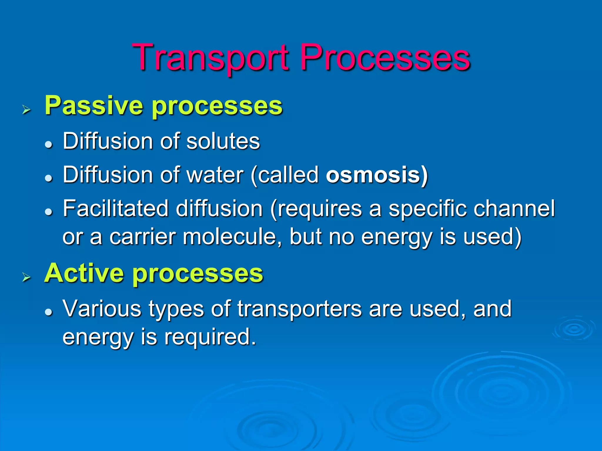
Passive processes involve substances
moving across the cell membranes without
the input of any energy - they are said to
move “with” or “down” their concentration
gradient ([gradient] , where [ ] indicates
“concentration”).
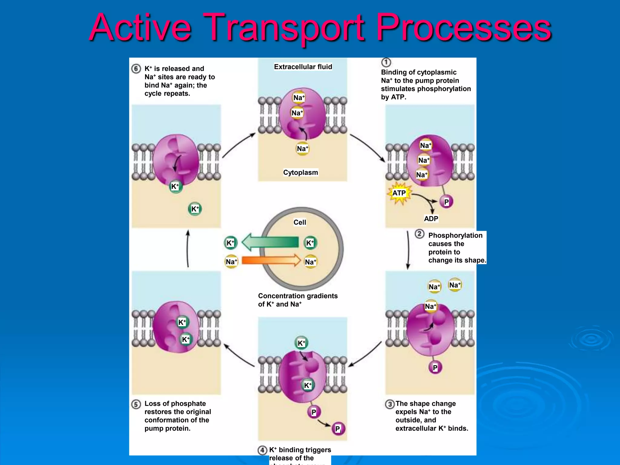
Active processes involve the use of energy,
primarily from the breakdown of ATP, to
move a substance against its [gradient].](https://image.slidesharecdn.com/chapter3-140731103318-phpapp01/75/Chapter-3-21-2048.jpg)
![Concentration gradients
Concentrations of some key ions are very
different on the inside versus the outside
of cells creating a gradient
IN:
[Na+] = low
[K+] = high
[Ca2+] = very
low
[Cl-] = low
OUT:
[Na+] = high
[K+] = low
[Ca2+] = low
[Cl-] = high
(blood,
interstitial fluid)](https://image.slidesharecdn.com/chapter3-140731103318-phpapp01/75/Chapter-3-23-2048.jpg)
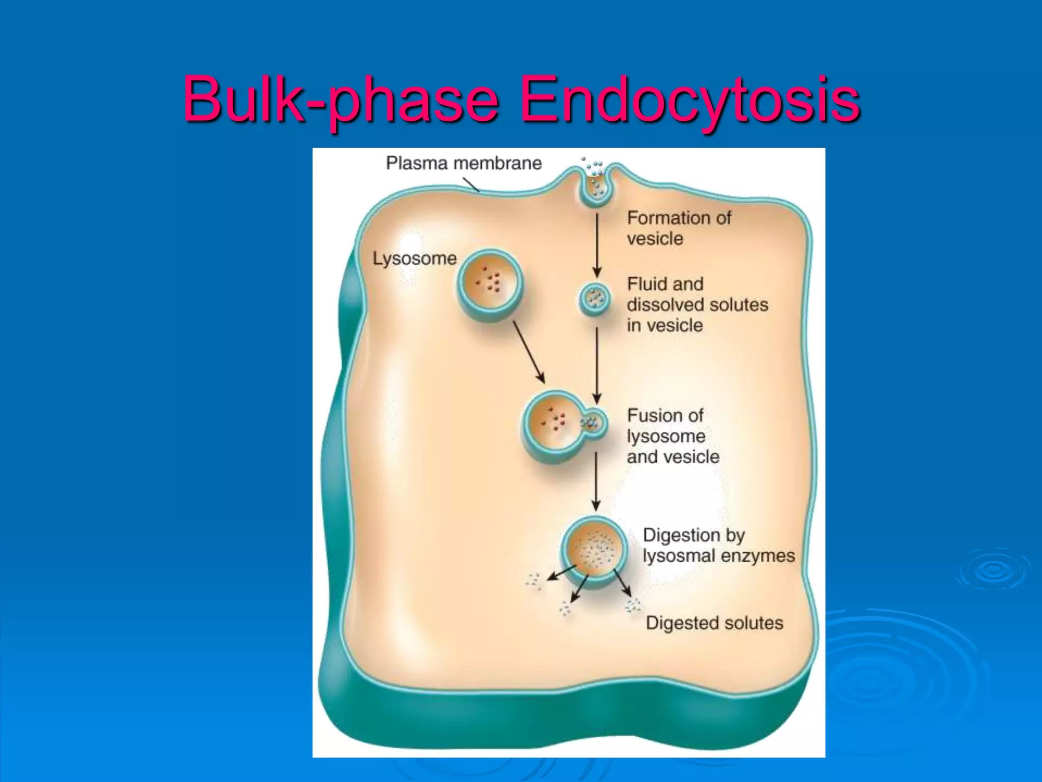
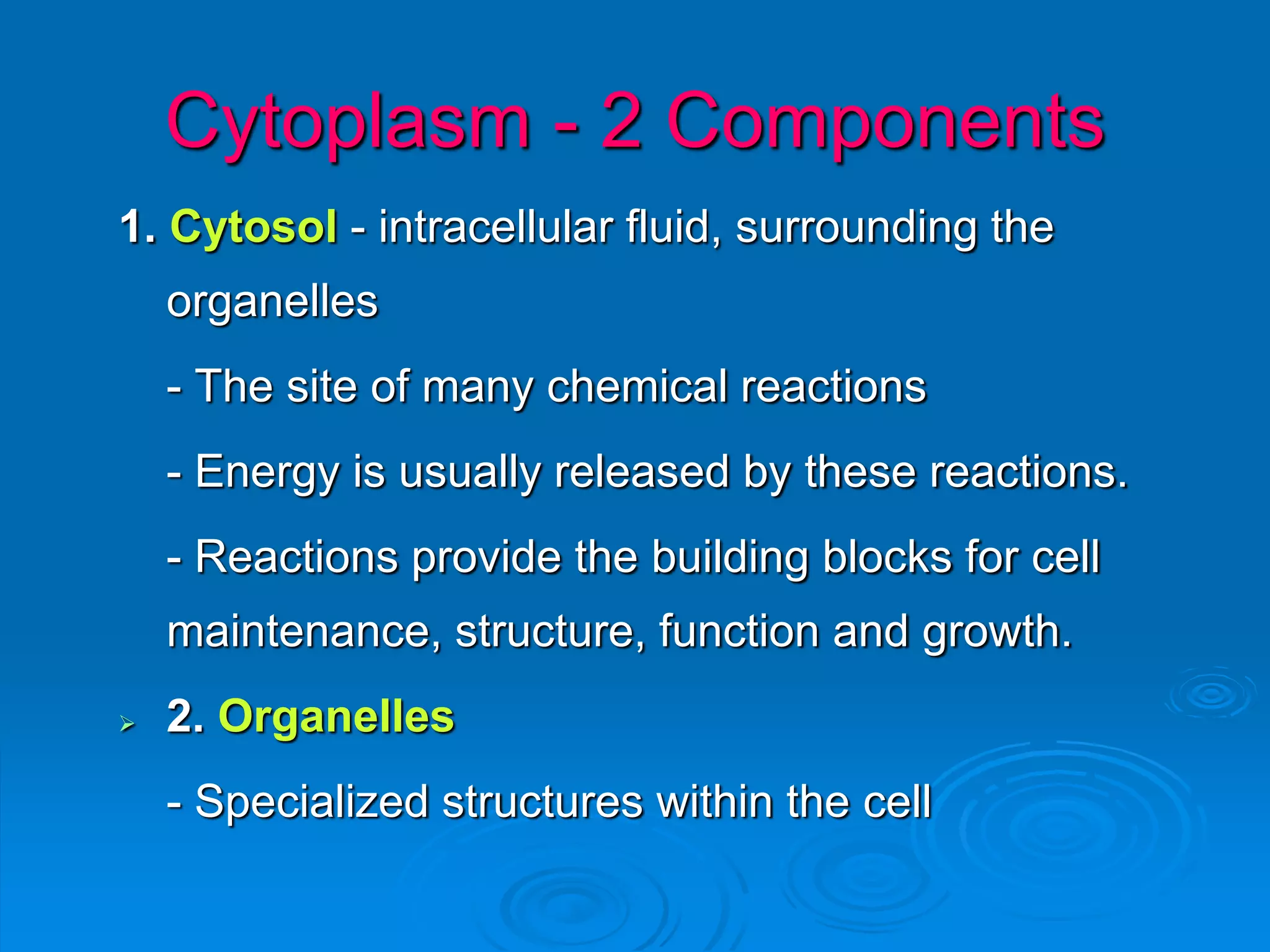
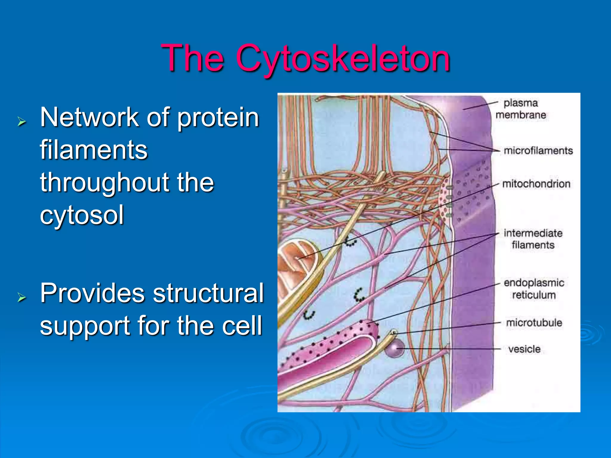
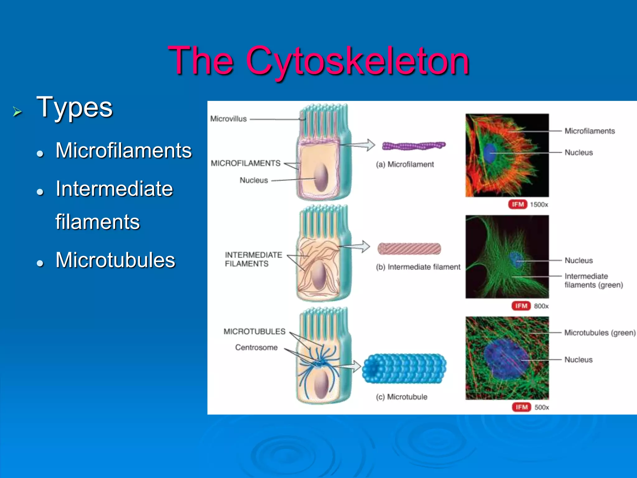
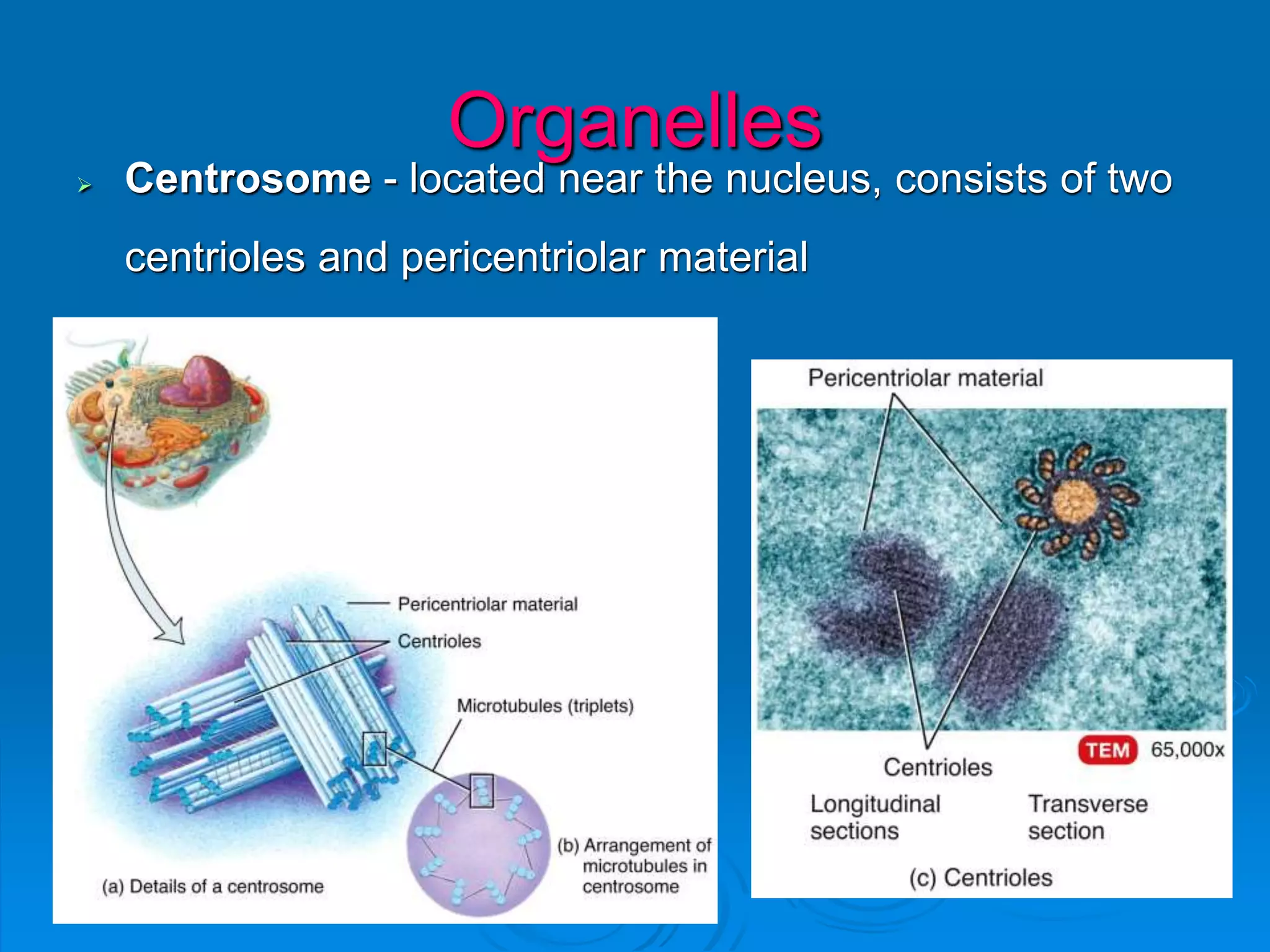
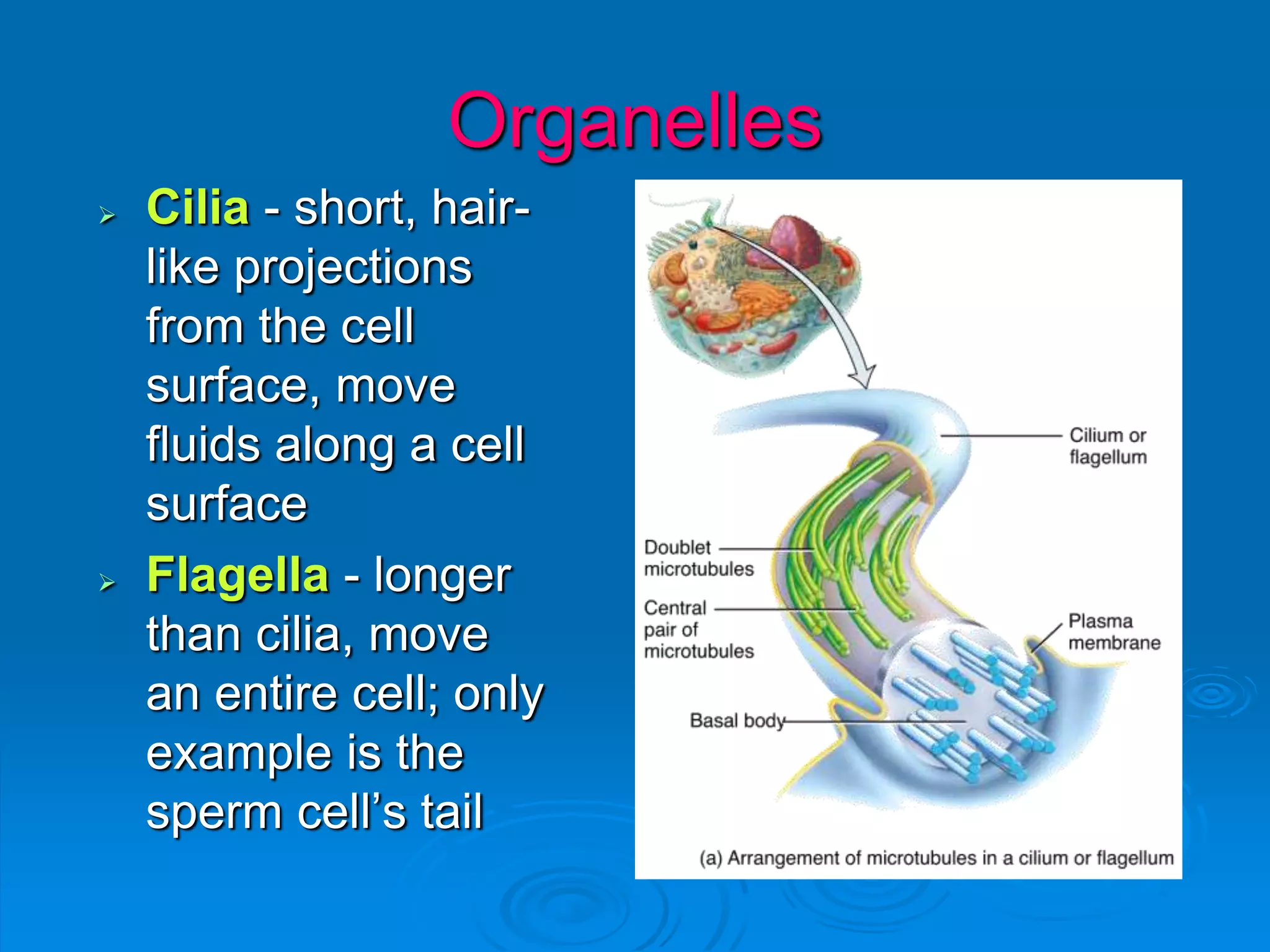
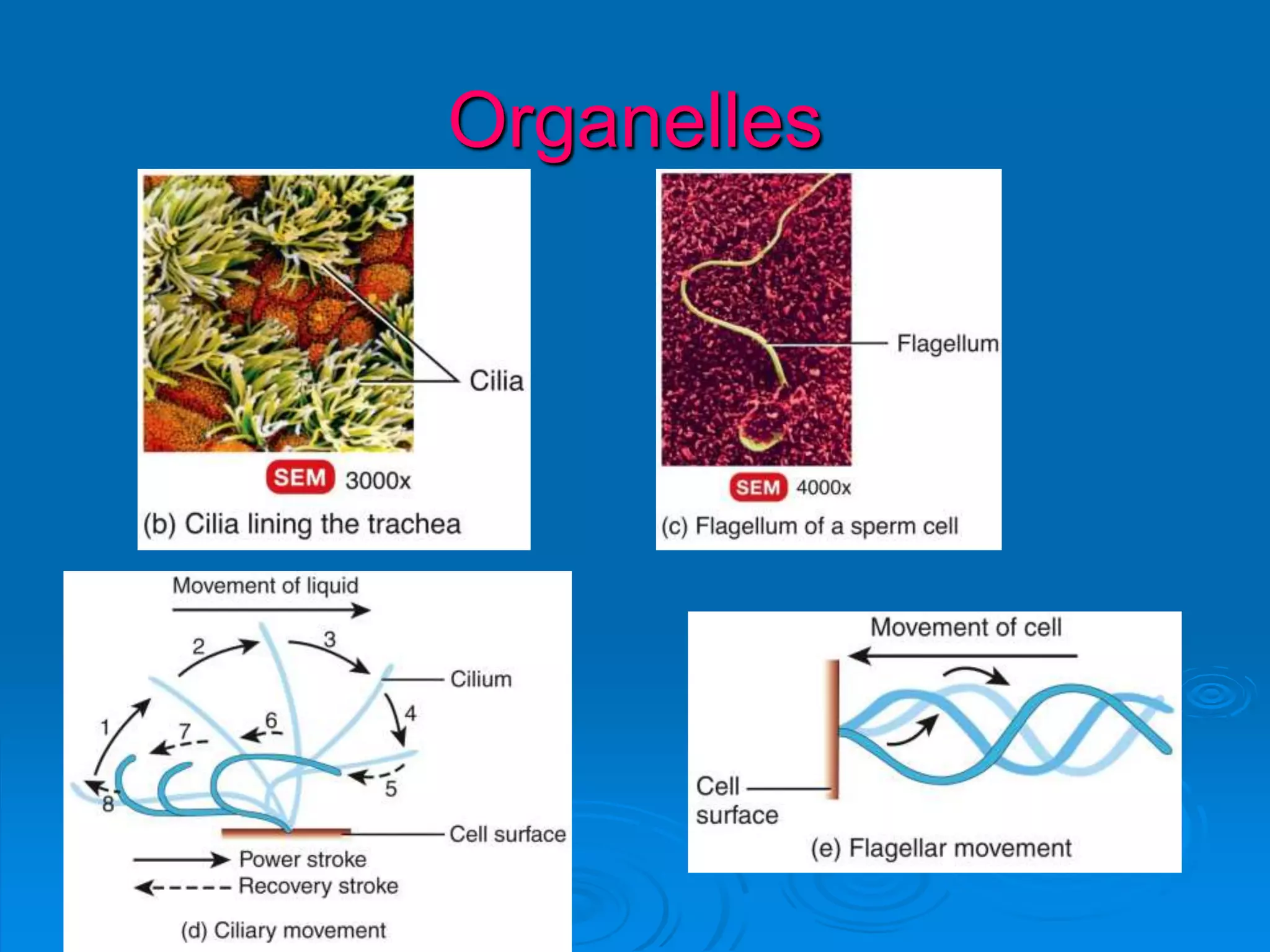
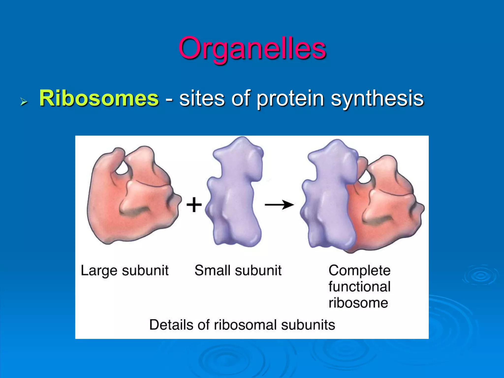
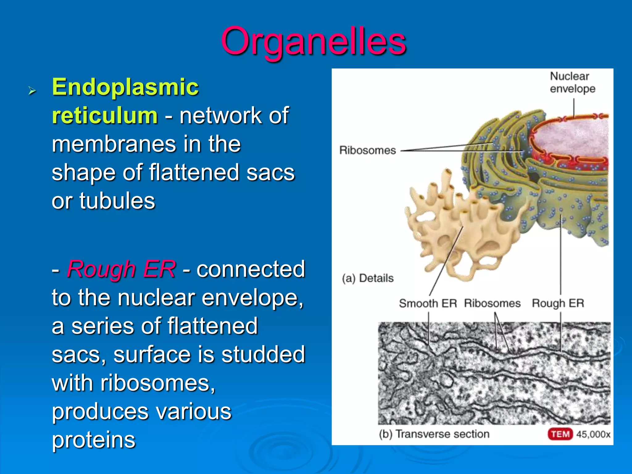
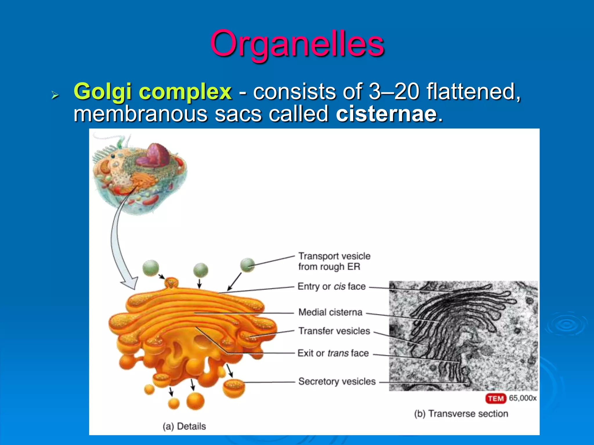
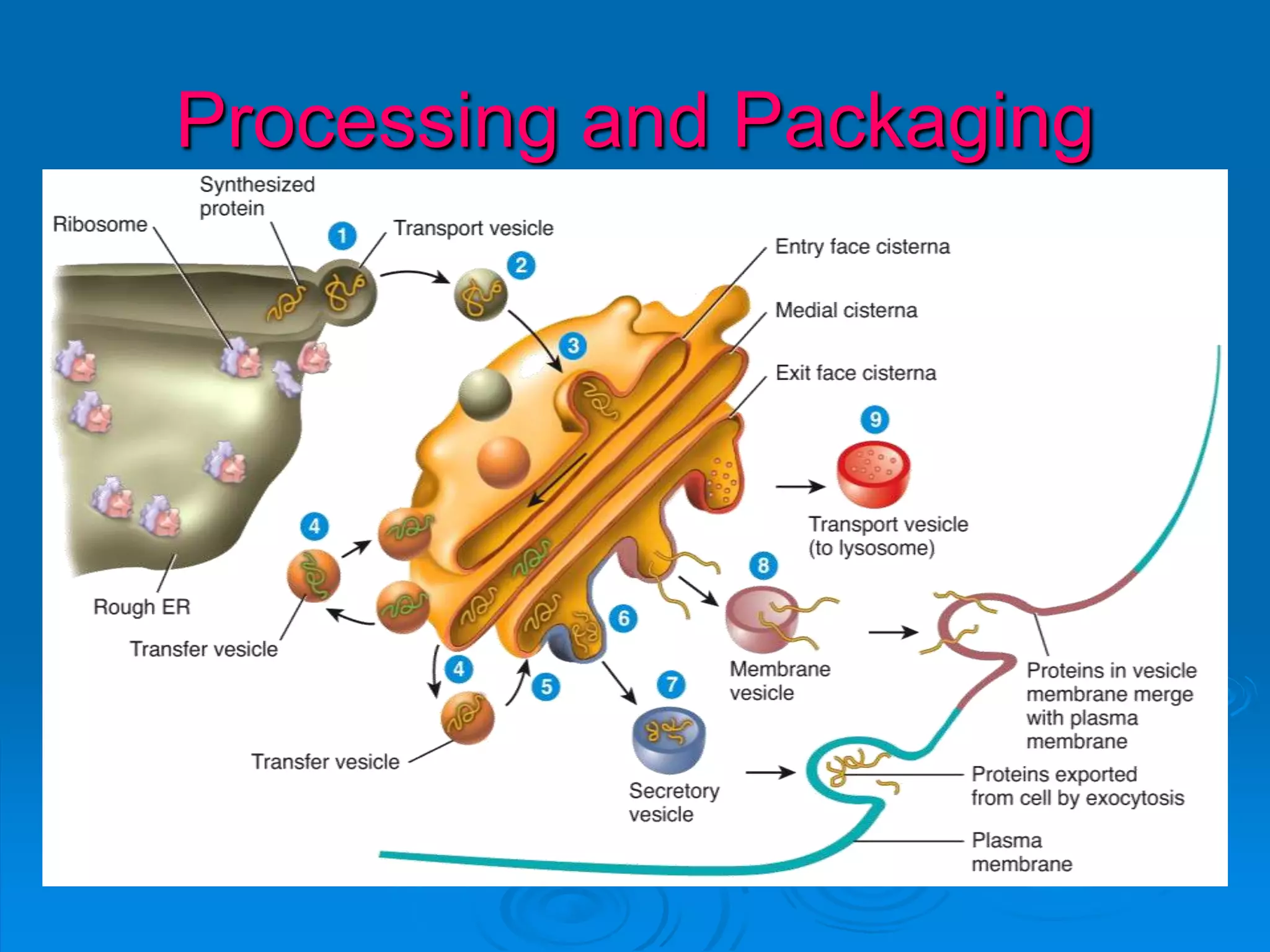
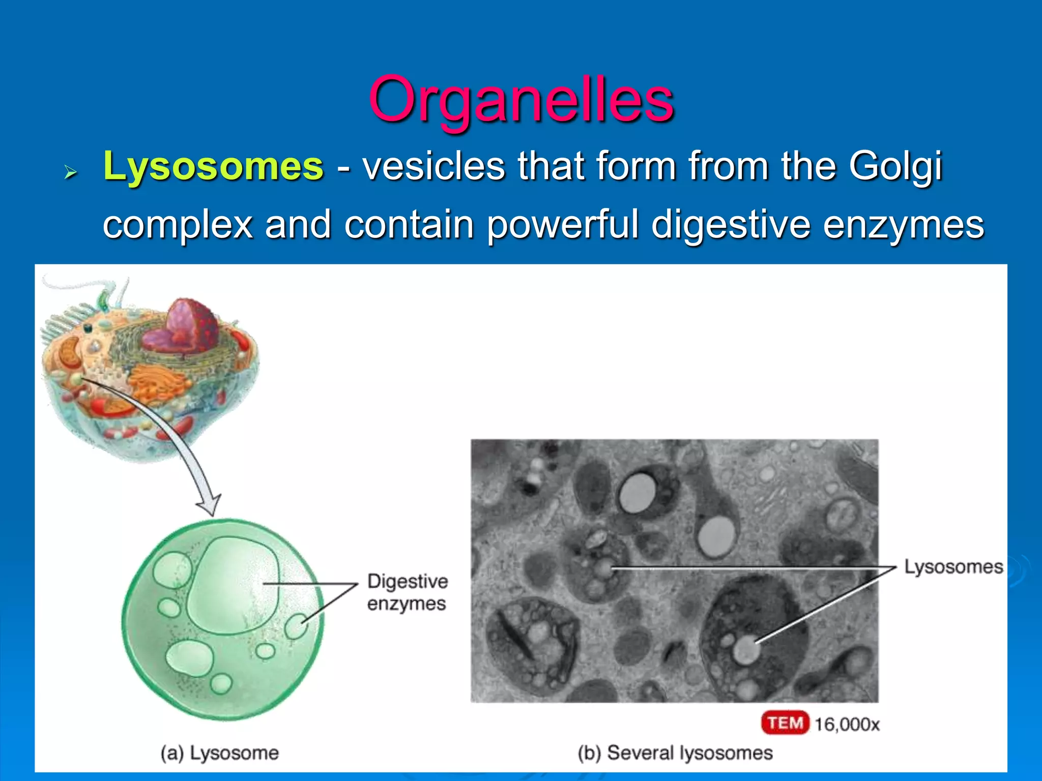
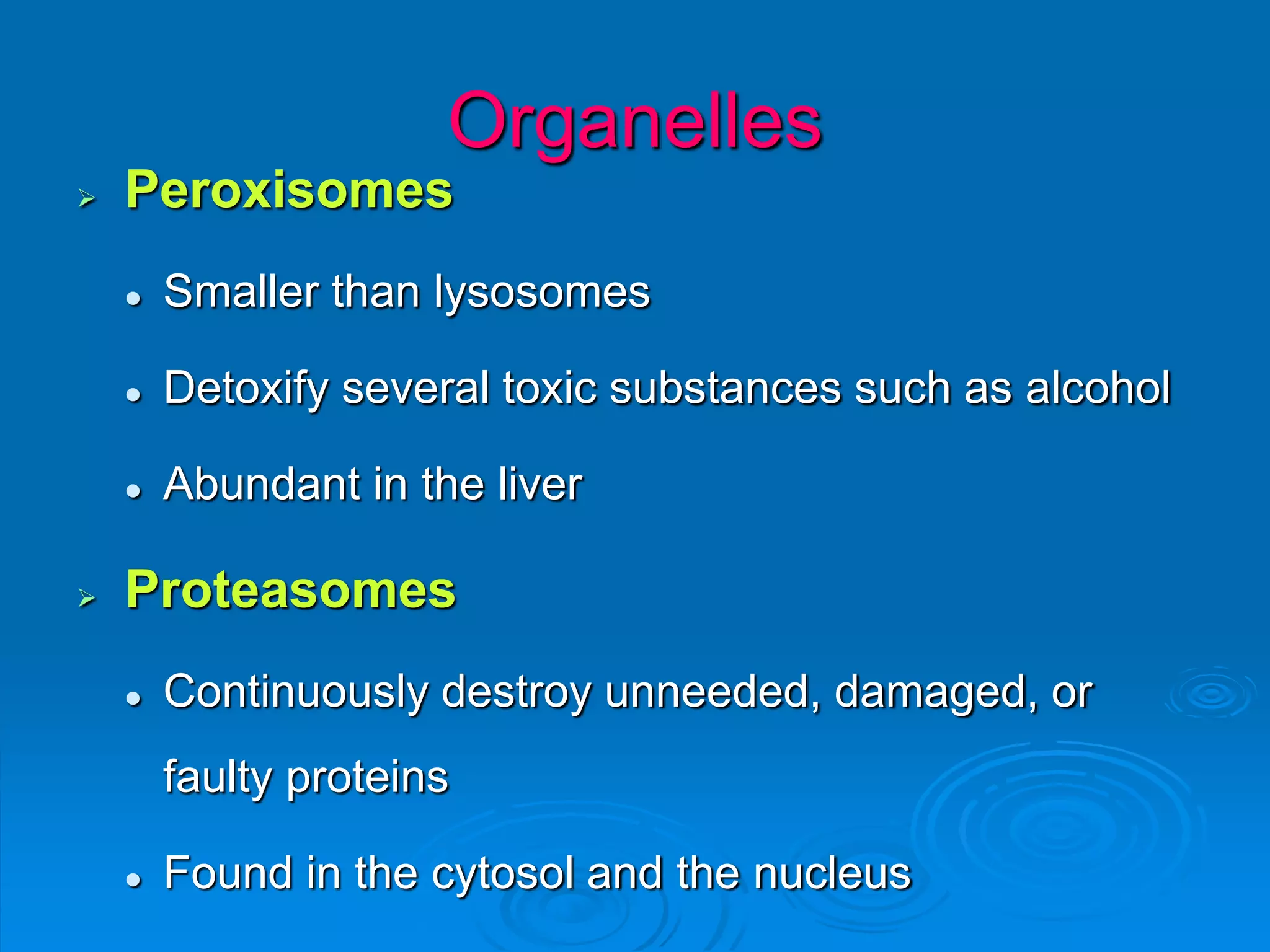
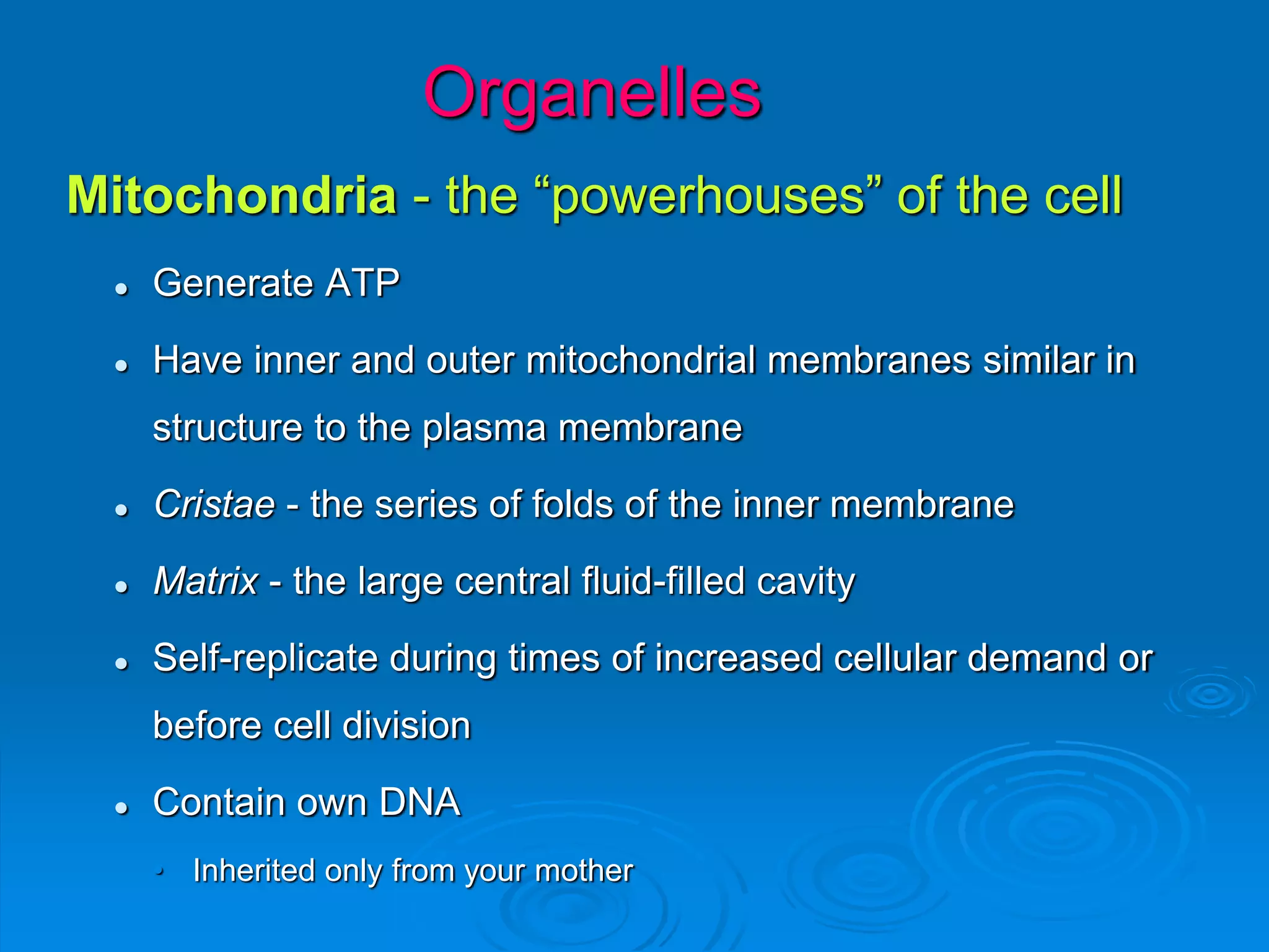
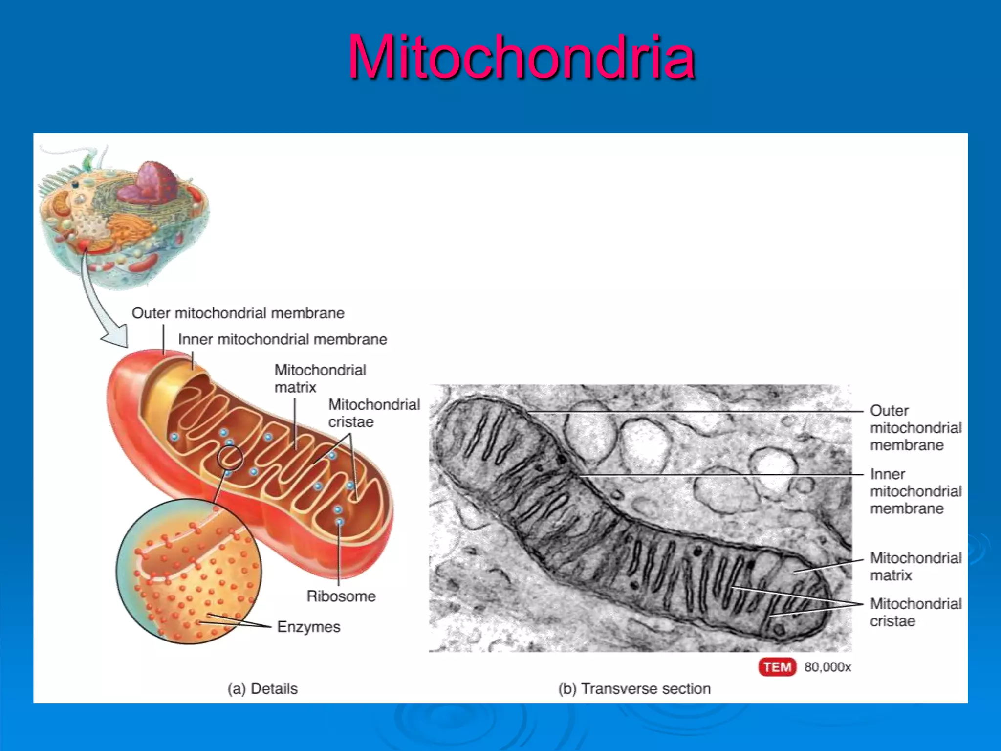
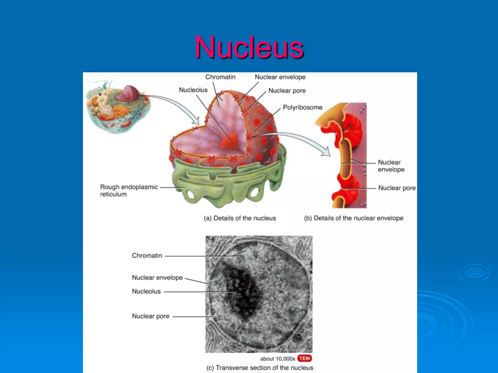
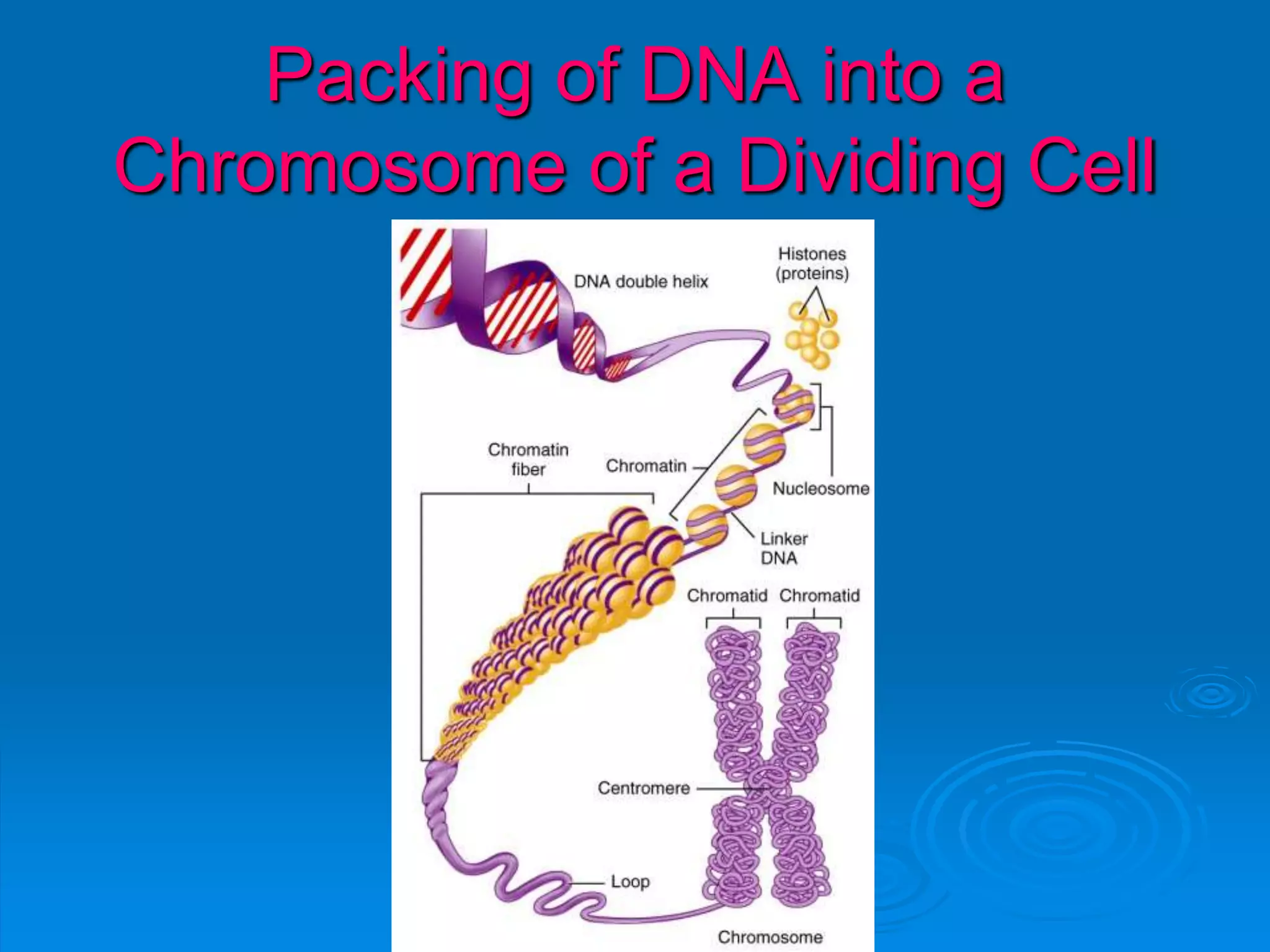
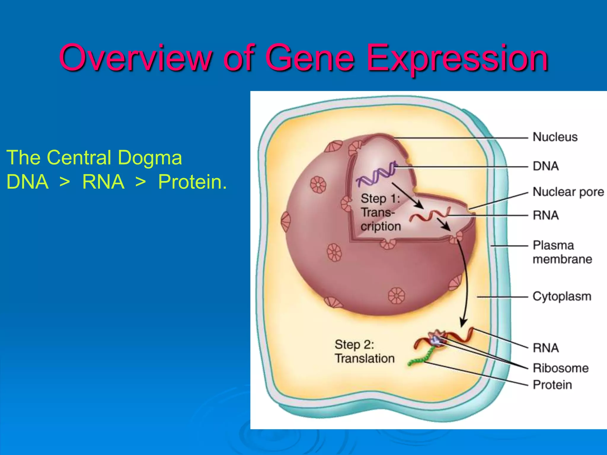
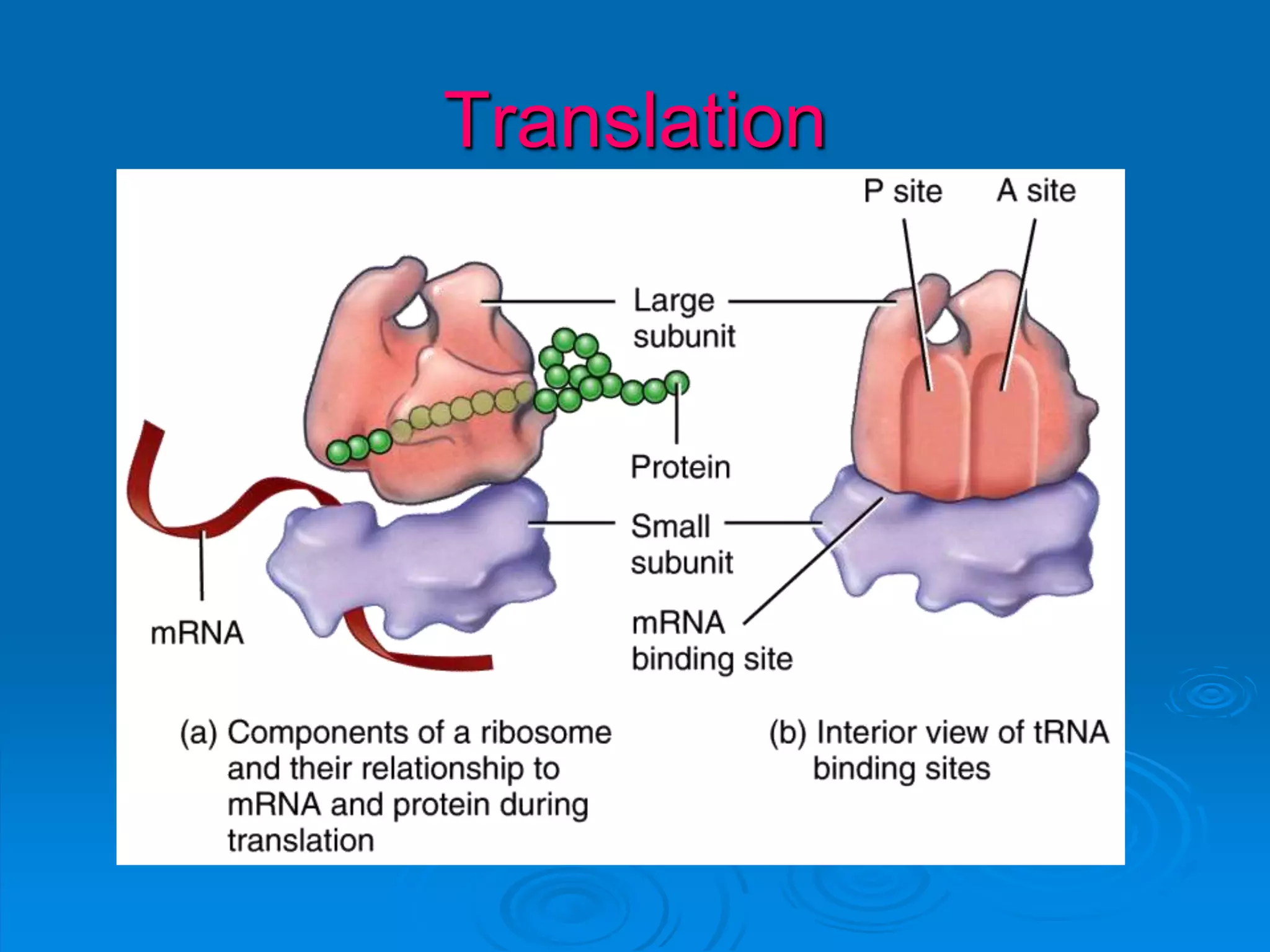
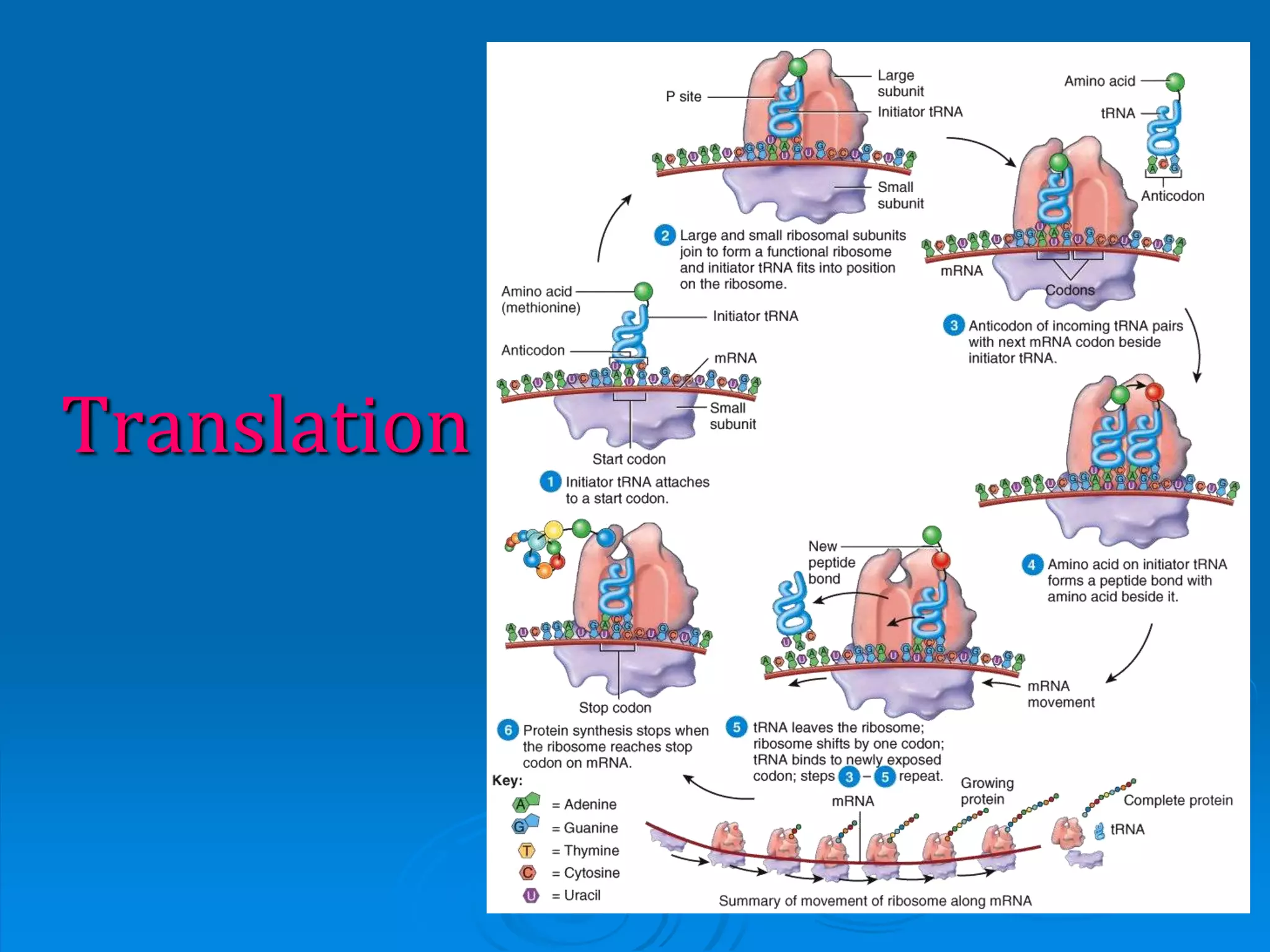
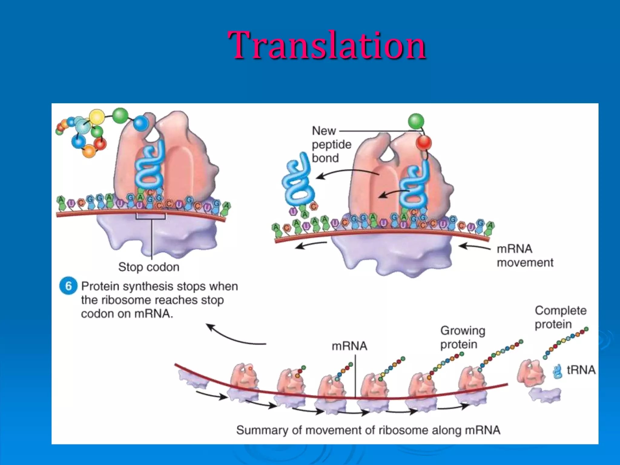
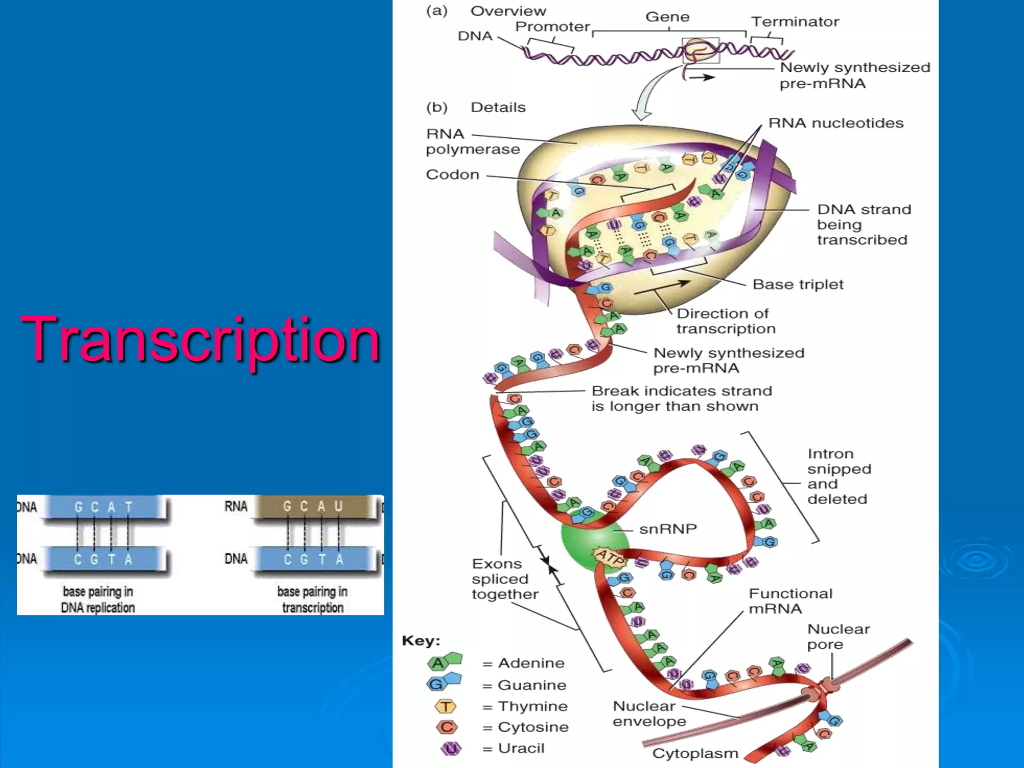

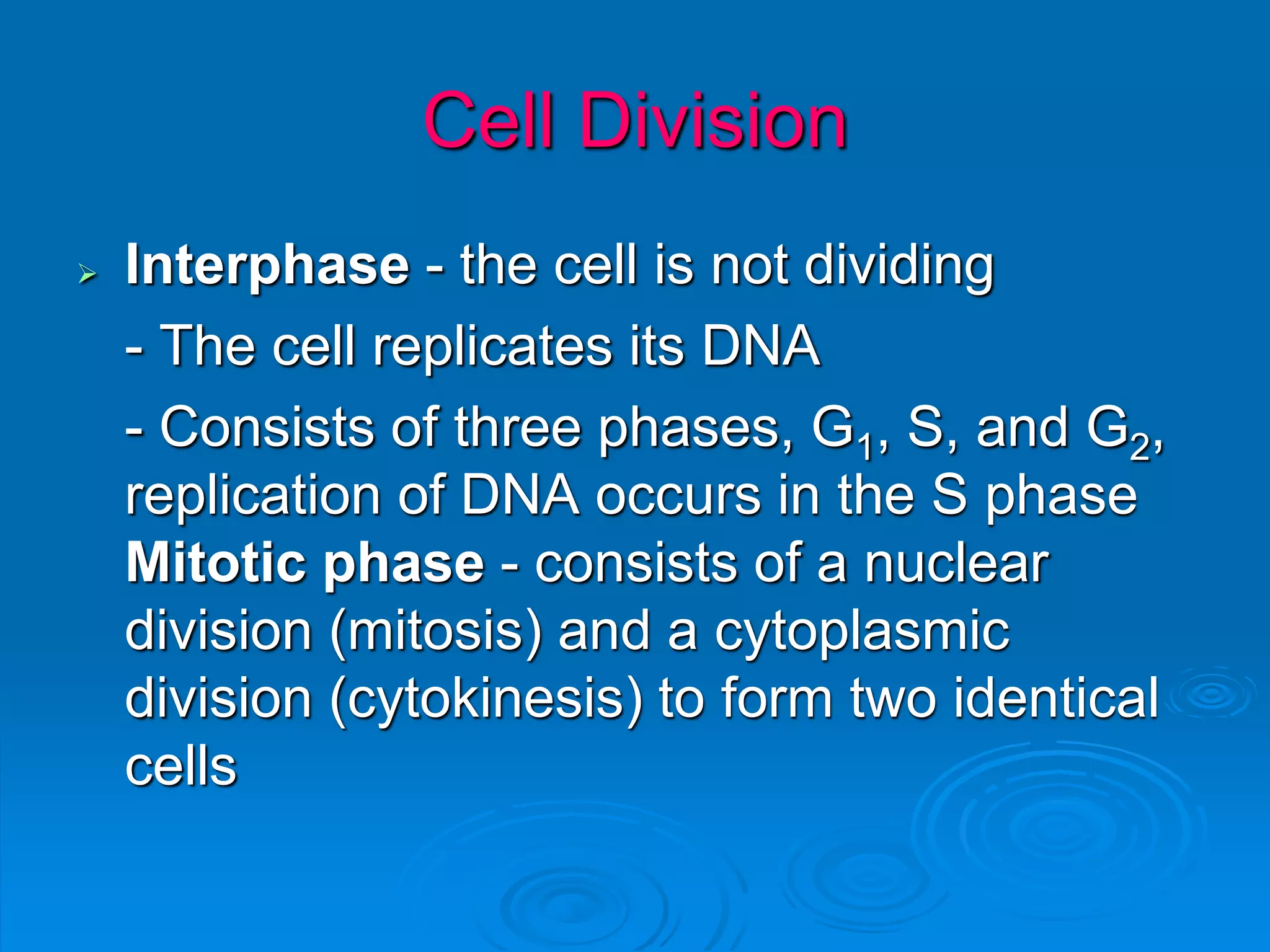
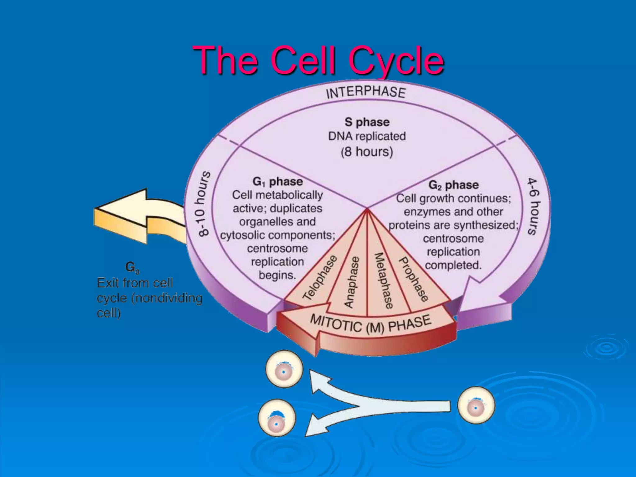
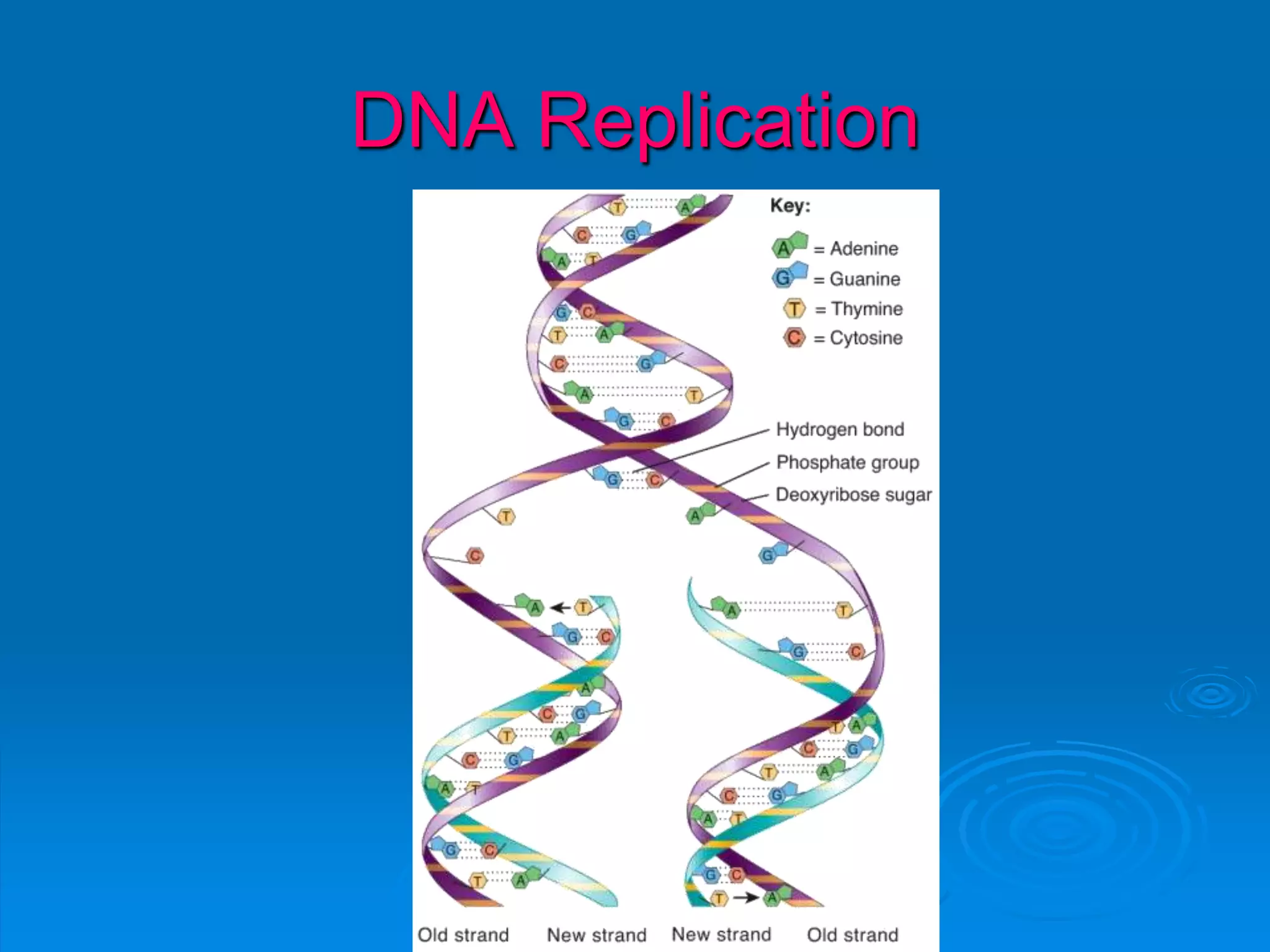
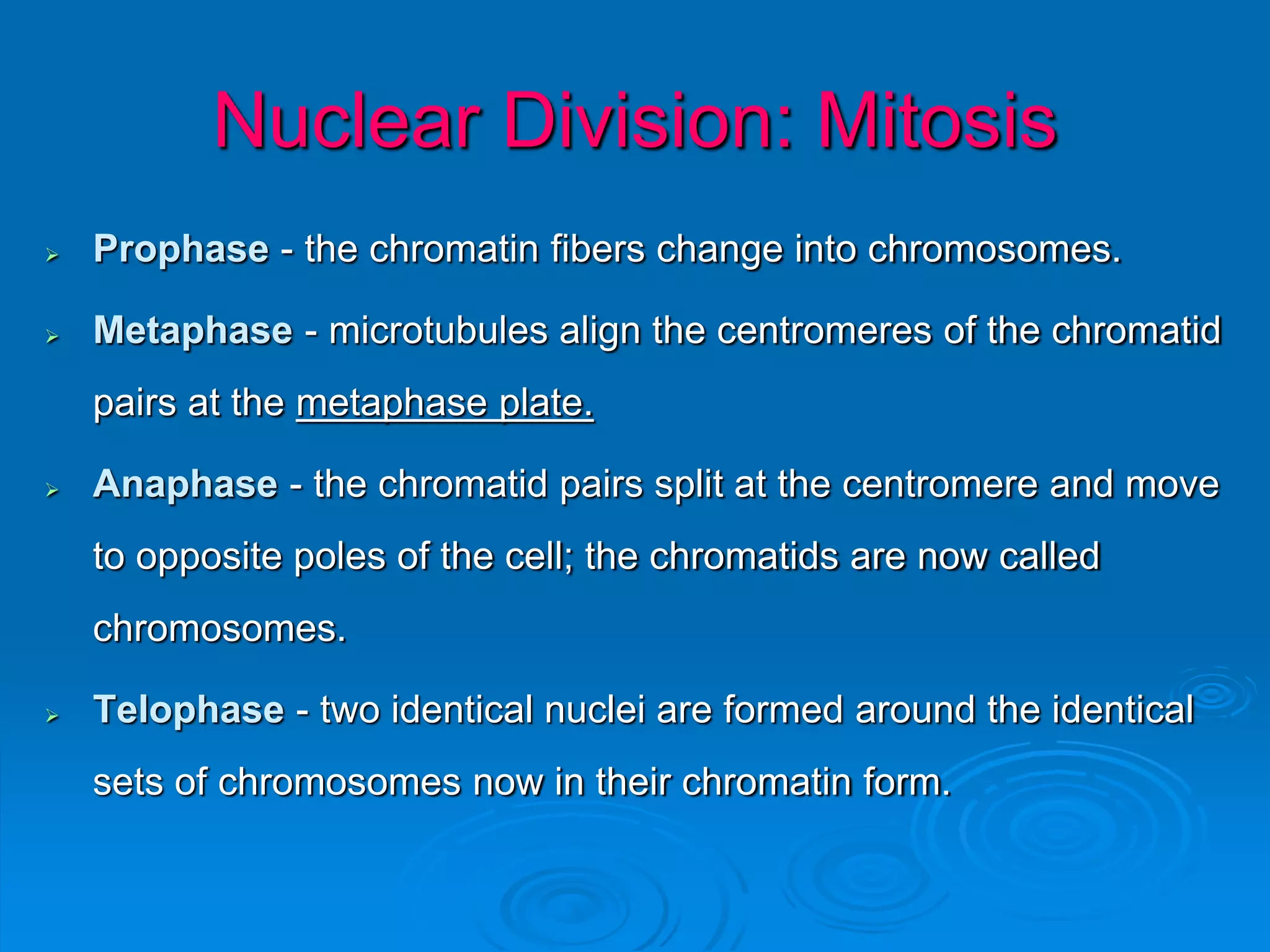
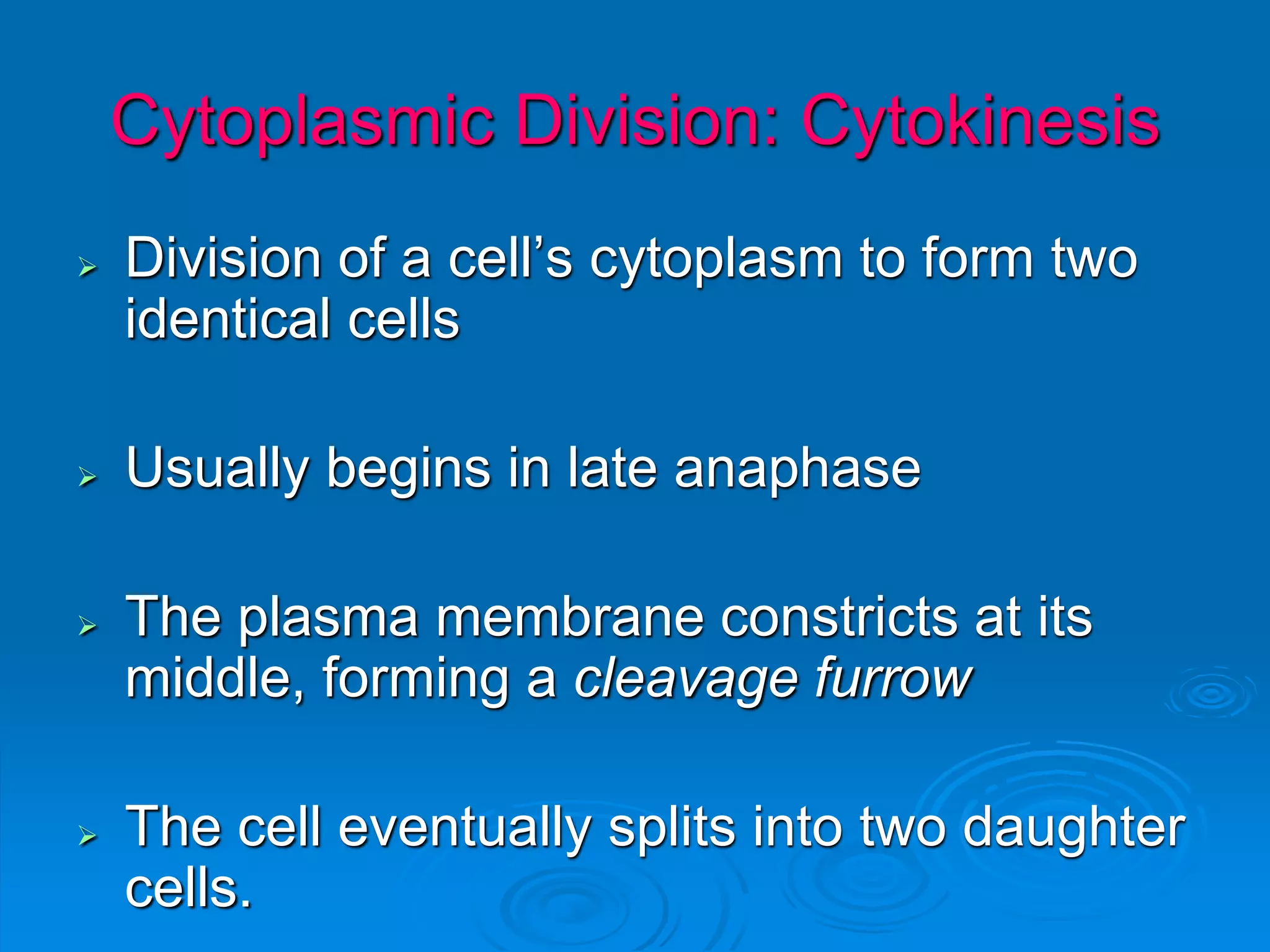
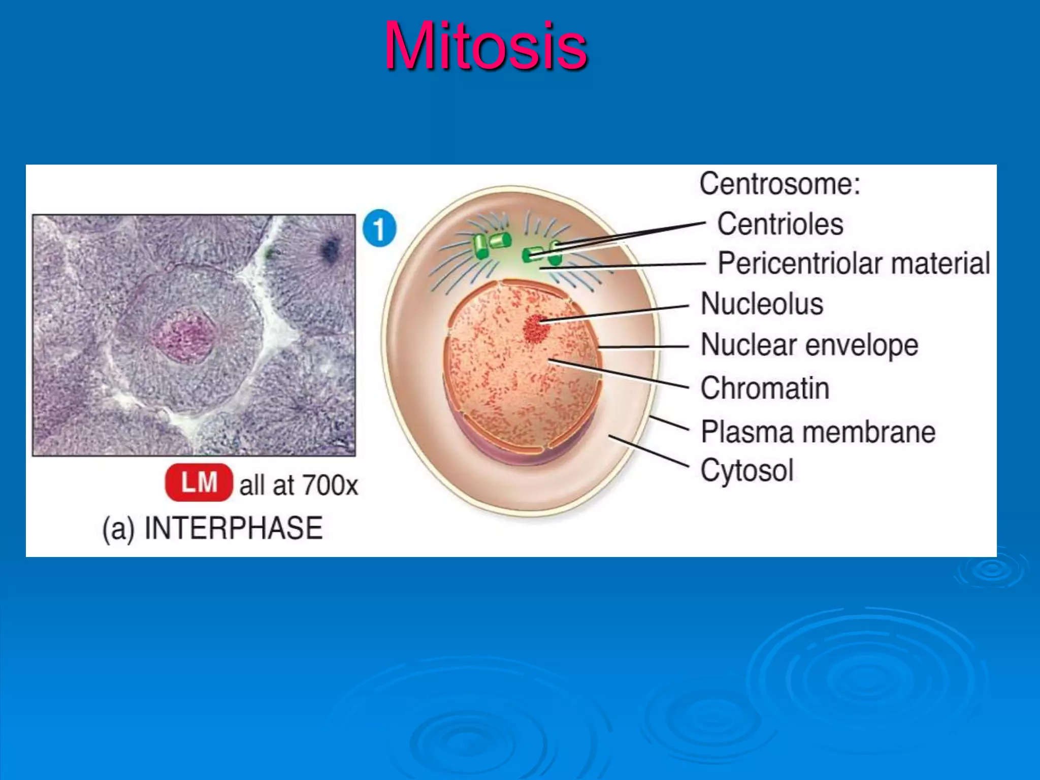
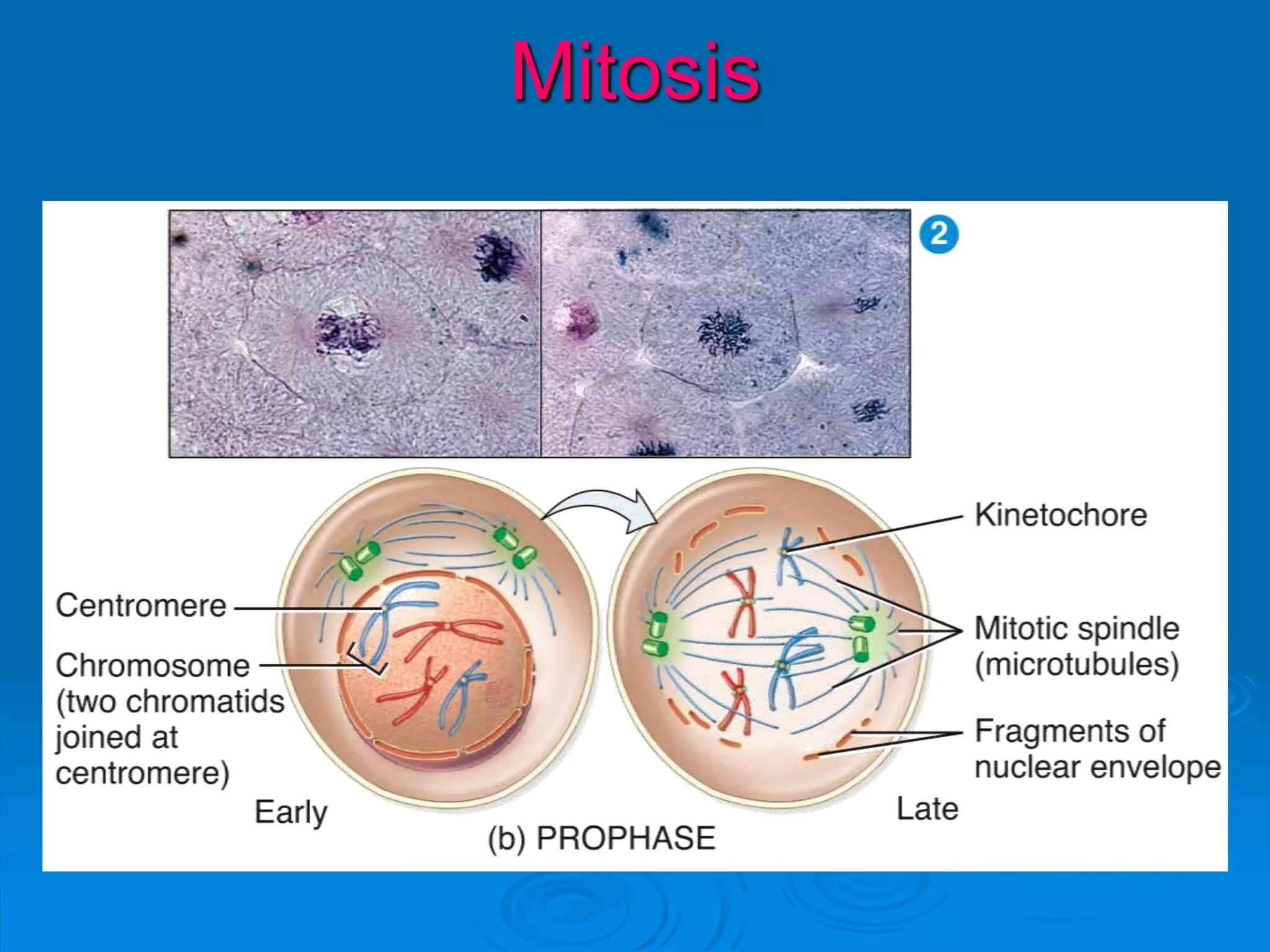
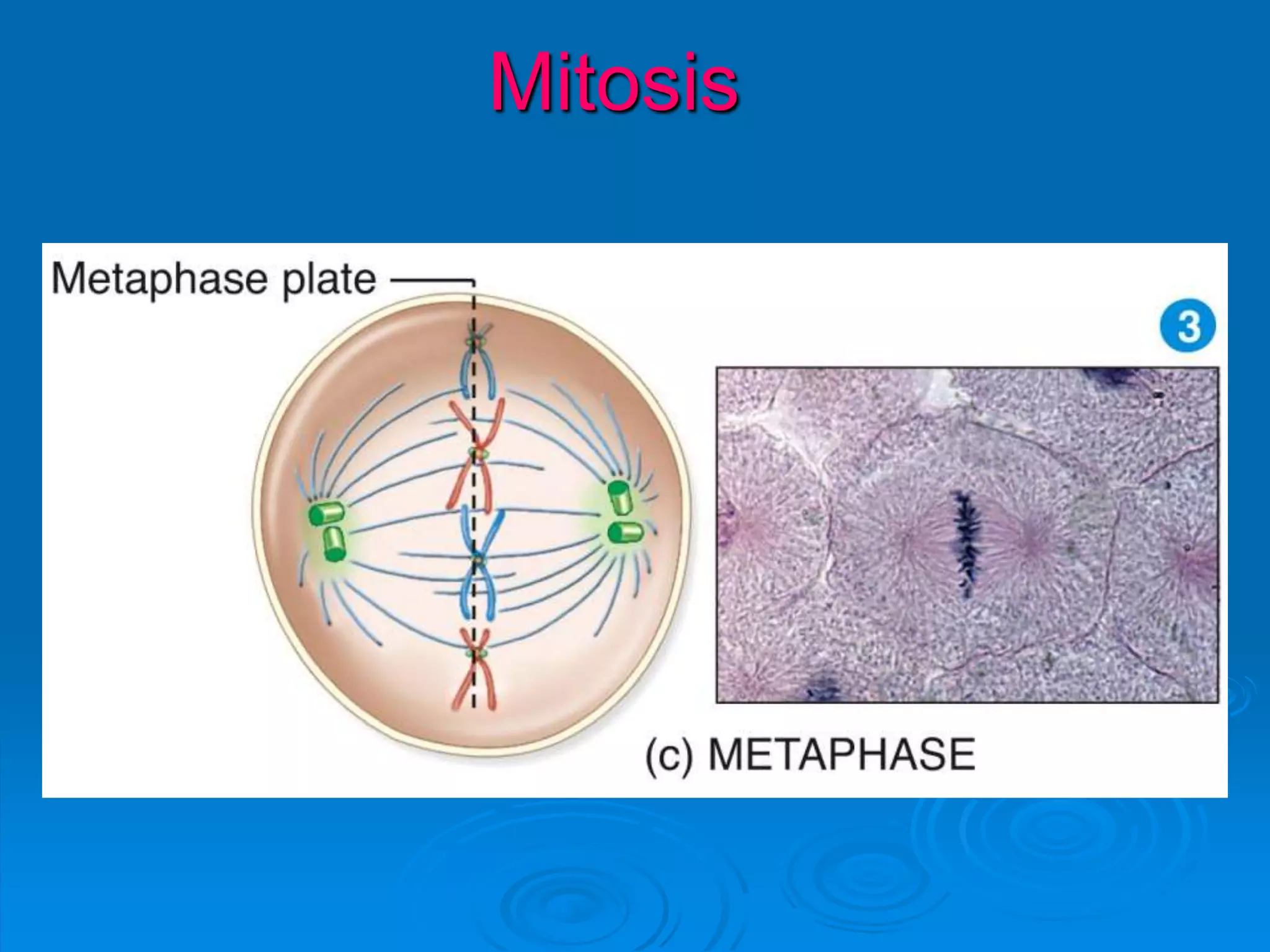
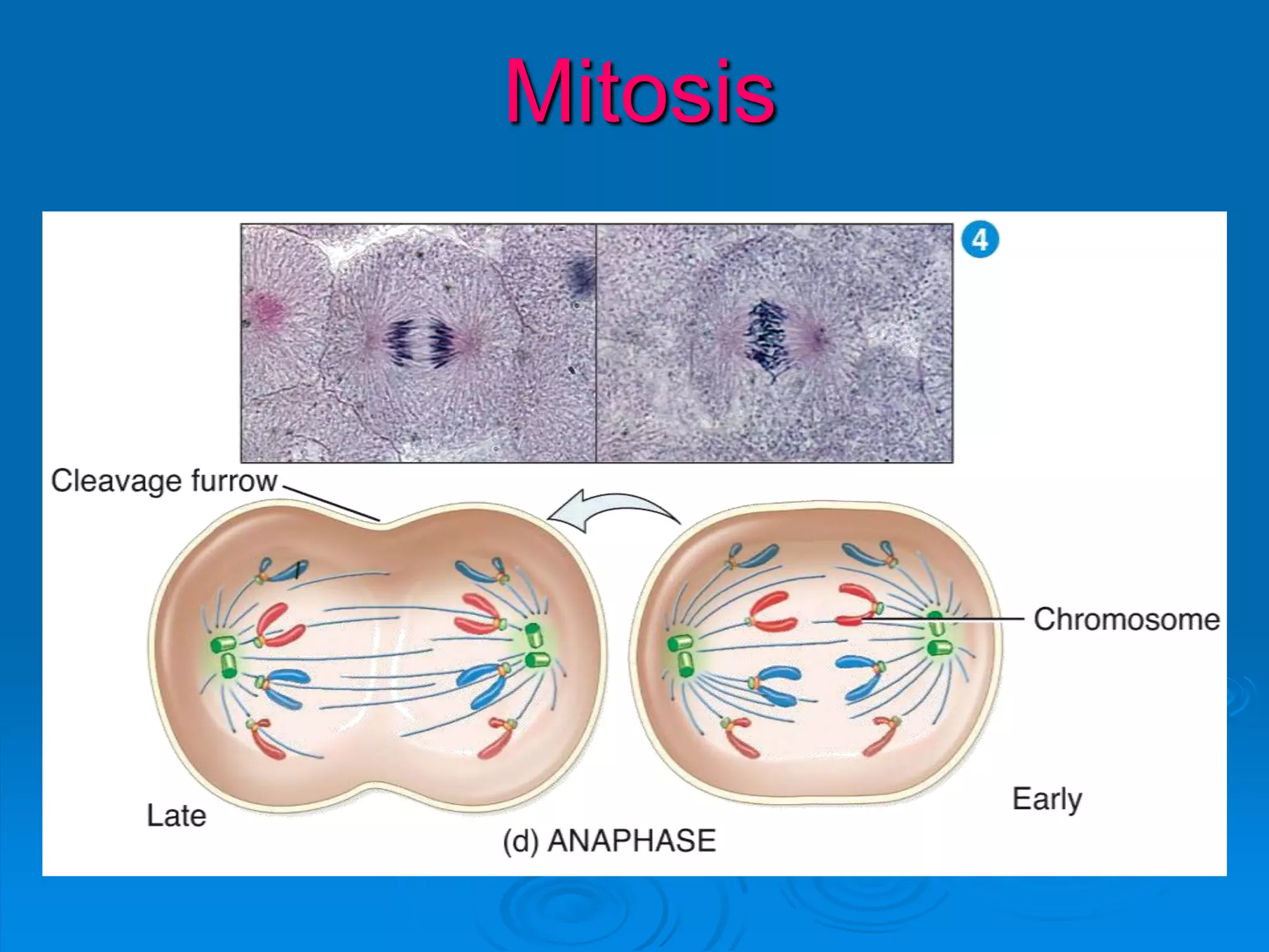
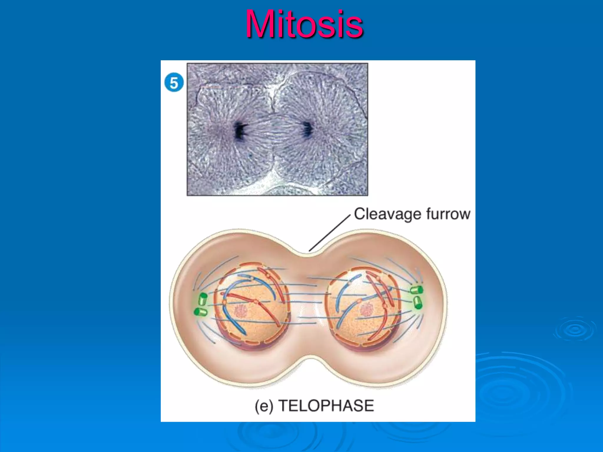
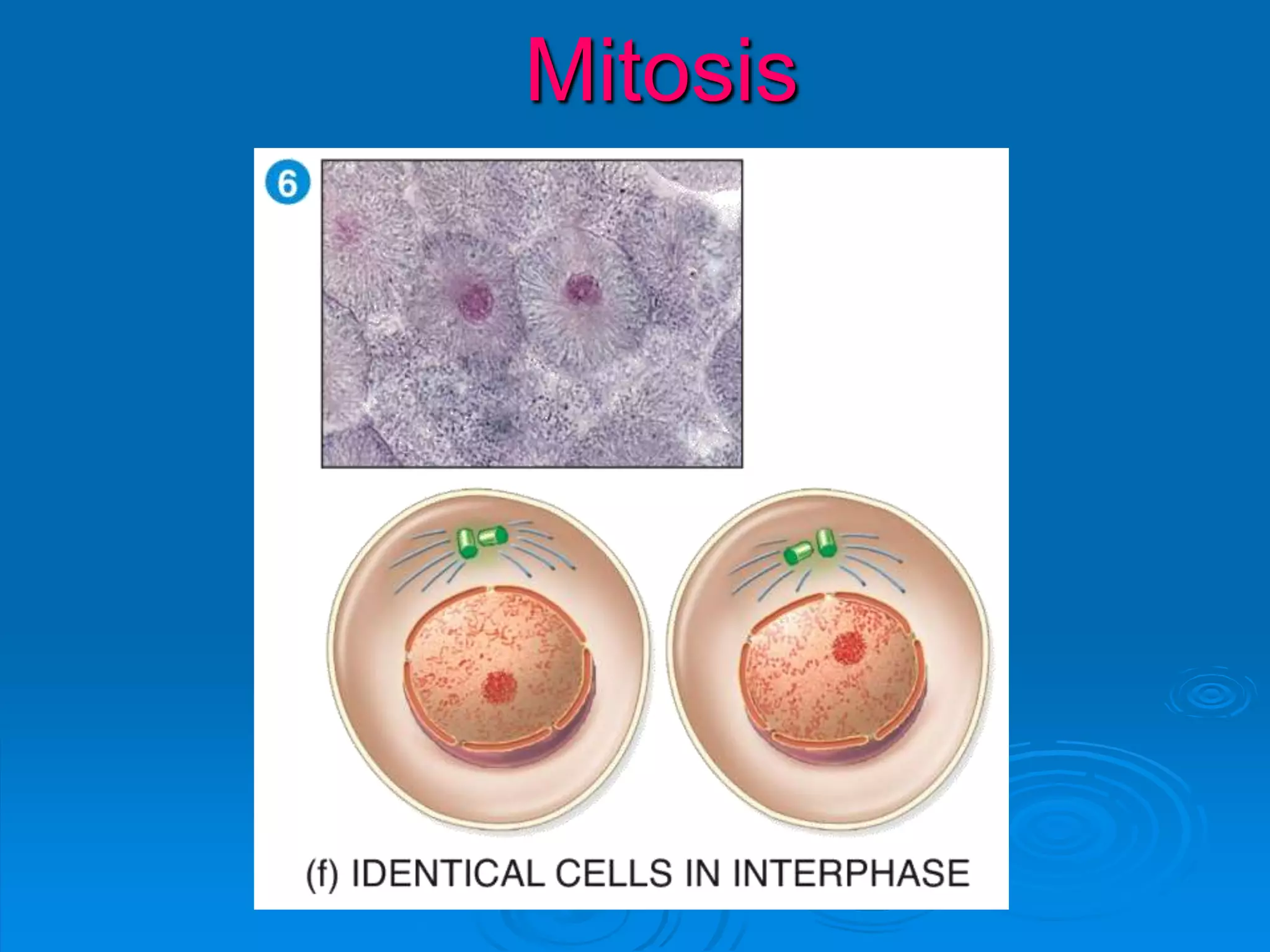
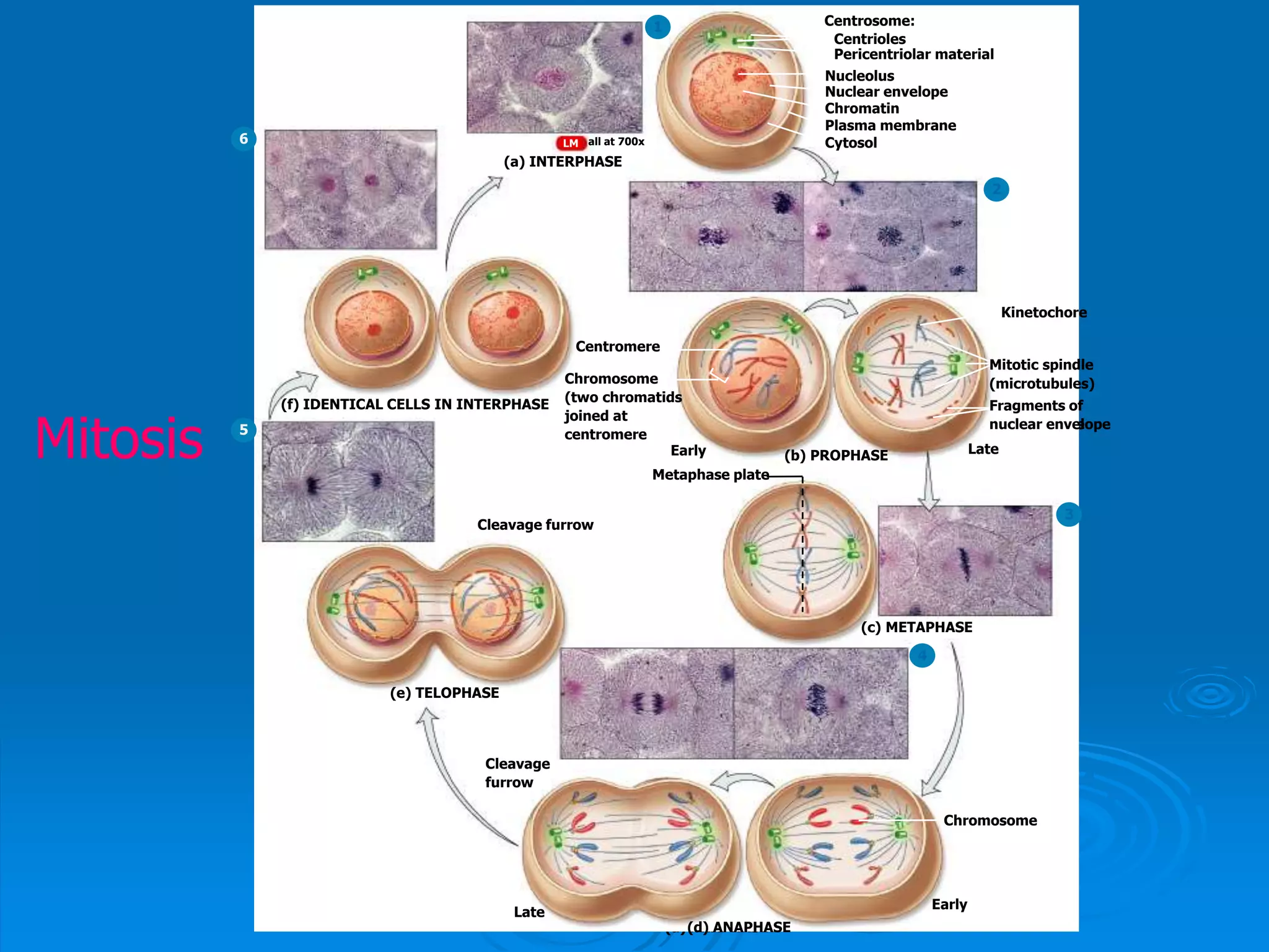

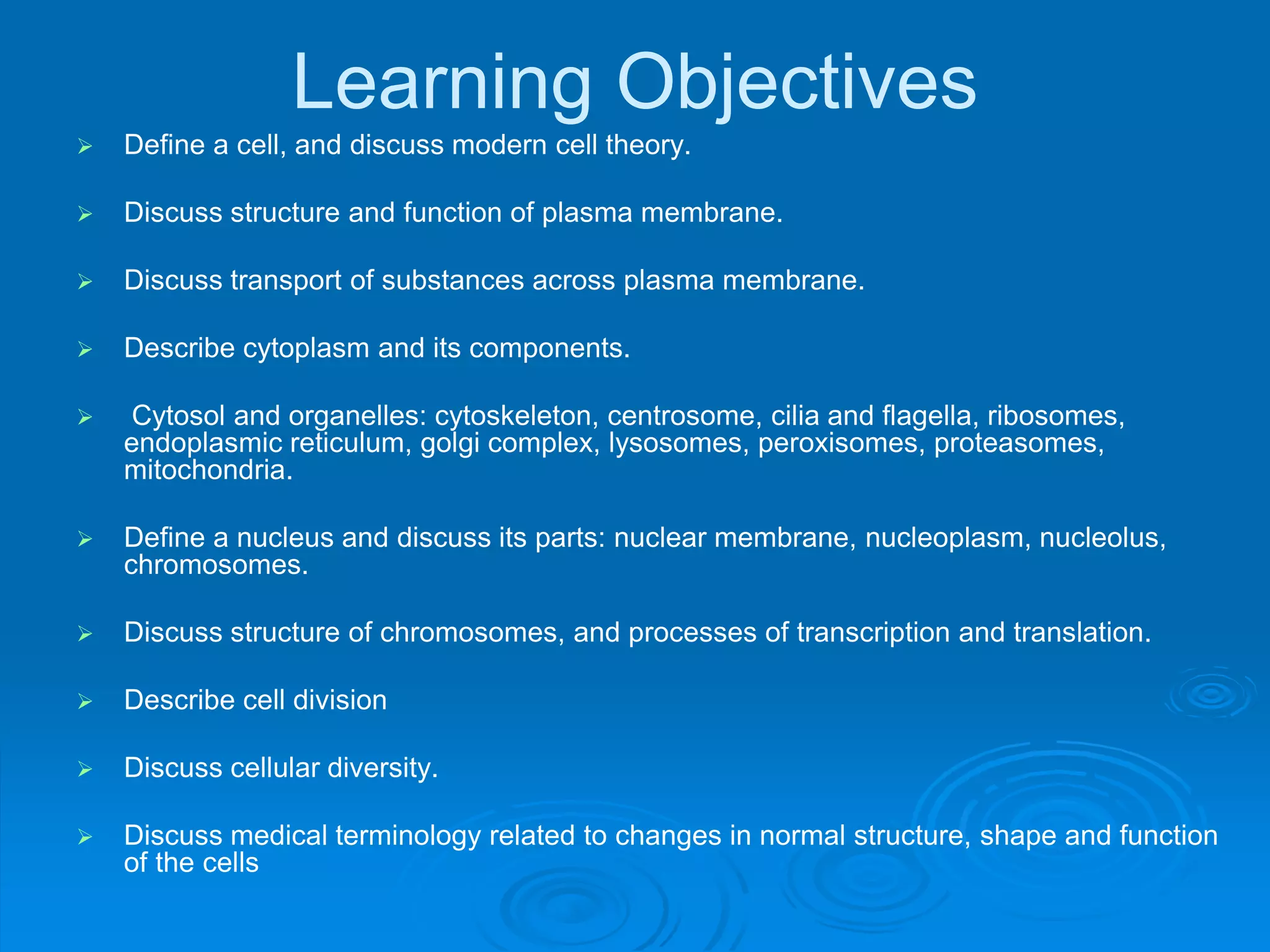
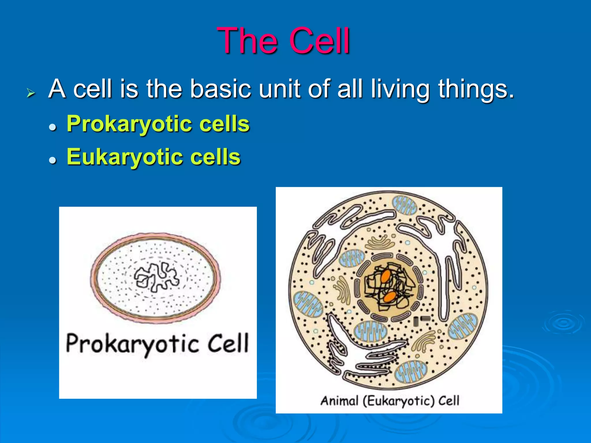
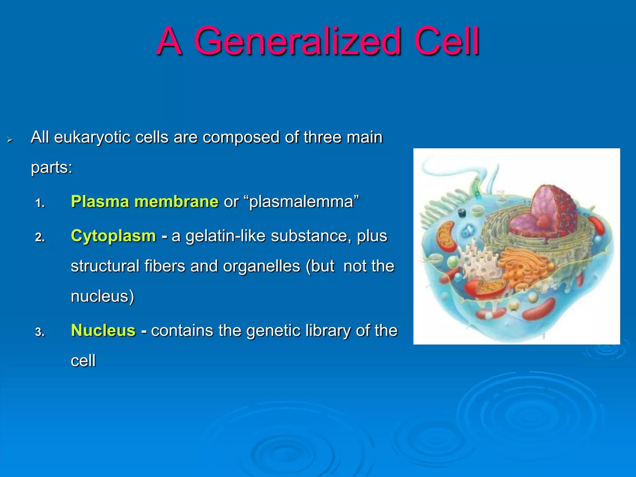
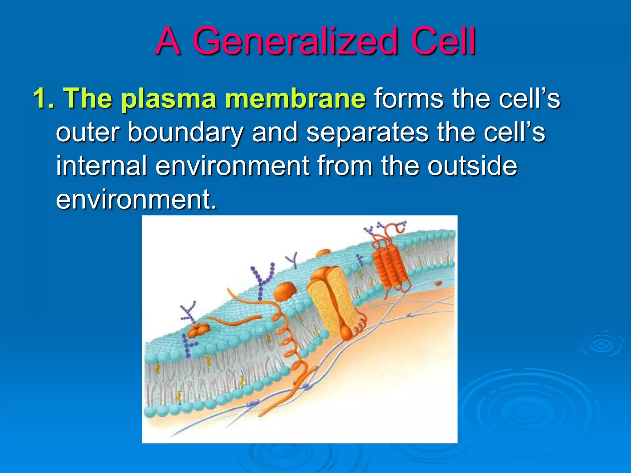
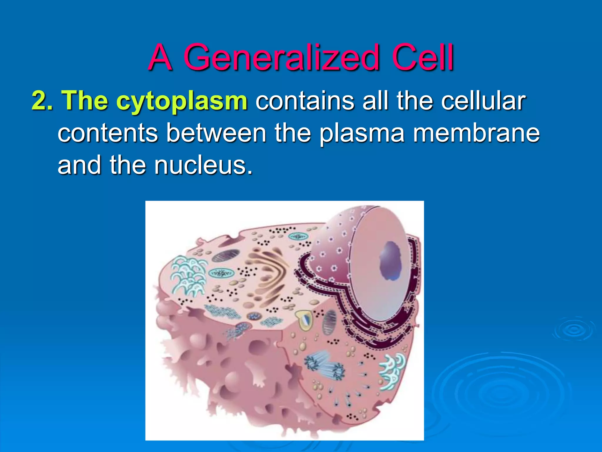
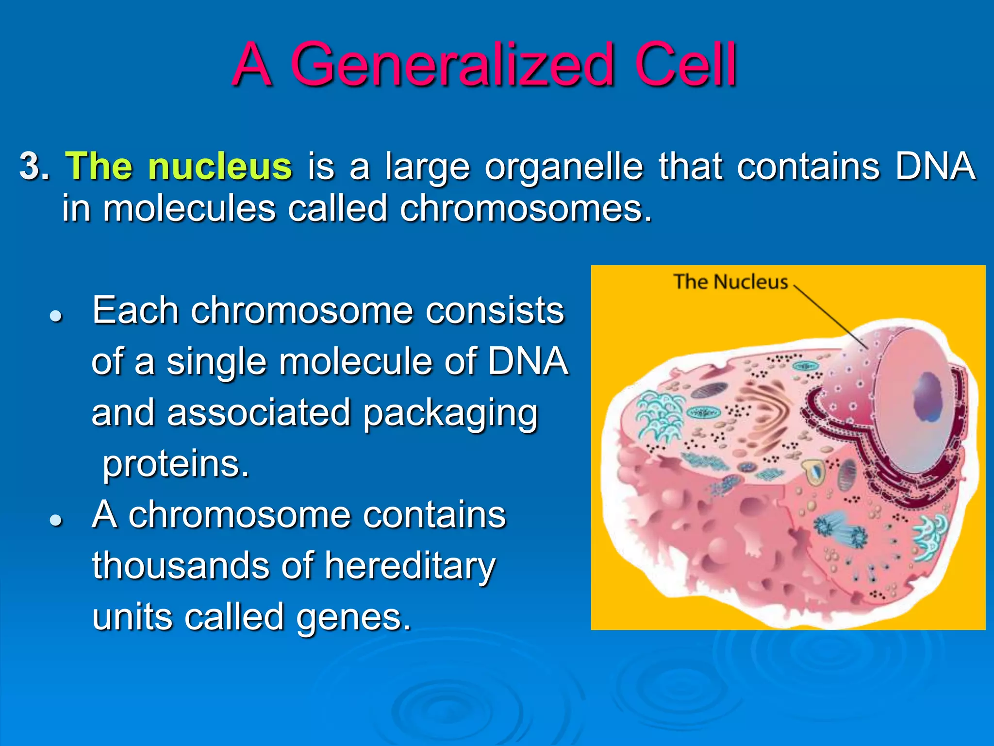
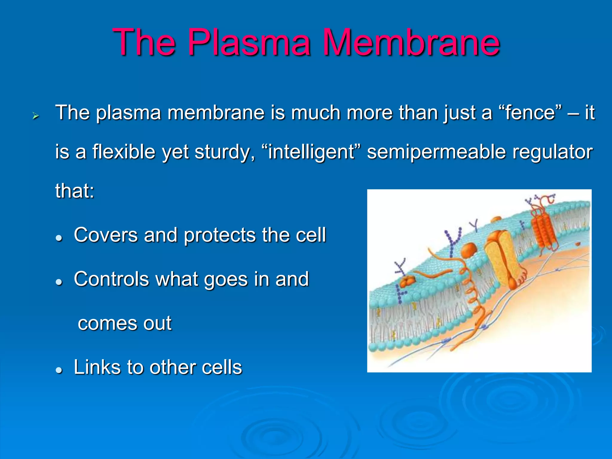
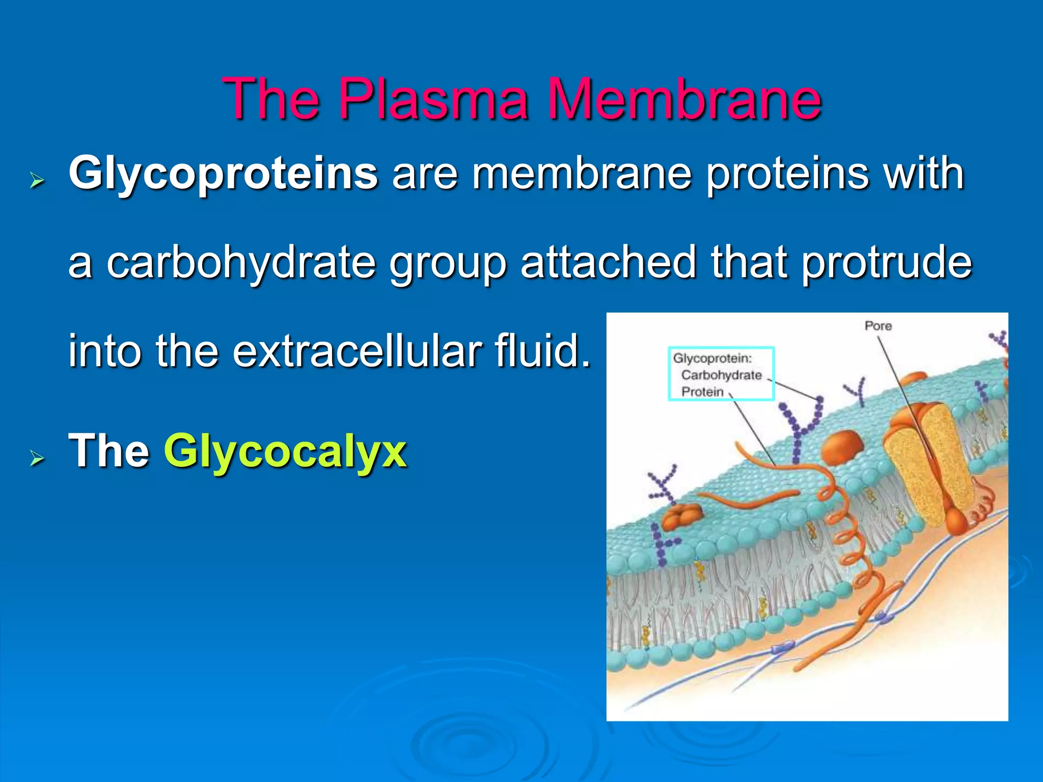
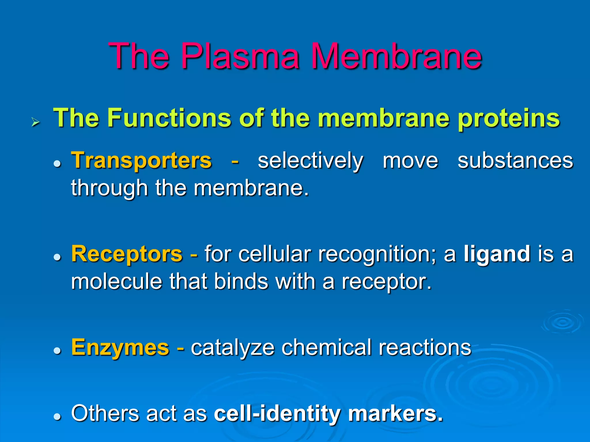
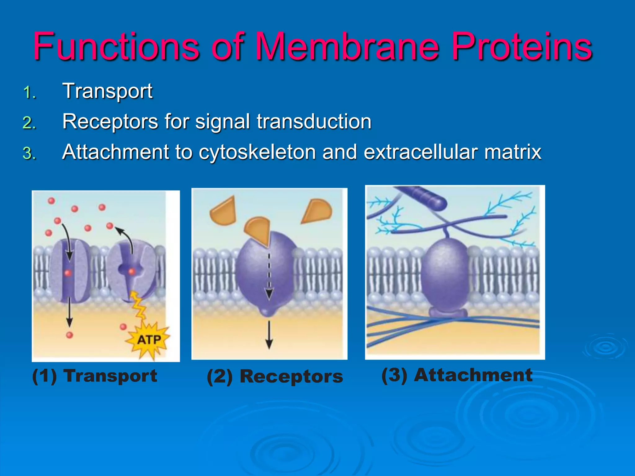
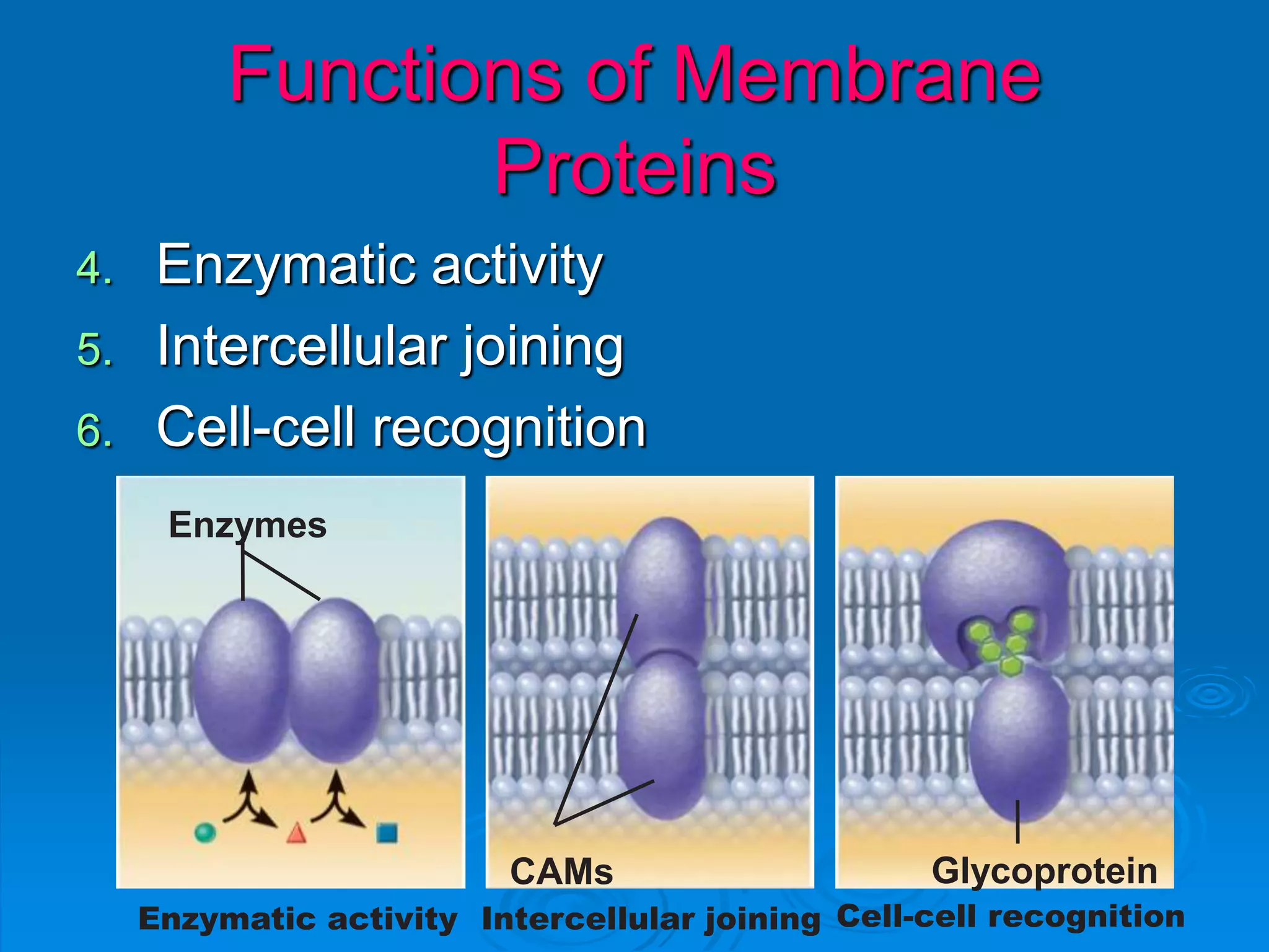
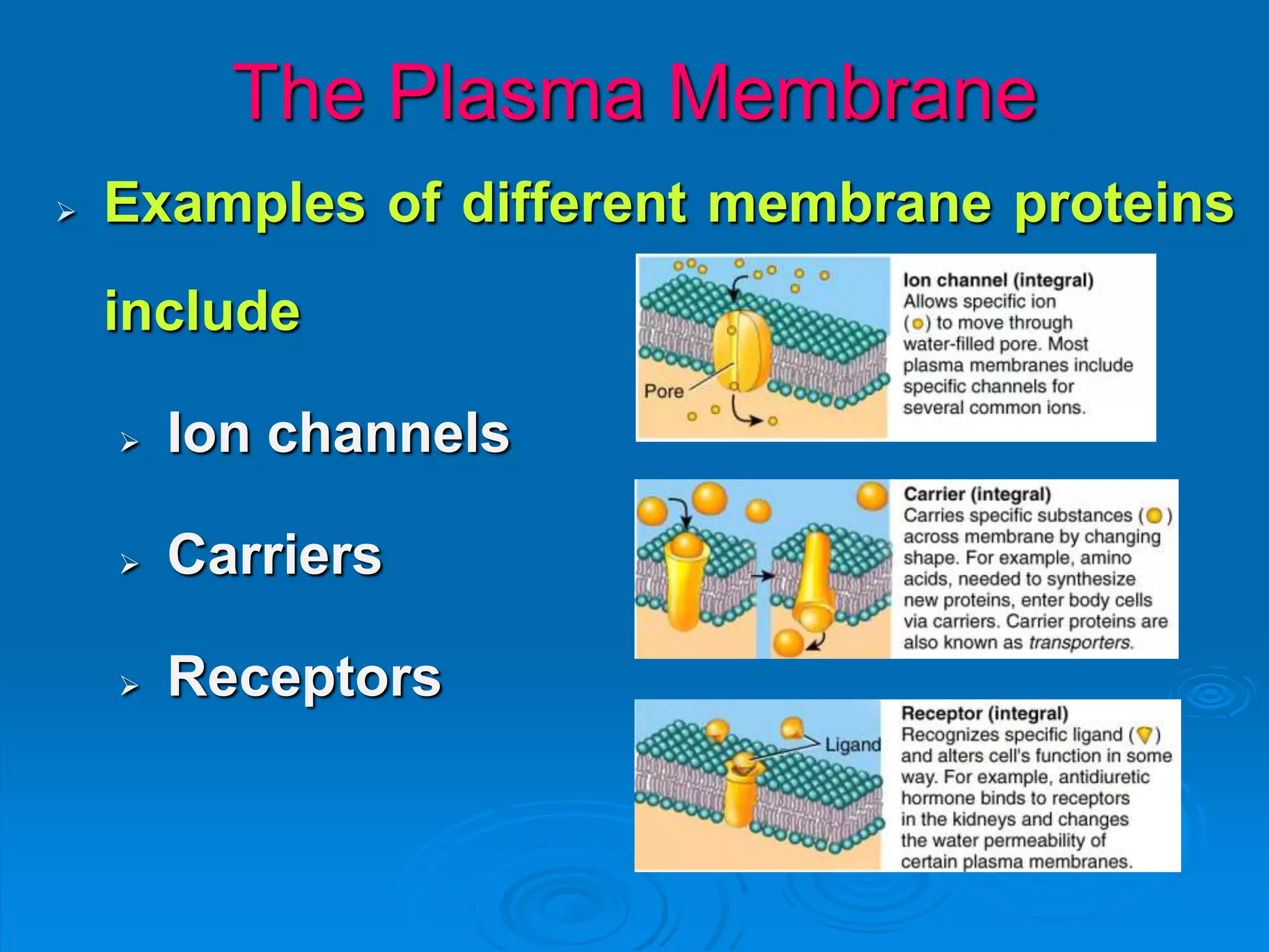
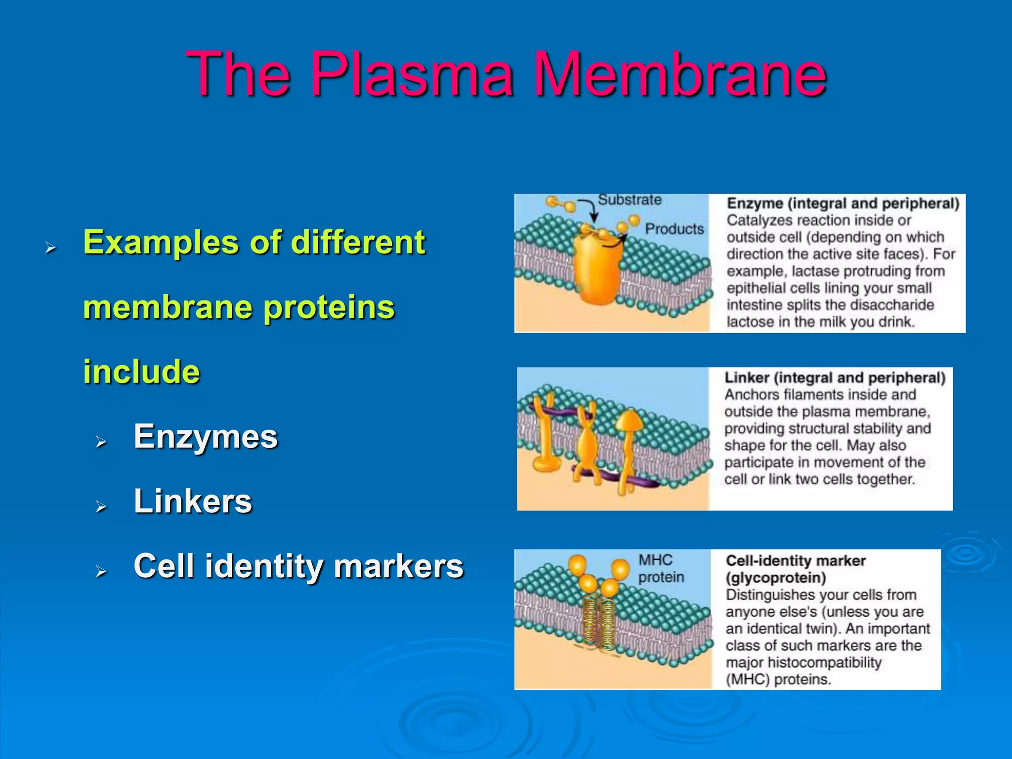
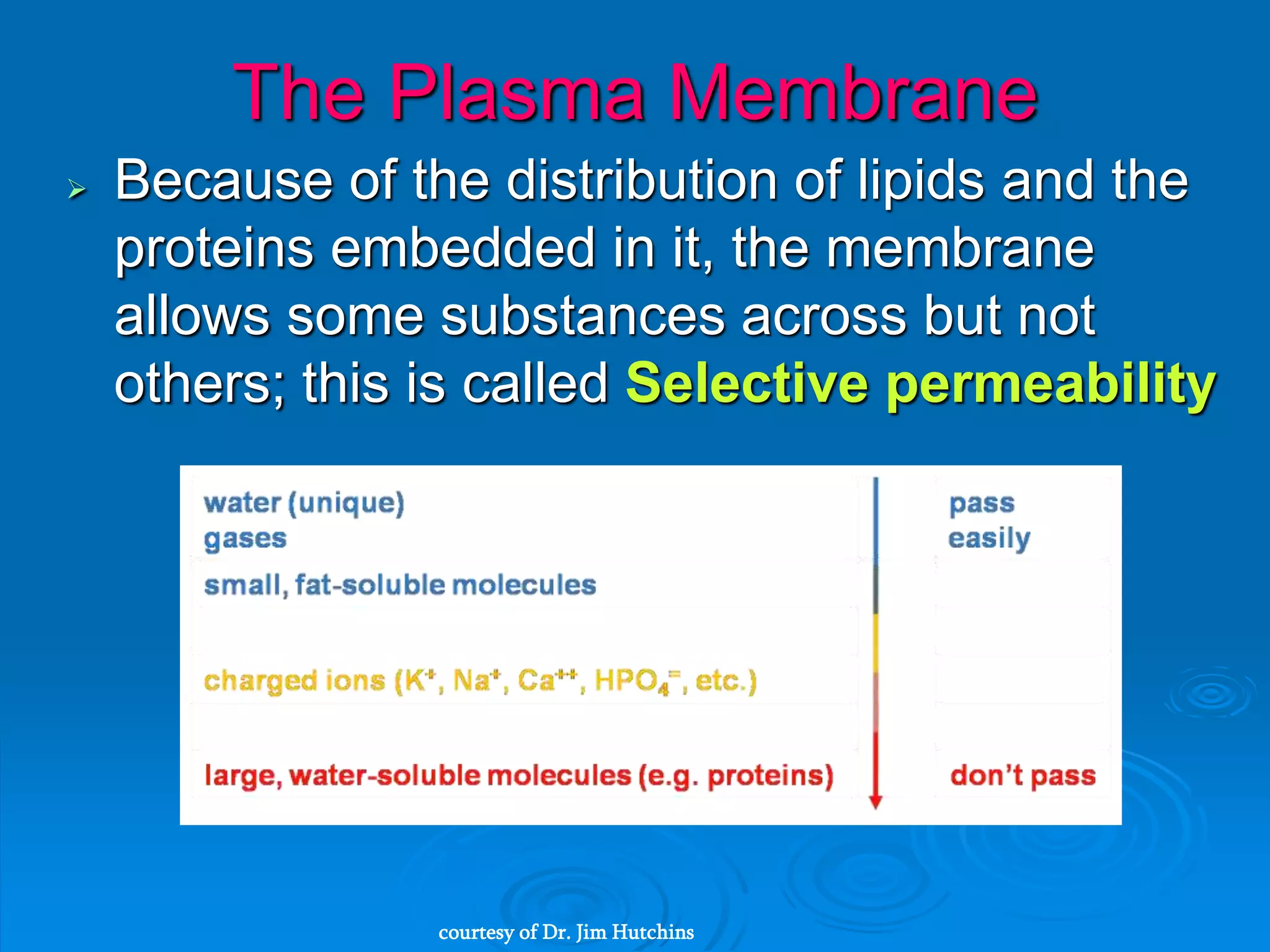
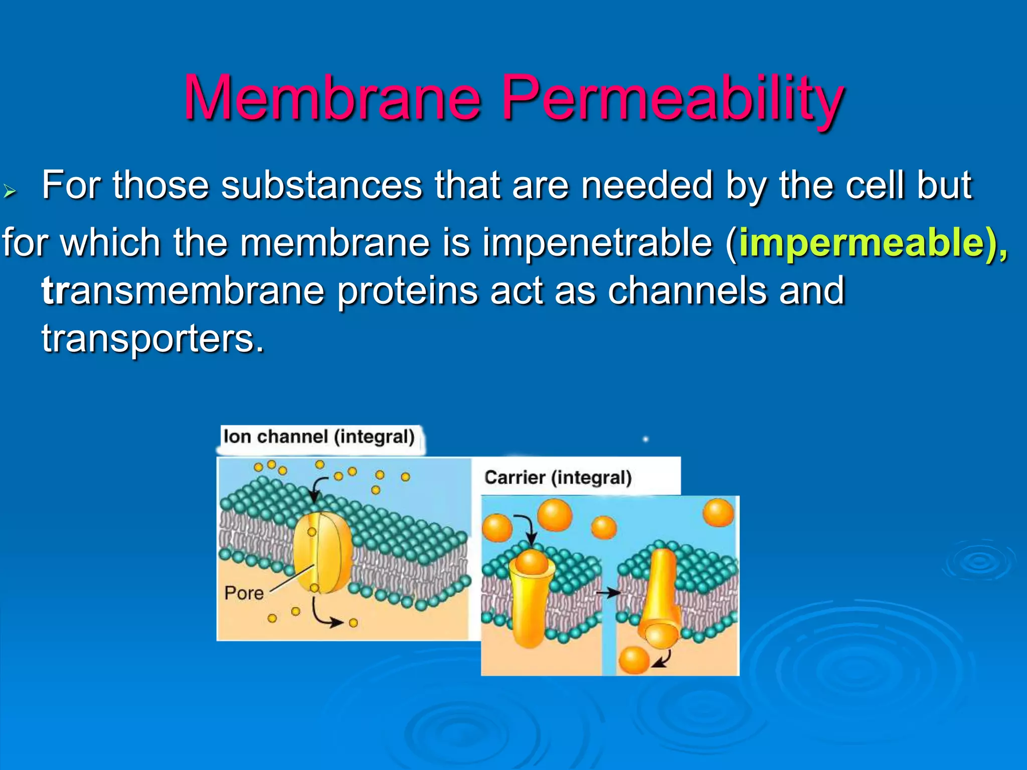
The document provides an overview of the cellular level of organization, including key concepts such as: - Cells are the basic unit of structure and function in living things, and all cells contain a plasma membrane, cytoplasm, and nucleus. - The plasma membrane is selectively permeable and regulates what enters and exits the cell. Transport across the membrane includes passive diffusion and active transport processes. - The cytoplasm contains cytosol and various organelles that carry out specialized functions. Organelles include the endoplasmic reticulum, Golgi complex, lysosomes, mitochondria and more. - The nucleus houses the cell's DNA within chromosomes and controls gene expression through transcription and translation. Cell division occurs through mitosis and