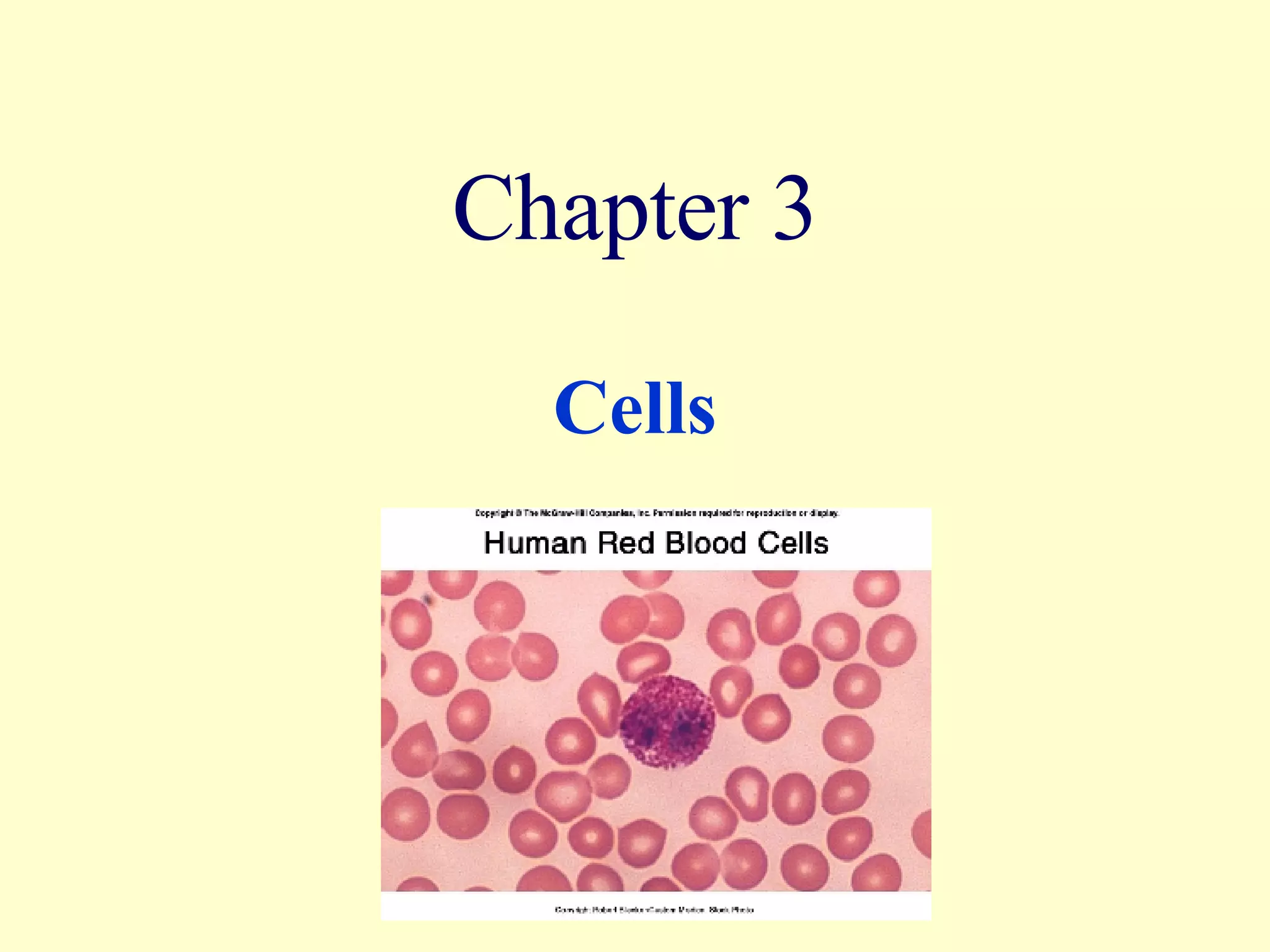
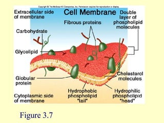
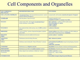
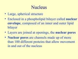
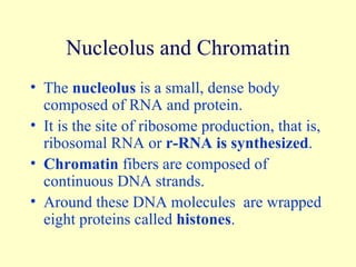
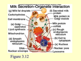
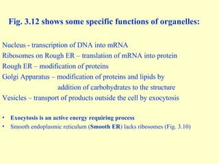
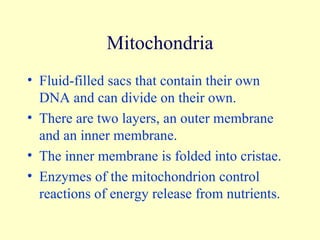
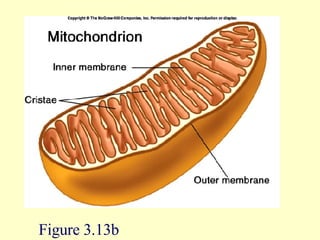
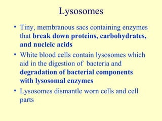
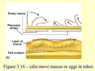
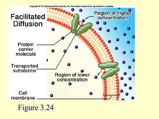
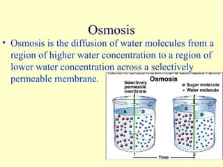
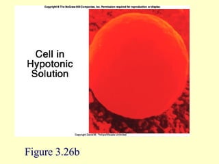
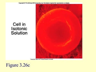
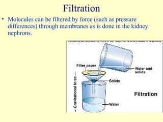
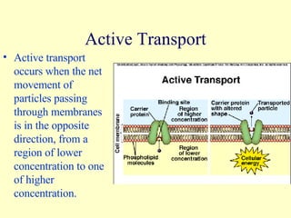
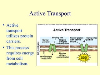
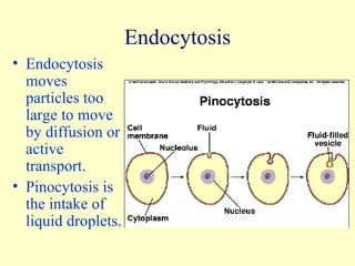
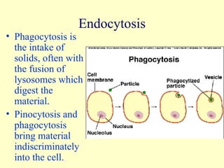
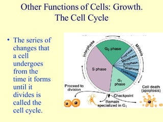
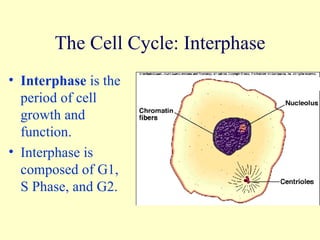
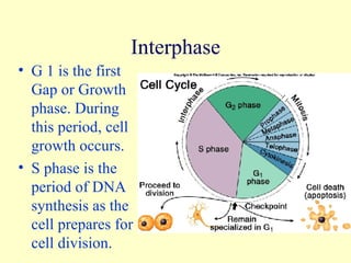
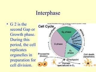
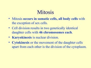
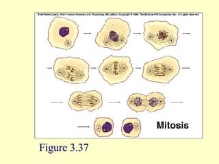
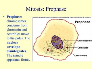
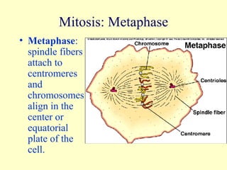
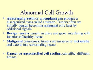
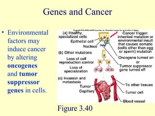
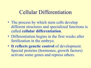
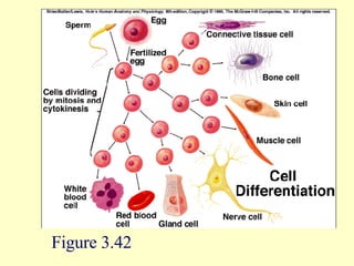
Chapter 3 Cells
You are responsible for the following figures and tables : Fig. 3.1 - The cells of the human body.  Fig. 3.2 - Specialized cells.  Fig. 3.42. Fig. 3.3 - Generalized diagram of a universal animal cell.  Fig. 3.6 - Phospholipid bilayer  Fig. 3.7 - Embedded in this cell membrane are transmembrane proteins. Fig. 3.8 - cells communicate with each other  Fig. 3.12 - representation of a cell.  Fig. 3.12, 3.29, 3.30, 3.31 – endocytosis and exocytosis. Fig. 3.21 - Passive transport does not require energy.  Tab. 3.4 - summary of transport mechanisms in and out of the cell. Fig. 3.36 - cell cycle - Name the phases. Fig. 3.37 - Name the phases of cell division, describe them. Define ' cancer'. Tab. 3.6.
Cells The adult human body consists of about 75 million cells, the basic units of the human body.  All precursor cells, also called  stem cells , are alike and  omnipotent . They only  differentiate into specialized cells  as they mature (Fig. 3.42). Whether precursor or specialized, all body or somatic cells have 46 chromosomes.  Mature cells vary in size, shape, and function (Figs. 3.1 and 3.2).
Cells –  They consist of: Cell membrane Cytoplasm Cytoplasmic organelles Nucleus
Cell Membrane Outermost limit of the cell consisting of a phospholipid bilayer  (Fig. 3.6). Thin, flexible, elastic Maintains integrity of the cell Controls entrance and exit of substances, selectively semipermeable Receives and responds to messages, signal transduction
Cell Membrane Structure It is a phospholipid bilayer. It contains lipids, proteins, and glycolipids or glycoproteins with some carbohydrates. The surface of the membrane  is formed by water-soluble heads made up of  phosphate groups ( hydrophilic or polar ) The interior of the membrane  is formed by water-insoluble tails composed of fatty acids ( hydrophobic or nonpolar )
Membrane Transport Molecules that are soluble in lipids can easily pass through the membrane. Molecules that are water-soluble do not move through the membrane. Cholesterol  molecules embedded inside of the lipid bilayer make the cell membrane even more  impermeable and make it inflexible.
Membrane Proteins Fibrous proteins  that span the membrane function as receptors. Globular, integral proteins  spanning the membrane allow passage of certain molecules or ions such as the cystic fibrosis transporter protein (CFTR) that transports chlorine across the cell membrane. Globular, peripheral proteins  that do not span the membrane function as enzymes or signal transducers. These often aid in cell recognition and cell binding of  ‘growth factors’.
Figure 3.7
Cytoplasm The cytoplasm is a clear gel called  cytosol . It contains a network of membranes around organelles that are suspended in the cytosol. Protein rods and microtubules form the  cytoskeleto n, a supportive framework. The many different organelles perform specific  cellular functions  that aid in the growth of cells and the body.
Cell Components and Organelles CELL COMPONENT/ ORGANELLES DESCRIPTION/STRUCTURE FUNCTION(S) CELL or PLASMA MEMBRANE a  phospholipid bilayer  with transmembranous proteins / protein channels dispersed throughout it semipermeable cell boundary ( controls active/passive   Transport ) CYTOPLASM jelly-like plasma / fluid (70% H 2 O) suspends organelles in cell;  translation of m-RNA  into  protein  using  t-RNA NUCLEUS control center of the  cell; bound by  phospholipid bilayer = nuclear membrane; contains  DNA DNA replication  = synthesis of DNA double strand;  DNA transcription  into  mRNA m-RNA synthesis   r-RNA synthesis NUCLEOLUS dense spherical body within nucleus; area of  r-RNA  synthesis / protein synthesis of ribosomes RIBOSOMES consist of r-RNAs / proteins; dispersed throughout cytoplasm and on RER protein synthesis  using m-RNA as template and t-RNA adding on amnio acids to the growing protein  = translation ROUGH ER ( RER ) Membranous network studded with ribosomes protein synthesis = translation SMOOTH ER  ( SER ) Membranous network lacking ribosomes lipid / cholesterol synthesis GOLGI Membrane stacks looking like “Stack of Pancakes”; cisternae posttranslational modification  transport and packaging of proteins in  vesicles  = endocytosis / exocytosis LYSOSOMES Membranous sac of  digestive enzymes degradation  of worn cell parts (“autolysis) and foreign particles PEROXISOMES Membranous sacs filled with  catalase enzymes  (catalase) detoxification  of harmful substances(i.e.ethanol,drugs) MITOCHONDRIA “ Powerhouse” kidney shaped; inner membrane is folded into “cristae”;  cellular respiration ; uses O 2 , releases CO 2 ;  makes 38  ATP = energy CYTOSKELETON protein filaments=microtubules intracellular transport along the protein filaments
Nucleus Large, spherical structure Enclosed in a phospholipid bilayer called  nuclear envelope , composed of an inner and outer lipid bilayer Layers are joined at openings, the  nuclear pores Nuclear pores  are channels made up of more than 100 different proteins that allow movement in and out of the nucleus
Nucleolus and Chromatin The  nucleolus  is a small, dense body composed of RNA and protein. It is the site of ribosome production, that is, ribosomal RNA or  r-RNA is synthesized . Chromatin  fibers are composed of continuous DNA strands.  Around these DNA molecules  are wrapped eight proteins called  histones .
Figure 3.12
Fig. 3.12 shows some specific functions of organelles: Nucleus - transcription of DNA into mRNA Ribosomes on Rough ER – translation of mRNA into protein Rough ER – modification of proteins Golgi Apparatus – modification of proteins and lipids by  addition of carbohydrates to the structure Vesicles – transport of products outside the cell by exocytosis Exocytosis is an active energy requiring process Smooth endoplasmic reticulum ( Smooth ER ) lacks ribosomes (Fig. 3.10)
Mitochondria Fluid-filled sacs that contain their own DNA and can divide on their own. There are two layers, an outer membrane and an inner membrane. The inner membrane is folded into cristae. Enzymes of the mitochondrion control reactions of energy release from nutrients.
Figure 3.13b
Lysosomes Tiny, membranous sacs containing enzymes that  break down proteins, carbohydrates, and nucleic acids White blood cells contain lysosomes which aid in the digestion of  bacteria and  degradation of bacterial components with lysosomal enzymes Lysosomes dismantle worn cells and cell parts
Peroxisomes Membranous sacs found in all cells, but abundant in liver and kidneys that  break down lipidic/fatty toxins, drugs and alcohol Enzymes, peroxidases, catalyze these metabolic  degradative reactions  that release hydrogen peroxide as a byproduct Catalase decomposes  hydrogen peroxide  (H 2 O 2 )
Other Cell Components Figure 3.3 – Hypothetical Composite Cell
Other Cell Components Figure 3.3 Figure 3.3 – Hypothetical Composite Cell
CELL COMPONENTS that modify cell surface Or are specialized during mitosis DESCRIPTION/STRUCTURE FUNCTION(S) FLAGELLA long, tail-like extension;  sperm locomotion CILIA short, eyelash-like extensions;  respiratory tract & fallopian tube to push substances through passageways MICROVILLI microscopic folds of cell membrane increase surface area CENTRIOLES paired cylinders of microtubules at right angles near nuclear poles; they function during cell division; synthesis and resorption of protein filaments called microtubules aid in chromosome movement during mitosis
Figure 3.16 – cilia move mucus or eggs in tubes
Microfilaments and Microtubules Threadlike structures in the cytoplasm Microfilaments are tiny rods of the protein actin.  They function in cellular movements Microtubules are long, slender tubes composed of the protein tubulin.  They form the cytoskeleton and help move organelles within the cells
Cells Also Contain Inclusions Chemicals stored temporarily in the cell. Nutrients such as glycogen and lipids can be stored. Pigments such as melanin can be stored.
Movements Into and Out of the Cell The cell membrane controls the movement of substances into and out of the cell. Movements involve physical or  passive processes  such as diffusion, facilitated diffusion, osmosis, and filtration. Movements can be physiological or  active processes  such as active transport, endocytosis, and exocytosis.
Diffusion Diffusion is the tendency of molecules, and ions in solution to move  from areas of higher concentration to areas of lower concentration . The difference in concentration is the  concentration gradient . Diffusion occurs because particles are in constant motion  attempting to equilibrate  across a membrane or inside an organelle.
Diffusion Diffusion progresses until there is no net movement any longer called  diffusional equilibrium .
Diffusion in Living Systems Diffusional equilibrium  is more correctly referred to as seeking of  a  physiological steady state . Diffusion of substances occurs if the membrane is permeable to that substance and if a concentration gradient exists Examples: oxygen, carbon dioxide, cholesterol
Facilitated Diffusion Substances that are insoluble in lipids and too large to move through pores move by facilitated diffusion. Facilitated diffusion includes protein channels and protein carriers. Molecules fit into the carrier and are transported across the membrane.  The number of carriers limits the rate of movement.
Figure 3.24
Osmosis Osmosis is the diffusion of water molecules from a region of higher water concentration to a region of lower water concentration across a selectively permeable membrane.
Osmosis One might also say : Osmosis is the diffusion of water molecules from a region of low solute concentration to a region of high solute concentration across a selectively permeable membrane. Red Blood Cells (RBC) exhibit how osmosis works  nicely: when in  isotonic  or plasma solution, they appear donut shaped. When they are placed into a  hypotonic  solution, they take up water and swell and might even burst. When they are placed into a  hypertonic  solution, they loose water and shrink in size.
Figure 3.26a
Figure 3.26b
Figure 3.26c
Filtration Molecules can be filtered by force (such as pressure differences) through membranes as is done in the kidney nephrons.
Active Transport Active transport occurs when the net movement of particles passing through membranes is in the opposite direction, from a region of lower concentration to one of higher concentration.
Active Transport Active transport utilizes protein carriers.  This process requires energy from cell metabolism.
Endocytosis Endocytosis moves particles too large to move by diffusion or active transport. Pinocytosis is the intake of liquid droplets.
Endocytosis Phagocytosis is the intake of solids, often with the fusion of lysosomes which digest the material. Pinocytosis and phagocytosis bring material indiscriminately into the cell.
Exocytosis Exocytosis often expels the residue after lysosomal enzymes have digested solids brought in through phagocytosis.  Exocytosis allows cells to secrete material produced by the cell, for example, neurotransmitters.
Other Functions of Cells: Growth. The Cell Cycle The series of changes that a cell undergoes from the time it forms until it divides is called the cell cycle.
The Cell Cycle: Interphase Interphase  is the period of cell growth and function. Interphase is composed of G1, S Phase, and G2.
Interphase G 1 is the first Gap or Growth phase. During this period, cell growth occurs. S phase is the period of DNA synthesis as the cell prepares for cell division.
G 2 is the second Gap or Growth phase.  During this period, the cell replicates organelles in preparation for cell division. Interphase
Mitosis Mitosis  occurs in somatic cells, all body cells  with the exception of sex cells. Cell division results in two genetically identical daughter cells with  46 chromosomes each . Karyokinesis  is nuclear division. Cytokinesis  or the movement of the daughter cells apart from each other is the division of the cytoplasm.
Figure 3.37
Mitosis: Prophase Prophase : chromosomes condense from chromatin and centrioles move to the poles. The  nuclear envelope disintegrates . The spindle apparatus forms.
Mitosis: Metaphase Metaphase : spindle fibers attach to centromeres and chromosomes align in the center or equatorial plate of the cell.
Mitosis: Anaphase Anaphase : Spindle fibers contract and the chromosomes move to the opposite poles of the future daughter cells.
Mitosis: Telophase Telophase :  Nuclear envelope reforms  and chromosomes unwind into chromatin.
Last Stage Of The Cell Cycle: Cytokinesis Cytokinesis or cytoplasmic division   begins in anaphase when the cell membrane starts to constrict in the middle. Cytoplasmic inclusions and organelles are divided between the two new cells.
Control of the Cell Cycle How often cells divide is strictly controlled. Cells appear to keep track of their divisions, the  “mitotic clock”. DNA at the tips of chromosomes, telomeres, wear down as the cell divides and may be a signal for the cell to stop dividing.  Size, space, hormones, and growth factors influence cell division.
Abnormal Cell Growth Abnormal growth or a neoplasm  can produce a disorganized mass called a  tumor . Tumors often are initially  benign  becoming  malignant  only later by additional signals Benign tumors  remain in place and grow, interfering with function of healthy tissue. Malignant  (cancerous) tumors are invasive or  metastatic  and extend into surrounding tissue. Cancer or uncontrolled cell cycling , can affect different tissues.
Genes and Cancer Environmental factors may induce cancer by altering  oncogenes  and  tumor suppressor genes  in cells. Figure 3.40
Cellular Differentiation The process by which stem cells develop different structures and specialized functions is called  cellular differentiation . Differentiation begins in the first weeks after fertilization in the embryo. It reflects genetic control  of development.  Special proteins (hormones, growth factors) activate some genes and repress others.
Figure 3.42
Clinical Applications 3.1 – Cloning to produce therapeutic stem cells In this as yet hypothetical scenario, embryonic cells can be altered to contain the normal unmutated genes. When these cells mature and start to specialize, they can then synthesize normal proteins. Cloning

Ch3 Ppt Lect

  • 1.
  • 2.
    You are responsiblefor the following figures and tables : Fig. 3.1 - The cells of the human body. Fig. 3.2 - Specialized cells. Fig. 3.42. Fig. 3.3 - Generalized diagram of a universal animal cell. Fig. 3.6 - Phospholipid bilayer Fig. 3.7 - Embedded in this cell membrane are transmembrane proteins. Fig. 3.8 - cells communicate with each other Fig. 3.12 - representation of a cell. Fig. 3.12, 3.29, 3.30, 3.31 – endocytosis and exocytosis. Fig. 3.21 - Passive transport does not require energy. Tab. 3.4 - summary of transport mechanisms in and out of the cell. Fig. 3.36 - cell cycle - Name the phases. Fig. 3.37 - Name the phases of cell division, describe them. Define ' cancer'. Tab. 3.6.
  • 3.
    Cells The adulthuman body consists of about 75 million cells, the basic units of the human body. All precursor cells, also called stem cells , are alike and omnipotent . They only differentiate into specialized cells as they mature (Fig. 3.42). Whether precursor or specialized, all body or somatic cells have 46 chromosomes. Mature cells vary in size, shape, and function (Figs. 3.1 and 3.2).
  • 4.
    Cells – They consist of: Cell membrane Cytoplasm Cytoplasmic organelles Nucleus
  • 5.
    Cell Membrane Outermostlimit of the cell consisting of a phospholipid bilayer (Fig. 3.6). Thin, flexible, elastic Maintains integrity of the cell Controls entrance and exit of substances, selectively semipermeable Receives and responds to messages, signal transduction
  • 6.
    Cell Membrane StructureIt is a phospholipid bilayer. It contains lipids, proteins, and glycolipids or glycoproteins with some carbohydrates. The surface of the membrane is formed by water-soluble heads made up of phosphate groups ( hydrophilic or polar ) The interior of the membrane is formed by water-insoluble tails composed of fatty acids ( hydrophobic or nonpolar )
  • 7.
    Membrane Transport Moleculesthat are soluble in lipids can easily pass through the membrane. Molecules that are water-soluble do not move through the membrane. Cholesterol molecules embedded inside of the lipid bilayer make the cell membrane even more impermeable and make it inflexible.
  • 8.
    Membrane Proteins Fibrousproteins that span the membrane function as receptors. Globular, integral proteins spanning the membrane allow passage of certain molecules or ions such as the cystic fibrosis transporter protein (CFTR) that transports chlorine across the cell membrane. Globular, peripheral proteins that do not span the membrane function as enzymes or signal transducers. These often aid in cell recognition and cell binding of ‘growth factors’.
  • 9.
  • 10.
    Cytoplasm The cytoplasmis a clear gel called cytosol . It contains a network of membranes around organelles that are suspended in the cytosol. Protein rods and microtubules form the cytoskeleto n, a supportive framework. The many different organelles perform specific cellular functions that aid in the growth of cells and the body.
  • 11.
    Cell Components andOrganelles CELL COMPONENT/ ORGANELLES DESCRIPTION/STRUCTURE FUNCTION(S) CELL or PLASMA MEMBRANE a phospholipid bilayer with transmembranous proteins / protein channels dispersed throughout it semipermeable cell boundary ( controls active/passive Transport ) CYTOPLASM jelly-like plasma / fluid (70% H 2 O) suspends organelles in cell; translation of m-RNA into protein using t-RNA NUCLEUS control center of the cell; bound by phospholipid bilayer = nuclear membrane; contains DNA DNA replication = synthesis of DNA double strand; DNA transcription into mRNA m-RNA synthesis r-RNA synthesis NUCLEOLUS dense spherical body within nucleus; area of r-RNA synthesis / protein synthesis of ribosomes RIBOSOMES consist of r-RNAs / proteins; dispersed throughout cytoplasm and on RER protein synthesis using m-RNA as template and t-RNA adding on amnio acids to the growing protein = translation ROUGH ER ( RER ) Membranous network studded with ribosomes protein synthesis = translation SMOOTH ER ( SER ) Membranous network lacking ribosomes lipid / cholesterol synthesis GOLGI Membrane stacks looking like “Stack of Pancakes”; cisternae posttranslational modification transport and packaging of proteins in vesicles = endocytosis / exocytosis LYSOSOMES Membranous sac of digestive enzymes degradation of worn cell parts (“autolysis) and foreign particles PEROXISOMES Membranous sacs filled with catalase enzymes (catalase) detoxification of harmful substances(i.e.ethanol,drugs) MITOCHONDRIA “ Powerhouse” kidney shaped; inner membrane is folded into “cristae”; cellular respiration ; uses O 2 , releases CO 2 ; makes 38 ATP = energy CYTOSKELETON protein filaments=microtubules intracellular transport along the protein filaments
  • 12.
    Nucleus Large, sphericalstructure Enclosed in a phospholipid bilayer called nuclear envelope , composed of an inner and outer lipid bilayer Layers are joined at openings, the nuclear pores Nuclear pores are channels made up of more than 100 different proteins that allow movement in and out of the nucleus
  • 13.
    Nucleolus and ChromatinThe nucleolus is a small, dense body composed of RNA and protein. It is the site of ribosome production, that is, ribosomal RNA or r-RNA is synthesized . Chromatin fibers are composed of continuous DNA strands. Around these DNA molecules are wrapped eight proteins called histones .
  • 14.
  • 15.
    Fig. 3.12 showssome specific functions of organelles: Nucleus - transcription of DNA into mRNA Ribosomes on Rough ER – translation of mRNA into protein Rough ER – modification of proteins Golgi Apparatus – modification of proteins and lipids by addition of carbohydrates to the structure Vesicles – transport of products outside the cell by exocytosis Exocytosis is an active energy requiring process Smooth endoplasmic reticulum ( Smooth ER ) lacks ribosomes (Fig. 3.10)
  • 16.
    Mitochondria Fluid-filled sacsthat contain their own DNA and can divide on their own. There are two layers, an outer membrane and an inner membrane. The inner membrane is folded into cristae. Enzymes of the mitochondrion control reactions of energy release from nutrients.
  • 17.
  • 18.
    Lysosomes Tiny, membranoussacs containing enzymes that break down proteins, carbohydrates, and nucleic acids White blood cells contain lysosomes which aid in the digestion of bacteria and degradation of bacterial components with lysosomal enzymes Lysosomes dismantle worn cells and cell parts
  • 19.
    Peroxisomes Membranous sacsfound in all cells, but abundant in liver and kidneys that break down lipidic/fatty toxins, drugs and alcohol Enzymes, peroxidases, catalyze these metabolic degradative reactions that release hydrogen peroxide as a byproduct Catalase decomposes hydrogen peroxide (H 2 O 2 )
  • 20.
    Other Cell ComponentsFigure 3.3 – Hypothetical Composite Cell
  • 21.
    Other Cell ComponentsFigure 3.3 Figure 3.3 – Hypothetical Composite Cell
  • 22.
    CELL COMPONENTS thatmodify cell surface Or are specialized during mitosis DESCRIPTION/STRUCTURE FUNCTION(S) FLAGELLA long, tail-like extension; sperm locomotion CILIA short, eyelash-like extensions; respiratory tract & fallopian tube to push substances through passageways MICROVILLI microscopic folds of cell membrane increase surface area CENTRIOLES paired cylinders of microtubules at right angles near nuclear poles; they function during cell division; synthesis and resorption of protein filaments called microtubules aid in chromosome movement during mitosis
  • 23.
    Figure 3.16 –cilia move mucus or eggs in tubes
  • 24.
    Microfilaments and MicrotubulesThreadlike structures in the cytoplasm Microfilaments are tiny rods of the protein actin. They function in cellular movements Microtubules are long, slender tubes composed of the protein tubulin. They form the cytoskeleton and help move organelles within the cells
  • 25.
    Cells Also ContainInclusions Chemicals stored temporarily in the cell. Nutrients such as glycogen and lipids can be stored. Pigments such as melanin can be stored.
  • 26.
    Movements Into andOut of the Cell The cell membrane controls the movement of substances into and out of the cell. Movements involve physical or passive processes such as diffusion, facilitated diffusion, osmosis, and filtration. Movements can be physiological or active processes such as active transport, endocytosis, and exocytosis.
  • 27.
    Diffusion Diffusion isthe tendency of molecules, and ions in solution to move from areas of higher concentration to areas of lower concentration . The difference in concentration is the concentration gradient . Diffusion occurs because particles are in constant motion attempting to equilibrate across a membrane or inside an organelle.
  • 28.
    Diffusion Diffusion progressesuntil there is no net movement any longer called diffusional equilibrium .
  • 29.
    Diffusion in LivingSystems Diffusional equilibrium is more correctly referred to as seeking of a physiological steady state . Diffusion of substances occurs if the membrane is permeable to that substance and if a concentration gradient exists Examples: oxygen, carbon dioxide, cholesterol
  • 30.
    Facilitated Diffusion Substancesthat are insoluble in lipids and too large to move through pores move by facilitated diffusion. Facilitated diffusion includes protein channels and protein carriers. Molecules fit into the carrier and are transported across the membrane. The number of carriers limits the rate of movement.
  • 31.
  • 32.
    Osmosis Osmosis isthe diffusion of water molecules from a region of higher water concentration to a region of lower water concentration across a selectively permeable membrane.
  • 33.
    Osmosis One mightalso say : Osmosis is the diffusion of water molecules from a region of low solute concentration to a region of high solute concentration across a selectively permeable membrane. Red Blood Cells (RBC) exhibit how osmosis works nicely: when in isotonic or plasma solution, they appear donut shaped. When they are placed into a hypotonic solution, they take up water and swell and might even burst. When they are placed into a hypertonic solution, they loose water and shrink in size.
  • 34.
  • 35.
  • 36.
  • 37.
    Filtration Molecules canbe filtered by force (such as pressure differences) through membranes as is done in the kidney nephrons.
  • 38.
    Active Transport Activetransport occurs when the net movement of particles passing through membranes is in the opposite direction, from a region of lower concentration to one of higher concentration.
  • 39.
    Active Transport Activetransport utilizes protein carriers. This process requires energy from cell metabolism.
  • 40.
    Endocytosis Endocytosis movesparticles too large to move by diffusion or active transport. Pinocytosis is the intake of liquid droplets.
  • 41.
    Endocytosis Phagocytosis isthe intake of solids, often with the fusion of lysosomes which digest the material. Pinocytosis and phagocytosis bring material indiscriminately into the cell.
  • 42.
    Exocytosis Exocytosis oftenexpels the residue after lysosomal enzymes have digested solids brought in through phagocytosis. Exocytosis allows cells to secrete material produced by the cell, for example, neurotransmitters.
  • 43.
    Other Functions ofCells: Growth. The Cell Cycle The series of changes that a cell undergoes from the time it forms until it divides is called the cell cycle.
  • 44.
    The Cell Cycle:Interphase Interphase is the period of cell growth and function. Interphase is composed of G1, S Phase, and G2.
  • 45.
    Interphase G 1is the first Gap or Growth phase. During this period, cell growth occurs. S phase is the period of DNA synthesis as the cell prepares for cell division.
  • 46.
    G 2 isthe second Gap or Growth phase. During this period, the cell replicates organelles in preparation for cell division. Interphase
  • 47.
    Mitosis Mitosis occurs in somatic cells, all body cells with the exception of sex cells. Cell division results in two genetically identical daughter cells with 46 chromosomes each . Karyokinesis is nuclear division. Cytokinesis or the movement of the daughter cells apart from each other is the division of the cytoplasm.
  • 48.
  • 49.
    Mitosis: Prophase Prophase: chromosomes condense from chromatin and centrioles move to the poles. The nuclear envelope disintegrates . The spindle apparatus forms.
  • 50.
    Mitosis: Metaphase Metaphase: spindle fibers attach to centromeres and chromosomes align in the center or equatorial plate of the cell.
  • 51.
    Mitosis: Anaphase Anaphase: Spindle fibers contract and the chromosomes move to the opposite poles of the future daughter cells.
  • 52.
    Mitosis: Telophase Telophase: Nuclear envelope reforms and chromosomes unwind into chromatin.
  • 53.
    Last Stage OfThe Cell Cycle: Cytokinesis Cytokinesis or cytoplasmic division begins in anaphase when the cell membrane starts to constrict in the middle. Cytoplasmic inclusions and organelles are divided between the two new cells.
  • 54.
    Control of theCell Cycle How often cells divide is strictly controlled. Cells appear to keep track of their divisions, the “mitotic clock”. DNA at the tips of chromosomes, telomeres, wear down as the cell divides and may be a signal for the cell to stop dividing. Size, space, hormones, and growth factors influence cell division.
  • 55.
    Abnormal Cell GrowthAbnormal growth or a neoplasm can produce a disorganized mass called a tumor . Tumors often are initially benign becoming malignant only later by additional signals Benign tumors remain in place and grow, interfering with function of healthy tissue. Malignant (cancerous) tumors are invasive or metastatic and extend into surrounding tissue. Cancer or uncontrolled cell cycling , can affect different tissues.
  • 56.
    Genes and CancerEnvironmental factors may induce cancer by altering oncogenes and tumor suppressor genes in cells. Figure 3.40
  • 57.
    Cellular Differentiation Theprocess by which stem cells develop different structures and specialized functions is called cellular differentiation . Differentiation begins in the first weeks after fertilization in the embryo. It reflects genetic control of development. Special proteins (hormones, growth factors) activate some genes and repress others.
  • 58.
  • 59.
    Clinical Applications 3.1– Cloning to produce therapeutic stem cells In this as yet hypothetical scenario, embryonic cells can be altered to contain the normal unmutated genes. When these cells mature and start to specialize, they can then synthesize normal proteins. Cloning