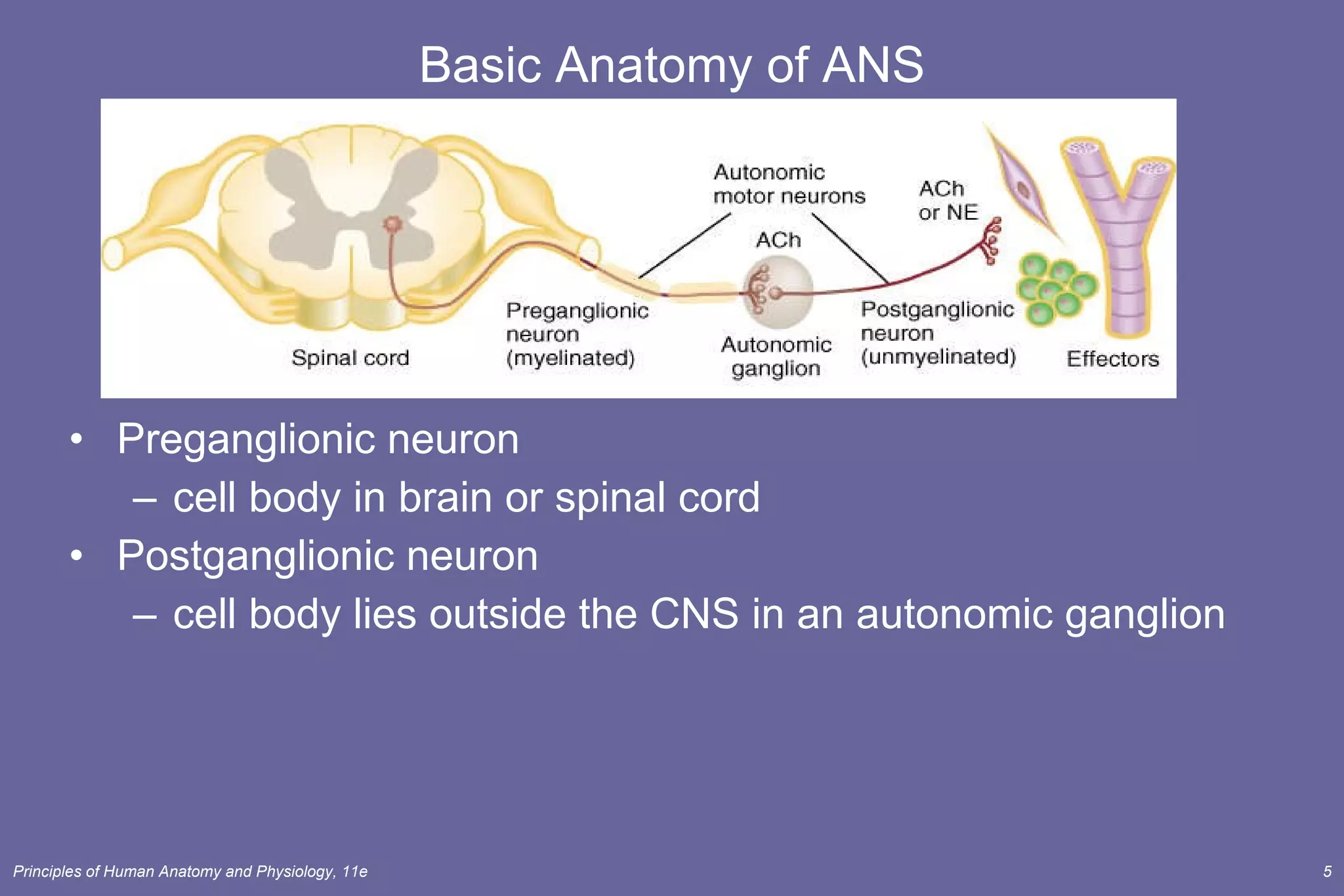
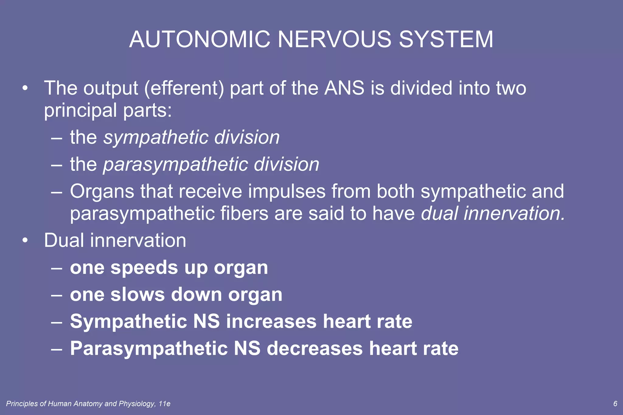
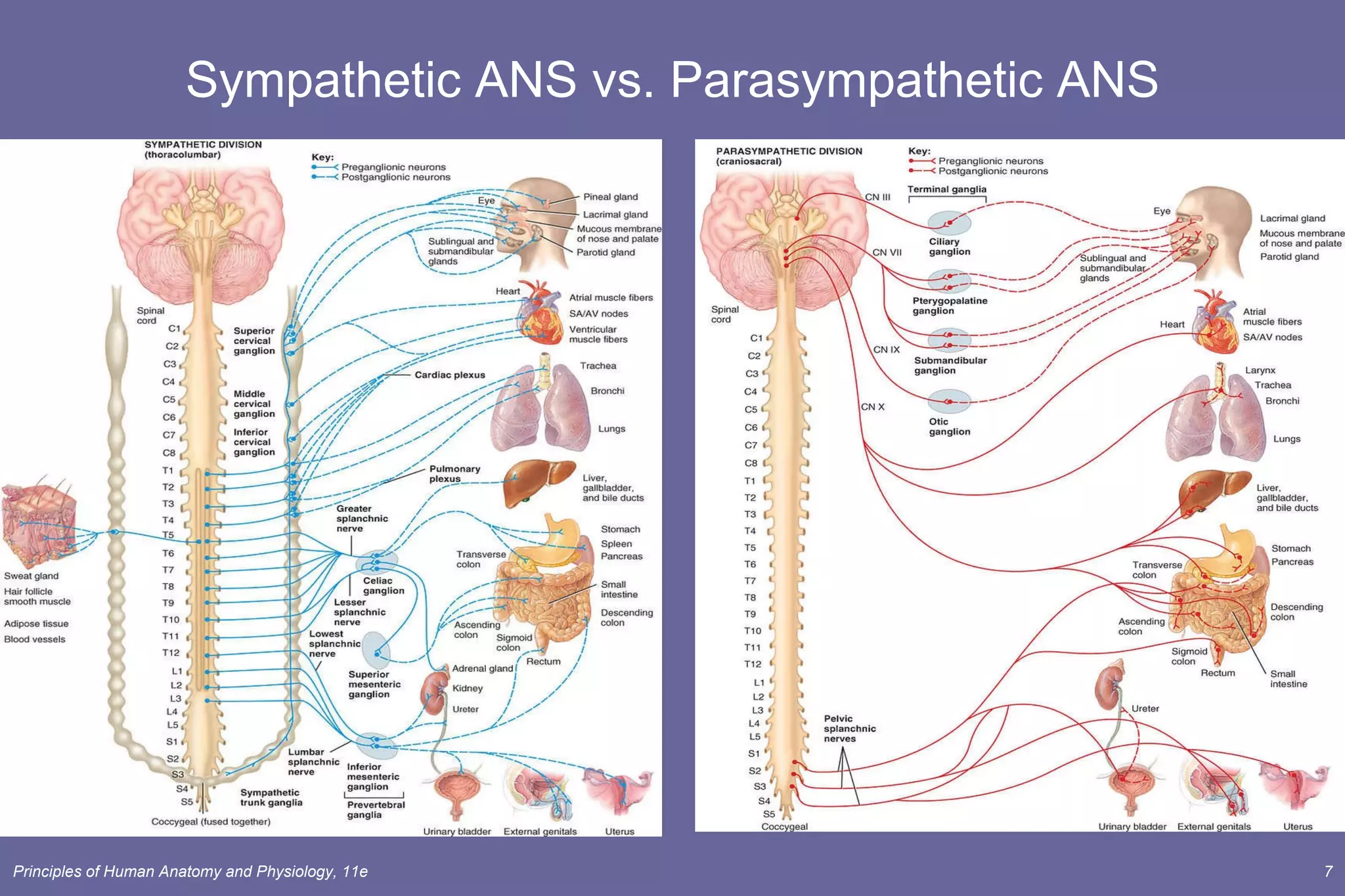
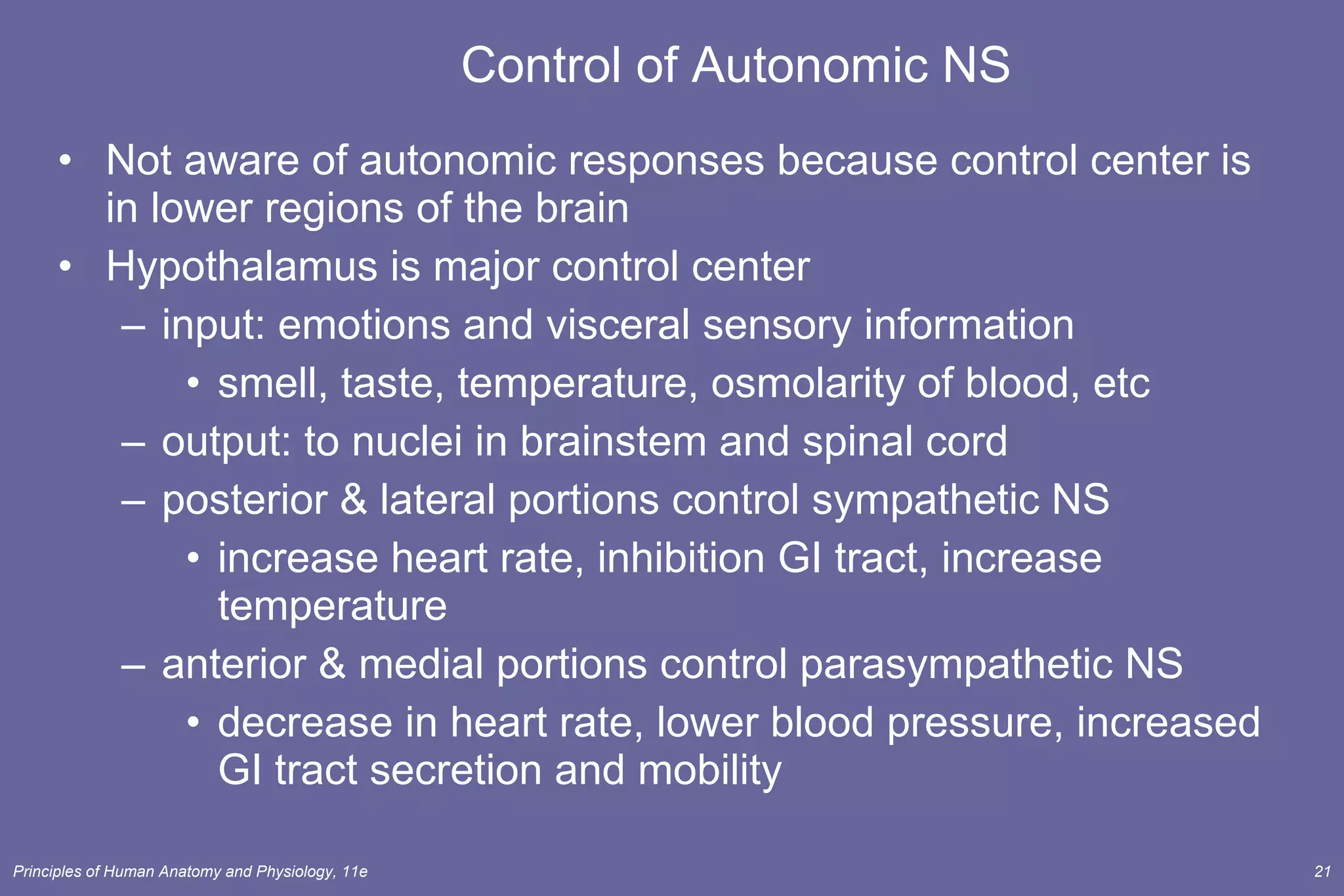
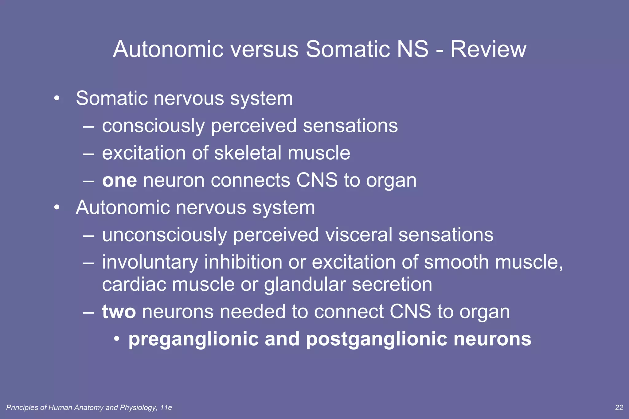
Chapter 15 The Autonomic Nervous System Lecture Outline
INTRODUCTION The  autonomic nervous system  ( ANS ) operates via reflex arcs. Regulate activity of smooth muscle, cardiac muscle & certain glands Receives input from limbic system and other regions of the cerebrum Operation of the ANS to maintain homeostasis, however, depends on a continual flow of sensory afferent input, from receptors in organs, and efferent motor output to the same effector organs. Structurally, the ANS includes autonomic sensory neurons, integrating centers in the CNS, and autonomic motor neurons. Functionally, the ANS usually operates without conscious control. The ANS is regulated by the hypothalamus and brain stem.
SOMATIC AND  AUTONOMIC  NERVOUS SYSTEMS The  somatic nervous system  contains both sensory and motor neurons. The somatic sensory neurons receive input from receptors of the special and somatic senses. Somatic motor neurons innervate skeletal muscle to produce conscious, voluntary movements. The  autonomic nervous system  contains both autonomic sensory and motor neurons. The ANS also receives sensory input from somatic senses and special sensory neurons. The autonomic motor neurons regulate visceral activities by either increasing (exciting) or decreasing (inhibiting) ongoing activities of cardiac muscle, smooth muscle, and glands.
Somatic versus Autonomic NS All somatic motor pathways consist of a  single  motor neuron Autonomic motor pathways consists of  two  motor neurons in series
Basic Anatomy of ANS Preganglionic neuron cell body in brain or spinal cord  Postganglionic neuron cell body lies outside the CNS in an autonomic ganglion
AUTONOMIC NERVOUS SYSTEM The output (efferent) part of the ANS is divided into two principal parts:  the  sympathetic division the  parasympathetic division Organs that receive impulses from both sympathetic and parasympathetic fibers are said to have  dual innervation. Dual innervation one speeds up organ one slows down organ Sympathetic NS increases heart rate Parasympathetic NS decreases heart rate
Sympathetic ANS vs. Parasympathetic ANS
Structures of Sympathetic NS Preganglionic cell bodies at  T1 to L2 Postganglionic cell bodies sympathetic chain ganglia along the spinal column prevertebral ganglia at a distance from spinal cord celiac ganglion superior mesenteric ganglion inferior mesenteric ganglion
Organs Innervated by Sympathetic NS Structures innervated by each spinal nerve sweat glands, arrector pili mm., blood vessels to skin & skeletal mm. Thoracic & cranial plexuses supply: heart, lungs, esophagus & thoracic blood vessels plexus around carotid artery to head structures Splanchnic nerves to prevertebral ganglia supply: GI tract from stomach to rectum, urinary & reproductive organs
Anatomy of Parasympathetic NS Preganglionic cell bodies found in 4 cranial nerve nuclei in brainstem  S2 to S4 spinal cord Postganglionic cell bodies very near or in the wall of the target organ in a terminal ganglia
Parasympathetic Sacral Nerve Fibers Form pelvic splanchnic nerves  Preganglionic fibers end on terminal ganglia in walls of target organs Innervate smooth muscle and glands in colon, ureters, bladder & reproductive organs
ANS Neurotransmitters Classified as either cholinergic or adrenergic neurons based upon the neurotransmitter released Adrenergic Cholinergic
Cholinergic Neurons and Receptors Cholinergic neurons release acetylcholine  all  preganglionic neurons all parasympathetic postganglionic neurons few sympathetic postganglionic neurons (to most sweat glands) Excitation or inhibition depending upon receptor subtype and organ involved.
Cholinergic Neurons and Receptors Cholinergic receptors are integral membrane proteins in the postsynaptic plasma membrane.  The two types of cholinergic receptors are  nicotinic  and  muscarinic receptors . Activation of nicotinic receptors causes excitation of the postsynaptic cell. Nicotinic receptors are found on dendrites & cell bodies of autonomic NS cells (and at NMJ.) Activation of muscarinic receptors can cause either excitation or inhibition depending on the cell that bears the receptors. Muscarinic receptors are found on plasma membranes of all parasympathetic effectors
Adrenergic Neurons and Receptors Adrenergic neurons release norepinephrine (NE) ) from postganglionic sympathetic neurons only Excites or inhibits organs depending on receptors NE lingers at the synapse until enzymatically inactivated by monoamine oxidase (MAO) or catechol-O-methyltransferase (COMT)
Physiological Effects of the ANS Most body organs receive dual innervation innervation by both sympathetic & parasympathetic Hypothalamus regulates balance (tone) between  sympathetic and parasympathetic activity levels  Some organs have only sympathetic innervation sweat glands, adrenal medulla, arrector pili mm & many blood vessels   controlled by regulation of the “tone” of the sympathetic system
Sympathetic Responses Dominance by the sympathetic system is caused by physical or emotional stress -- “E situations” emergency, embarrassment, excitement, exercise   Alarm reaction = flight or fight response dilation of pupils increase of heart rate, force of contraction & BP decrease in blood flow to nonessential organs increase in blood flow to skeletal & cardiac muscle airways dilate & respiratory rate increases blood glucose level increase Long lasting due to lingering of NE in synaptic gap and release of norepinephrine by the adrenal gland
Parasympathetic Responses Enhance “rest-and-digest” activities Mechanisms that help conserve and restore body energy during times of rest Normally dominate over sympathetic impulses SLUDD type responses =  salivation, lacrimation, urination, digestion & defecation and 3 “decreases”--- decreased HR, diameter of airways and diameter of pupil Paradoxical fear when there is no escape route or no way to win causes massive activation of parasympathetic division loss of control over urination and defecation
PHYSIOLOGICAL EFFECTS OF THE ANS - Summary The sympathetic responses prepare the body for emergency situations (the fight-or-flight responses).  The parasympathetic division regulates activities that conserve and restore body energy (energy conservation-restorative system).
Autonomic or Visceral Reflexes A visceral autonomic reflex adjusts the activity of a visceral effector, often unconsciously. changes in blood pressure, digestive functions etc filling & emptying of bladder or defecation Autonomic reflexes occur over autonomic reflex arcs. Components of that reflex arc: sensory receptor sensory neuron integrating center pre & postganglionic motor neurons visceral effectors
Control of Autonomic NS Not aware of autonomic responses because control center is in lower regions of the brain Hypothalamus is major control center input: emotions and visceral sensory information  smell, taste, temperature, osmolarity of blood, etc output: to nuclei in brainstem and spinal cord posterior & lateral portions control sympathetic NS increase heart rate, inhibition GI tract, increase temperature anterior & medial portions control parasympathetic NS decrease in heart rate, lower blood pressure, increased GI tract secretion and mobility
Autonomic versus Somatic NS - Review Somatic nervous system consciously perceived sensations excitation of skeletal muscle one  neuron connects CNS to organ Autonomic nervous system unconsciously perceived visceral sensations  involuntary inhibition or excitation of smooth muscle, cardiac muscle or glandular secretion two  neurons needed to connect CNS to organ preganglionic and postganglionic neurons

Lecture 5 ans

  • 1.
    Chapter 15 TheAutonomic Nervous System Lecture Outline
  • 2.
    INTRODUCTION The autonomic nervous system ( ANS ) operates via reflex arcs. Regulate activity of smooth muscle, cardiac muscle & certain glands Receives input from limbic system and other regions of the cerebrum Operation of the ANS to maintain homeostasis, however, depends on a continual flow of sensory afferent input, from receptors in organs, and efferent motor output to the same effector organs. Structurally, the ANS includes autonomic sensory neurons, integrating centers in the CNS, and autonomic motor neurons. Functionally, the ANS usually operates without conscious control. The ANS is regulated by the hypothalamus and brain stem.
  • 3.
    SOMATIC AND AUTONOMIC NERVOUS SYSTEMS The somatic nervous system contains both sensory and motor neurons. The somatic sensory neurons receive input from receptors of the special and somatic senses. Somatic motor neurons innervate skeletal muscle to produce conscious, voluntary movements. The autonomic nervous system contains both autonomic sensory and motor neurons. The ANS also receives sensory input from somatic senses and special sensory neurons. The autonomic motor neurons regulate visceral activities by either increasing (exciting) or decreasing (inhibiting) ongoing activities of cardiac muscle, smooth muscle, and glands.
  • 4.
    Somatic versus AutonomicNS All somatic motor pathways consist of a single motor neuron Autonomic motor pathways consists of two motor neurons in series
  • 5.
    Basic Anatomy ofANS Preganglionic neuron cell body in brain or spinal cord Postganglionic neuron cell body lies outside the CNS in an autonomic ganglion
  • 6.
    AUTONOMIC NERVOUS SYSTEMThe output (efferent) part of the ANS is divided into two principal parts: the sympathetic division the parasympathetic division Organs that receive impulses from both sympathetic and parasympathetic fibers are said to have dual innervation. Dual innervation one speeds up organ one slows down organ Sympathetic NS increases heart rate Parasympathetic NS decreases heart rate
  • 7.
    Sympathetic ANS vs.Parasympathetic ANS
  • 8.
    Structures of SympatheticNS Preganglionic cell bodies at T1 to L2 Postganglionic cell bodies sympathetic chain ganglia along the spinal column prevertebral ganglia at a distance from spinal cord celiac ganglion superior mesenteric ganglion inferior mesenteric ganglion
  • 9.
    Organs Innervated bySympathetic NS Structures innervated by each spinal nerve sweat glands, arrector pili mm., blood vessels to skin & skeletal mm. Thoracic & cranial plexuses supply: heart, lungs, esophagus & thoracic blood vessels plexus around carotid artery to head structures Splanchnic nerves to prevertebral ganglia supply: GI tract from stomach to rectum, urinary & reproductive organs
  • 10.
    Anatomy of ParasympatheticNS Preganglionic cell bodies found in 4 cranial nerve nuclei in brainstem S2 to S4 spinal cord Postganglionic cell bodies very near or in the wall of the target organ in a terminal ganglia
  • 11.
    Parasympathetic Sacral NerveFibers Form pelvic splanchnic nerves Preganglionic fibers end on terminal ganglia in walls of target organs Innervate smooth muscle and glands in colon, ureters, bladder & reproductive organs
  • 12.
    ANS Neurotransmitters Classifiedas either cholinergic or adrenergic neurons based upon the neurotransmitter released Adrenergic Cholinergic
  • 13.
    Cholinergic Neurons andReceptors Cholinergic neurons release acetylcholine all preganglionic neurons all parasympathetic postganglionic neurons few sympathetic postganglionic neurons (to most sweat glands) Excitation or inhibition depending upon receptor subtype and organ involved.
  • 14.
    Cholinergic Neurons andReceptors Cholinergic receptors are integral membrane proteins in the postsynaptic plasma membrane. The two types of cholinergic receptors are nicotinic and muscarinic receptors . Activation of nicotinic receptors causes excitation of the postsynaptic cell. Nicotinic receptors are found on dendrites & cell bodies of autonomic NS cells (and at NMJ.) Activation of muscarinic receptors can cause either excitation or inhibition depending on the cell that bears the receptors. Muscarinic receptors are found on plasma membranes of all parasympathetic effectors
  • 15.
    Adrenergic Neurons andReceptors Adrenergic neurons release norepinephrine (NE) ) from postganglionic sympathetic neurons only Excites or inhibits organs depending on receptors NE lingers at the synapse until enzymatically inactivated by monoamine oxidase (MAO) or catechol-O-methyltransferase (COMT)
  • 16.
    Physiological Effects ofthe ANS Most body organs receive dual innervation innervation by both sympathetic & parasympathetic Hypothalamus regulates balance (tone) between sympathetic and parasympathetic activity levels Some organs have only sympathetic innervation sweat glands, adrenal medulla, arrector pili mm & many blood vessels controlled by regulation of the “tone” of the sympathetic system
  • 17.
    Sympathetic Responses Dominanceby the sympathetic system is caused by physical or emotional stress -- “E situations” emergency, embarrassment, excitement, exercise Alarm reaction = flight or fight response dilation of pupils increase of heart rate, force of contraction & BP decrease in blood flow to nonessential organs increase in blood flow to skeletal & cardiac muscle airways dilate & respiratory rate increases blood glucose level increase Long lasting due to lingering of NE in synaptic gap and release of norepinephrine by the adrenal gland
  • 18.
    Parasympathetic Responses Enhance“rest-and-digest” activities Mechanisms that help conserve and restore body energy during times of rest Normally dominate over sympathetic impulses SLUDD type responses = salivation, lacrimation, urination, digestion & defecation and 3 “decreases”--- decreased HR, diameter of airways and diameter of pupil Paradoxical fear when there is no escape route or no way to win causes massive activation of parasympathetic division loss of control over urination and defecation
  • 19.
    PHYSIOLOGICAL EFFECTS OFTHE ANS - Summary The sympathetic responses prepare the body for emergency situations (the fight-or-flight responses). The parasympathetic division regulates activities that conserve and restore body energy (energy conservation-restorative system).
  • 20.
    Autonomic or VisceralReflexes A visceral autonomic reflex adjusts the activity of a visceral effector, often unconsciously. changes in blood pressure, digestive functions etc filling & emptying of bladder or defecation Autonomic reflexes occur over autonomic reflex arcs. Components of that reflex arc: sensory receptor sensory neuron integrating center pre & postganglionic motor neurons visceral effectors
  • 21.
    Control of AutonomicNS Not aware of autonomic responses because control center is in lower regions of the brain Hypothalamus is major control center input: emotions and visceral sensory information smell, taste, temperature, osmolarity of blood, etc output: to nuclei in brainstem and spinal cord posterior & lateral portions control sympathetic NS increase heart rate, inhibition GI tract, increase temperature anterior & medial portions control parasympathetic NS decrease in heart rate, lower blood pressure, increased GI tract secretion and mobility
  • 22.
    Autonomic versus SomaticNS - Review Somatic nervous system consciously perceived sensations excitation of skeletal muscle one neuron connects CNS to organ Autonomic nervous system unconsciously perceived visceral sensations involuntary inhibition or excitation of smooth muscle, cardiac muscle or glandular secretion two neurons needed to connect CNS to organ preganglionic and postganglionic neurons