Structure and function of plasma membrane 2ICHHA PURAK
The presentation consists of 72 slides,describes following heads
DEFINITION : STRUCTURE OF PLASMA MEMBRANE
COMPONENTS OF PLASMA MEMBRANE ( (BIOCHEMICAL PROPERTIES)
LIPID BILAYER
PROTEINS
CARBOHYDRATES
CHOLESTEROL
MODELS EXPLAINING STRUCTURE OF BIO MEMBRANE
FLUID MOSAIC MODEL
MOBILITY OF MEMBRANE
GLYCOCALYX : GLYCOPROTEINS AND GLYCOLIPIDS
TRANSPORT OF IONS AND MOLECULES ACROSS PLASMA MEMBRANE
FUNCTIONS OF PLASMA MEMBRANE
DIVERSITY OF CELL MEMBRANES
SITE OF ATPASE ION CARRIER CHANNELS AND PUMPS-RECEPTORS
General overview of Plasma/ Cell membrane.
Definition of Plasma/ Cell membrane
Structure of Plasma membrane
1. Sandwitch model ORDanielli- Davson Model
2. Fluid mosaic model
Plasma Membrane Proteins
Chemical Composition of Plasma/ Cell Membrane
Movement across the Cell Membrane
Channels through cell membrane
Describes the plasma membrane in detail, explains the each major component with its functions.
Transport mechanism across the cell is covered with detailed explanation with examples.
by Dr. N.Sivaranjani, MD
A membrane protein is a protein molecule that is attached to, or associated with the membrane of a cell or an organelle.
More than half of all proteins interact with membranes.
Structure and functions of MitochondriaICHHA PURAK
This Power Point Presentation (PPT) entitled “Structure and Functions of Mitochondria” consists of 118 slides with following sub-heads
INTRODUCTION
HISTORY
ORIGIN AND EVOLUTION OF MITOCHONDRIA
SYNTHESIS OF MITOCHONDRIA
ISOLATION OF MITOCHNDRIA
SHAPE , SIZE AND NUMBER OF MITOCHONDRIA
STRUCTURE OF MITOCHONDRIA
CHEMICAL COMPOSITION OF MITOCHONDRIA
FUNCTIONS OF MITOCHONDRIA
MITOCHONDRIA –POWER HOUSE OF CELL
MITOCHONDRIAL DNA/ GENOME
TRANSPORT OF PROTEINS INTO MITOCHONDRIA
MITOCHONDRIAL INHERITANCE
MITOCHONDRIAL DISEASES IN HUMAN
SUMMARY
QUESTIONS
BOOKS CONSULTED
REFERENCES
This Power Point Presentation (PPT) entitled “ Structure and Function of Lysosome”includes 43 slides with following sub- heads.
DEFINITION
INTRODUCTION/ STRUCTURE OF LYSOSOME
DISCOVERY OF LYSOSOME
DISTRIBUTION/LOCATION OF LYSOSOME
ORIGIN/ SYNTHESIS OF LYSOSOME
SHAPE AND SIZE OF LYSOSOME
CHEMICAL COMPOSITION OF LYSOSOME
LYSOSOMES ARE KNOWN AS SUICIDE BAGS
HOW THE CELL IS PROTECTED FROM LYSOSOME RUPTURE
COMMON FUNCTION OF LYSOSOME
TYPES OF LYSOSOME
DISORDERS IN HUMAN RELATED WITH LYSOSOME
SUMMARY
QUESTIONS
BOOKS CONSULTED
REFERENCES
TRANSPORT PROCESS ACROSS CELL MEMBRANE.pptxPoojaDahale3
For a drug to get absorbed after oral administration, it must first pass through this biological barrier. Such a movement of drug across the membrane is called as drug transport.
It is the boundary that separates the cell from its surroundings.
It regulate the exchange of substances between inside and outside of the cell. It is semi-permeable.
It is made up of lipids, proteins, and carbohydrates.
• The membrane enclosing a cell is called cell membrane or plasma membrane (animal cells) and plasma lemma (plant cells).
• It contains proteins and lipids in the ratio of 80 : 20 in bacteria on one extreme and on the other extreme 20 : 80 in some nerve cells.
• The over all composition of most of the cell membranes is 40-50% protein and 50-60% lipids; both the components vary in their composition.
Gene Therapy, Somatic cell gene therapy, germ line gene therapy, classical gene therapy, non-classical gene therapy, targets of gene therapy, barriers of gene therapy, ex vivo gene therapy, in vivo gene therapy, vectors for gene delivery, antisense therapy
Explore natural remedies for syphilis treatment in Singapore. Discover alternative therapies, herbal remedies, and lifestyle changes that may complement conventional treatments. Learn about holistic approaches to managing syphilis symptoms and supporting overall health.
- Video recording of this lecture in English language: https://youtu.be/lK81BzxMqdo
- Video recording of this lecture in Arabic language: https://youtu.be/Ve4P0COk9OI
- Link to download the book free: https://nephrotube.blogspot.com/p/nephrotube-nephrology-books.html
- Link to NephroTube website: www.NephroTube.com
- Link to NephroTube social media accounts: https://nephrotube.blogspot.com/p/join-nephrotube-on-social-media.html
Pulmonary Thromboembolism - etilogy, types, medical- Surgical and nursing man...VarunMahajani
Disruption of blood supply to lung alveoli due to blockage of one or more pulmonary blood vessels is called as Pulmonary thromboembolism. In this presentation we will discuss its causes, types and its management in depth.
Ozempic: Preoperative Management of Patients on GLP-1 Receptor Agonists Saeid Safari
Preoperative Management of Patients on GLP-1 Receptor Agonists like Ozempic and Semiglutide
ASA GUIDELINE
NYSORA Guideline
2 Case Reports of Gastric Ultrasound
These lecture slides, by Dr Sidra Arshad, offer a quick overview of physiological basis of a normal electrocardiogram.
Learning objectives:
1. Define an electrocardiogram (ECG) and electrocardiography
2. Describe how dipoles generated by the heart produce the waveforms of the ECG
3. Describe the components of a normal electrocardiogram of a typical bipolar leads (limb II)
4. Differentiate between intervals and segments
5. Enlist some common indications for obtaining an ECG
Study Resources:
1. Chapter 11, Guyton and Hall Textbook of Medical Physiology, 14th edition
2. Chapter 9, Human Physiology - From Cells to Systems, Lauralee Sherwood, 9th edition
3. Chapter 29, Ganong’s Review of Medical Physiology, 26th edition
4. Electrocardiogram, StatPearls - https://www.ncbi.nlm.nih.gov/books/NBK549803/
5. ECG in Medical Practice by ABM Abdullah, 4th edition
6. ECG Basics, http://www.nataliescasebook.com/tag/e-c-g-basics
ARTIFICIAL INTELLIGENCE IN HEALTHCARE.pdfAnujkumaranit
Artificial intelligence (AI) refers to the simulation of human intelligence processes by machines, especially computer systems. It encompasses tasks such as learning, reasoning, problem-solving, perception, and language understanding. AI technologies are revolutionizing various fields, from healthcare to finance, by enabling machines to perform tasks that typically require human intelligence.
Flu Vaccine Alert in Bangalore Karnatakaaddon Scans
As flu season approaches, health officials in Bangalore, Karnataka, are urging residents to get their flu vaccinations. The seasonal flu, while common, can lead to severe health complications, particularly for vulnerable populations such as young children, the elderly, and those with underlying health conditions.
Dr. Vidisha Kumari, a leading epidemiologist in Bangalore, emphasizes the importance of getting vaccinated. "The flu vaccine is our best defense against the influenza virus. It not only protects individuals but also helps prevent the spread of the virus in our communities," he says.
This year, the flu season is expected to coincide with a potential increase in other respiratory illnesses. The Karnataka Health Department has launched an awareness campaign highlighting the significance of flu vaccinations. They have set up multiple vaccination centers across Bangalore, making it convenient for residents to receive their shots.
To encourage widespread vaccination, the government is also collaborating with local schools, workplaces, and community centers to facilitate vaccination drives. Special attention is being given to ensuring that the vaccine is accessible to all, including marginalized communities who may have limited access to healthcare.
Residents are reminded that the flu vaccine is safe and effective. Common side effects are mild and may include soreness at the injection site, mild fever, or muscle aches. These side effects are generally short-lived and far less severe than the flu itself.
Healthcare providers are also stressing the importance of continuing COVID-19 precautions. Wearing masks, practicing good hand hygiene, and maintaining social distancing are still crucial, especially in crowded places.
Protect yourself and your loved ones by getting vaccinated. Together, we can help keep Bangalore healthy and safe this flu season. For more information on vaccination centers and schedules, residents can visit the Karnataka Health Department’s official website or follow their social media pages.
Stay informed, stay safe, and get your flu shot today!
New Directions in Targeted Therapeutic Approaches for Older Adults With Mantl...i3 Health
i3 Health is pleased to make the speaker slides from this activity available for use as a non-accredited self-study or teaching resource.
This slide deck presented by Dr. Kami Maddocks, Professor-Clinical in the Division of Hematology and
Associate Division Director for Ambulatory Operations
The Ohio State University Comprehensive Cancer Center, will provide insight into new directions in targeted therapeutic approaches for older adults with mantle cell lymphoma.
STATEMENT OF NEED
Mantle cell lymphoma (MCL) is a rare, aggressive B-cell non-Hodgkin lymphoma (NHL) accounting for 5% to 7% of all lymphomas. Its prognosis ranges from indolent disease that does not require treatment for years to very aggressive disease, which is associated with poor survival (Silkenstedt et al, 2021). Typically, MCL is diagnosed at advanced stage and in older patients who cannot tolerate intensive therapy (NCCN, 2022). Although recent advances have slightly increased remission rates, recurrence and relapse remain very common, leading to a median overall survival between 3 and 6 years (LLS, 2021). Though there are several effective options, progress is still needed towards establishing an accepted frontline approach for MCL (Castellino et al, 2022). Treatment selection and management of MCL are complicated by the heterogeneity of prognosis, advanced age and comorbidities of patients, and lack of an established standard approach for treatment, making it vital that clinicians be familiar with the latest research and advances in this area. In this activity chaired by Michael Wang, MD, Professor in the Department of Lymphoma & Myeloma at MD Anderson Cancer Center, expert faculty will discuss prognostic factors informing treatment, the promising results of recent trials in new therapeutic approaches, and the implications of treatment resistance in therapeutic selection for MCL.
Target Audience
Hematology/oncology fellows, attending faculty, and other health care professionals involved in the treatment of patients with mantle cell lymphoma (MCL).
Learning Objectives
1.) Identify clinical and biological prognostic factors that can guide treatment decision making for older adults with MCL
2.) Evaluate emerging data on targeted therapeutic approaches for treatment-naive and relapsed/refractory MCL and their applicability to older adults
3.) Assess mechanisms of resistance to targeted therapies for MCL and their implications for treatment selection
Report Back from SGO 2024: What’s the Latest in Cervical Cancer?bkling
Are you curious about what’s new in cervical cancer research or unsure what the findings mean? Join Dr. Emily Ko, a gynecologic oncologist at Penn Medicine, to learn about the latest updates from the Society of Gynecologic Oncology (SGO) 2024 Annual Meeting on Women’s Cancer. Dr. Ko will discuss what the research presented at the conference means for you and answer your questions about the new developments.
TEST BANK for Operations Management, 14th Edition by William J. Stevenson, Ve...kevinkariuki227
TEST BANK for Operations Management, 14th Edition by William J. Stevenson, Verified Chapters 1 - 19, Complete Newest Version.pdf
Acute scrotum is a general term referring to an emergency condition affecting the contents or the wall of the scrotum.
There are a number of conditions that present acutely, predominantly with pain and/or swelling
A careful and detailed history and examination, and in some cases, investigations allow differentiation between these diagnoses. A prompt diagnosis is essential as the patient may require urgent surgical intervention
Testicular torsion refers to twisting of the spermatic cord, causing ischaemia of the testicle.
Testicular torsion results from inadequate fixation of the testis to the tunica vaginalis producing ischemia from reduced arterial inflow and venous outflow obstruction.
The prevalence of testicular torsion in adult patients hospitalized with acute scrotal pain is approximately 25 to 50 percent
Recomendações da OMS sobre cuidados maternos e neonatais para uma experiência pós-natal positiva.
Em consonância com os ODS – Objetivos do Desenvolvimento Sustentável e a Estratégia Global para a Saúde das Mulheres, Crianças e Adolescentes, e aplicando uma abordagem baseada nos direitos humanos, os esforços de cuidados pós-natais devem expandir-se para além da cobertura e da simples sobrevivência, de modo a incluir cuidados de qualidade.
Estas diretrizes visam melhorar a qualidade dos cuidados pós-natais essenciais e de rotina prestados às mulheres e aos recém-nascidos, com o objetivo final de melhorar a saúde e o bem-estar materno e neonatal.
Uma “experiência pós-natal positiva” é um resultado importante para todas as mulheres que dão à luz e para os seus recém-nascidos, estabelecendo as bases para a melhoria da saúde e do bem-estar a curto e longo prazo. Uma experiência pós-natal positiva é definida como aquela em que as mulheres, pessoas que gestam, os recém-nascidos, os casais, os pais, os cuidadores e as famílias recebem informação consistente, garantia e apoio de profissionais de saúde motivados; e onde um sistema de saúde flexível e com recursos reconheça as necessidades das mulheres e dos bebês e respeite o seu contexto cultural.
Estas diretrizes consolidadas apresentam algumas recomendações novas e já bem fundamentadas sobre cuidados pós-natais de rotina para mulheres e neonatos que recebem cuidados no pós-parto em unidades de saúde ou na comunidade, independentemente dos recursos disponíveis.
É fornecido um conjunto abrangente de recomendações para cuidados durante o período puerperal, com ênfase nos cuidados essenciais que todas as mulheres e recém-nascidos devem receber, e com a devida atenção à qualidade dos cuidados; isto é, a entrega e a experiência do cuidado recebido. Estas diretrizes atualizam e ampliam as recomendações da OMS de 2014 sobre cuidados pós-natais da mãe e do recém-nascido e complementam as atuais diretrizes da OMS sobre a gestão de complicações pós-natais.
O estabelecimento da amamentação e o manejo das principais intercorrências é contemplada.
Recomendamos muito.
Vamos discutir essas recomendações no nosso curso de pós-graduação em Aleitamento no Instituto Ciclos.
Esta publicação só está disponível em inglês até o momento.
Prof. Marcus Renato de Carvalho
www.agostodourado.com
Prix Galien International 2024 Forum ProgramLevi Shapiro
June 20, 2024, Prix Galien International and Jerusalem Ethics Forum in ROME. Detailed agenda including panels:
- ADVANCES IN CARDIOLOGY: A NEW PARADIGM IS COMING
- WOMEN’S HEALTH: FERTILITY PRESERVATION
- WHAT’S NEW IN THE TREATMENT OF INFECTIOUS,
ONCOLOGICAL AND INFLAMMATORY SKIN DISEASES?
- ARTIFICIAL INTELLIGENCE AND ETHICS
- GENE THERAPY
- BEYOND BORDERS: GLOBAL INITIATIVES FOR DEMOCRATIZING LIFE SCIENCE TECHNOLOGIES AND PROMOTING ACCESS TO HEALTHCARE
- ETHICAL CHALLENGES IN LIFE SCIENCES
- Prix Galien International Awards Ceremony
These simplified slides by Dr. Sidra Arshad present an overview of the non-respiratory functions of the respiratory tract.
Learning objectives:
1. Enlist the non-respiratory functions of the respiratory tract
2. Briefly explain how these functions are carried out
3. Discuss the significance of dead space
4. Differentiate between minute ventilation and alveolar ventilation
5. Describe the cough and sneeze reflexes
Study Resources:
1. Chapter 39, Guyton and Hall Textbook of Medical Physiology, 14th edition
2. Chapter 34, Ganong’s Review of Medical Physiology, 26th edition
3. Chapter 17, Human Physiology by Lauralee Sherwood, 9th edition
4. Non-respiratory functions of the lungs https://academic.oup.com/bjaed/article/13/3/98/278874
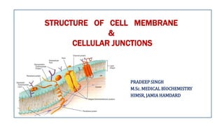
1. STRUCTURE OF CELL MEMBRANE
&
CELLULAR JUNCTIONS
PRADEEP SINGH
M.Sc. MEDICAL BIOCHEMISTRY
HIMSR, JAMIA HAMDARD
2. CONTENTS
I. Introduction of Plasma Membrane
II. History
III. Fluid Mosaic Model
IV. Chemical composition of plasma membrane
V. Function of Plasma Membrane
VI. Cellular Junctions
3. INTRODUCTION
• Selectively permeable & helps in transport of substances with the help of
proteins such as integral and peripheral proteins.
• Involved in a variety of cellular process such as Cell adhesion, Ion conductivity
and Cell Signaling.
• Provide mechanical strength to the cell.
• Cell Membrane or Plasma membrane
separates the interior of cells from the
outside environment.
• Composed of a lipid bilayer, including
cholesterol which maintain their fluidity
under various temperature.
4. HISTORY
• The cell theory was proposed by Theodor Schwann and Matthias Jakob
Schleiden in the 1830s.
1. All living organisms are composed of one or more cells.
2. The cell is the most basic unit of life.
3. All cells arise only from pre-existing cells.
• The lipid bilayer hypothesis was proposed in 1925 by Gorter and Grendel.
• The most accepted biological model of cell membrane was given by SJ
Singer and G. L. Nicolson in 1972.
• First cell was discovered by Robert Hooke in
1665 using a microscope.
5. FLUID MOSAIC MODEL
• In 1972 SJ Singer and GL Nicolson proposed fluid mosaic model.
• Fluid – Individual phospholipids and some proteins can move
sideways (laterally) in each layer-therefore FLUID
• Mosaic – Range of different proteins resting on the surface on
through the phospholipid layer gives it a mosaic appearance.
7. • Cell membranes contain a variety
of biological molecules, mainly
lipids and proteins.
• Carbohydrates are present in very
small amount, predominately as
Glycoproteins.
• Composition is not set, but
constantly changing for fluidity and
changes in the environment.
CHEMICAL COMPOSITION OF PLASMA MEMBRANE
Plasma
Membrane
Lipids
40-45%
Proteins
50-55%
Carbohydrates
1-5%
8. • The cell membranes consists of
three class of amphipathic lipids:
phospholipids, glycolipids and
sterols.
• Carbohydrates are present in very
small amount, predominately as
Glycoproteins.
• Composition is not set, but
constantly changing for fluidity and
changes in the environment.
1. LIPIDS
Lipids
Phospholipids
> 55%
Glycoslipids
2%
Cholesterol
40-45%
9. • Phospholipids are the most abundant
lipids in the cell membranes.
• Phospholipids consists of two classes
based on the types of alcohol moiety:
Glycerophospholipids, Sphingolipids
• Plasma membrane is asymmetric I
nature i.e., contains different types of
phospholipids in the outer and inner
leaflet (result in increase in fluidity)
• Outer leaflet: Phosphatidylcholine and
Phosphatidylethanolamine
• Inner leaflet: Phosphatidylserine,
Phosphatidylinositol & Sphingomylein
Phospholipids Phospholipds
Glycerophospholipids
Phosphatidylcholine
Phosphatidylserine
Phosphatidylethanolamine
Phosphatidylinsositol
Sphingolipids
Sphingomylein
10. • Phospholipids are amphipathic molecules consist of a
polar head and unsaturated fatty acid tails.
• The unsaturation in the fatty acid chains prevents the
close packing of the plasma membrane.
11. • Glycolipids only accounts for 2% of the total lipids.
• The fatty acids in the glycolipids usually contain even number of carbon atoms, typically between 16 and
20.
Glycolipids
Cholesterol
• Cholesterol is normally found dispersed between the hydrophobic tails of the membrane phospholipids.
• Cholesterol regulates the fluidity of the plasma membrane.
• At high temperatures, cholesterol inhibits the movement of phospholipid fatty acid and reduced membrane
fluidity.
• At cold temperatures, cholesterol interferes with fatty acid chain interactions. Acting as antifreeze,
cholesterol maintains the fluidity of the membrane.
12. Fluidity Of Lipid Bilayer
Low temperature High temperature
Phase transition
• Fluid like organization.
• Polar head loosely packed
• Tails disordered.
• Membrane thinner.
• Gel like organization.
• Polar head tightly packed.
• Tails regular
• Membrane thicker
13. • Plasma membranes also contain carbohydrates,
predominantly glycoproteins.
• Carbohydrates are located on the surface of the
cell where they recognize host cells and share
information.
• Viruses and other parasites bind to these
receptors cause an infection.
2. Carbohydrates
14. • Plasma membrane has large content of proteins, typically around 50% of membrane volume.
3. Proteins
Type Description Examples
Integral proteins or
transmembrane proteins
Span the membrane and have a
hydrophilic cytosolic domain, which interacts with internal
molecules, a hydrophobic membrane-spanning domain
that anchors it within the cell membrane, and a hydrophilic
extracellular domain that interacts with external molecules.
The hydrophobic domain consists of one, multiple, or a
combination of α-helices and β sheet protein motifs.
Ion channels, proton
pumps, G protein-
coupled receptor
Lipid anchored proteins
Covalently bound to single or multiple lipid molecules;
hydrophobically insert into the cell membrane and anchor
the protein. The protein itself is not in contact with the
membrane.
G proteins
Peripheral proteins
Attached to integral membrane proteins, or associated with
peripheral regions of the lipid bilayer. These proteins tend
to have only temporary interactions with biological
membranes, and once reacted, the molecule dissociates to
carry on its work in the cytoplasm.
Some enzymes, some
hormones
15. Integral protein Peripheral protein
• Integral proteins are permanently
attached to the membrane.
• Embedded in the whole membrane.
• Serve as carrier proteins, channels,
&enzymes.
• Detergents should be used to
remove integral proteins.
• Glycophorin are the example of
integral proteins.
• Peripheral proteins are temporarily
attached to the membrane.
• Located on the inner or outer surface of
the phospholipid bilayer.
• Serve as receptors and surface antigens.
• Peripheral proteins removed by salt, pH
changes
• Erythrocyte spectrin are the example of
peripheral proteins.
16. • Ion channels allow inorganic ions such as sodium,
potassium, calcium, or chlorine to diffuse down their
electrochemical gradient across the lipid bilayer.
• Ion channels plays an important role in controlling the
electrical behavior of cells (i.e. nerve cells).
• A G-protein coupled receptor is a single polypeptide
chain that crosses the lipid bilayer seven times
responding to signal molecules (i.e. hormones and
neurotransmitters).
• G-protein coupled receptors are used in processes
such as cell to cell signaling, the regulation of the
production of cAMP, and the regulation of ion
channels.
17. Asymmetry Of Lipid Bilayer
Outer leaflets
• Lots of carbohydrates.
• Sphingomyelin and
phosphotidylcholine.
• Floppase is an outward-directed ATP-
dependent transporter.
Inner leaflets
• Carbohydrates does not have
significant role.
• Phosphotidylserine and
phosphotidylethanolamine.
• Flippase is an inward-directed ATP-
dependent lipid class of transporters.
18. FUNCTIONS OF PLASMA MEMBRANE
• Protective:- Forms outermost boundary of the cells.
• Digestive:-Takes in food and excretes waste products.
• Selective Permeability:-Helps in transport across the membrane.
• Contains cell surface receptors (e.g: Glycoprotein receptors present on RBCs).
• Cell Adhesion Molecules (Cadherins) present on the plasma membrane of certain
cells plays an important role in the process of inflammation.
• Junctions: Helps in formation of various types of junction (Adherens &
Anchoring) along with the help of cytoskeleton elements.
20. CELL JUNCTIONS
• The cell junction is a cell-cell or cell-
extracellular matrix contact within a
tissue of a multicellular organism,
especially abundant in epithelia.
• Combined with cell adhesion
molecules and extracellular matrix,
cell junctions help hold animal cells
together.
21. • Eukaryotic cells contain protein
filaments that are collectively called as
cytoskeleton.
• These cytoskeleton filaments plays an
important role in the establishment of
Cellular Junctions.
• These cytoskeleton elements also
helps in establishing cell shape,
provide mechanical shape, help in
locomotion of cell, chromosome
separation, intracellular transport of
organelles
22.
23. Cellular Junctions
There are two main ways in
which animal cells are
bound together.
1. Cell – Cell Junction
2. Cell – Matrix Junction
24. ANCHORING JUNCTIONS
• All four types of Anchoring junctions depends on cell adhesion molecules
(CAMs).
• These proteins span the plasma membrane with one end linking to the
cytoskeleton {cell-cell or cell – matrix}and other is exposed outside the
membrane.
• The primary function of Anchoring junction is to resist the external forces that
pull the cells apart.
• Cytoskeleton linked transmembrane protiens falls into four superfamilies.
1. Cadherins <cell-cell>
2. Integrins
3. Immunoglobulins
4. selectins
28. 1. Adherens Junction
• Cadherins (named for "calcium-dependent adhesion") are a
type of (CAM) that is important in the formation of adherens
junctions to bind cells with each other.
• Cadherins are a class of type-1 transmembrane proteins.
• Cadherins depends on calcium (Ca2+) ions for their function.
29. Classification of cadherins
• Classical cadherins
1) E-cadherin {Epithelial cells}
2) N- cadherin {Nerve cells and the lens cells}
3) P- cadherin {placental and epidermal cells}
• Non-classical cadherins
1) Protocadherins {found in brain}
2) Desmocollins and Desmogleins { Desmosomes}
31. 2. DESMOSOMES
• Desmos means ‘bound’ , Soma means ‘body’.
• It is also called Macula Adherens.
• Provide strong mechanical strength between the epithelial and
muscle cells.
• These junctions are small disk shaped ‘spot welds’ between
adjacent cells.
33. TIGHT JUNCTION
• Occupies the most apical
position
• Closely associated areas of two
cells.
• Form a seal b/w cells and a
fence between plasma
membrane domains.
• Selectively limits the diffusion
of water , ions, and larger
solutes as well as migration of
cells.
DIAGRAMATIC REPRESENTATION OF TIGHT JUNCTION
35. Gap junctions
• Gap junctions couple cells both electrically and
metabolically.
• It bridges gaps between adjacent cells to create direct
channels.
• Present in most animal tissues, including connective ,
epithelia and heart muscle.
• Half channels in each membrane called connexons.
• Connexons consists of six protein subunits, called
connexins.
( gases like O2,CO2,N2,lipids, steroid hormones, alcohol) can dissolve in the polar region of the membrane and move rapidly across the membrane.
( gases like O2, CO2, N2, lipids, steroid hormones, alcohol) can dissolve in the polar region of the membrane and move rapidly across the membrane.
Attachment to cell and matrix control the orientation and behaviour of cells cytoskeleton , therby allowing cells to sense and respond to changes in the mechanical features of their environment. Thus the apparatus of cell junctions and extracellular matrix are critical for every aspects of organisation ,fxn and dynamics of multicellular structures.
There is specialization within each family : some cadherins link to actin and form adherens junction and some linked to intermediate and form desmosomes.
But there are some exceptions to these rules in case of integrins. Eg. Some mediate cell-cell rather than matrix.
The large no. of non classical cadherins more than 50 found in brain alone.
Together both cadherins constitute about 180 members in humans.
The cadherins are coupled indirectly to actin filaments through an adaptor protein complex containing p120-catenin, β-catenin, and α-catenin. Other proteins, including vinculin, associate with α-catenin and help provide the linkage to actin. β-Catenin has a second, and very important, function in intracellular signaling
These cells are specialized for absorption of nutrients; at their apex, facing the lumen of the gut, they have many microvilli (protrusions that increase the absorptive surface area).