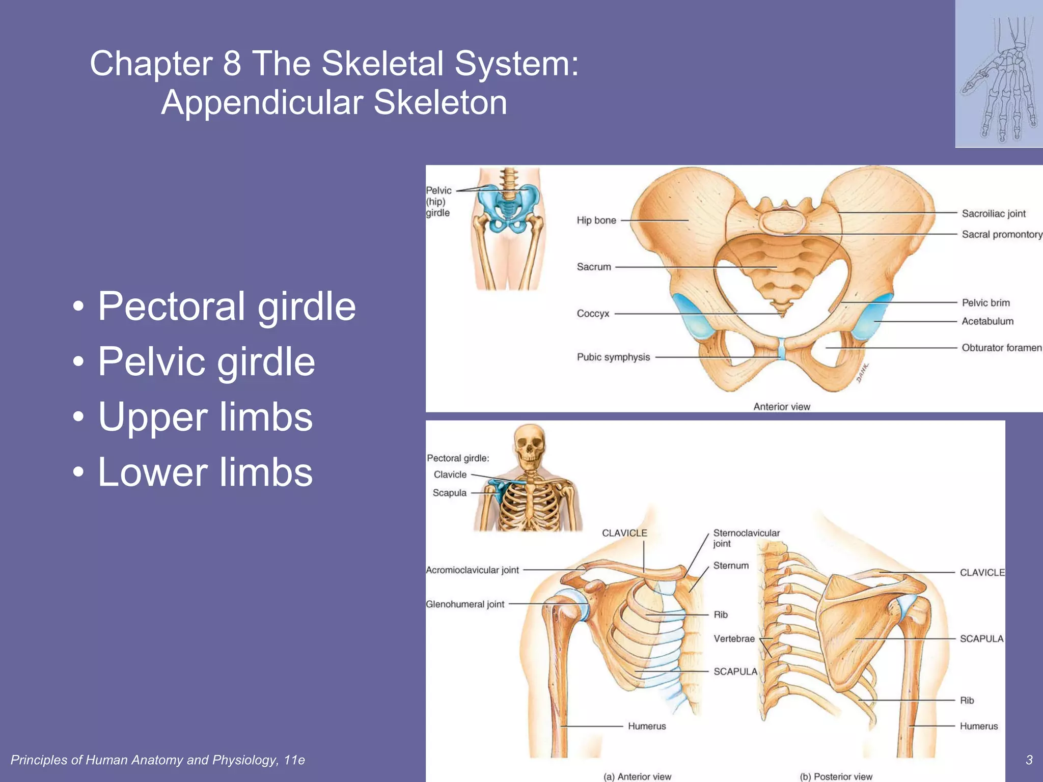
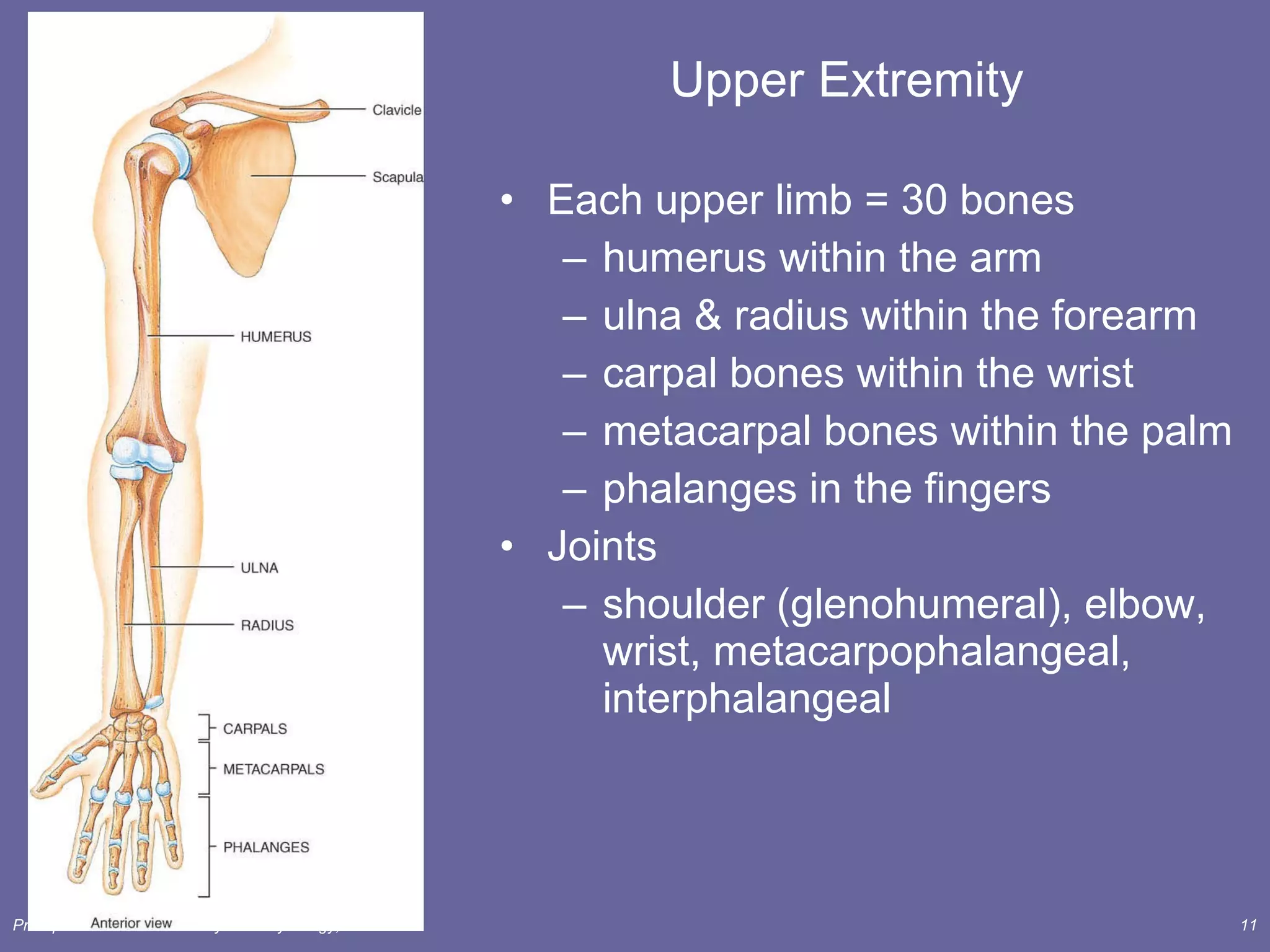
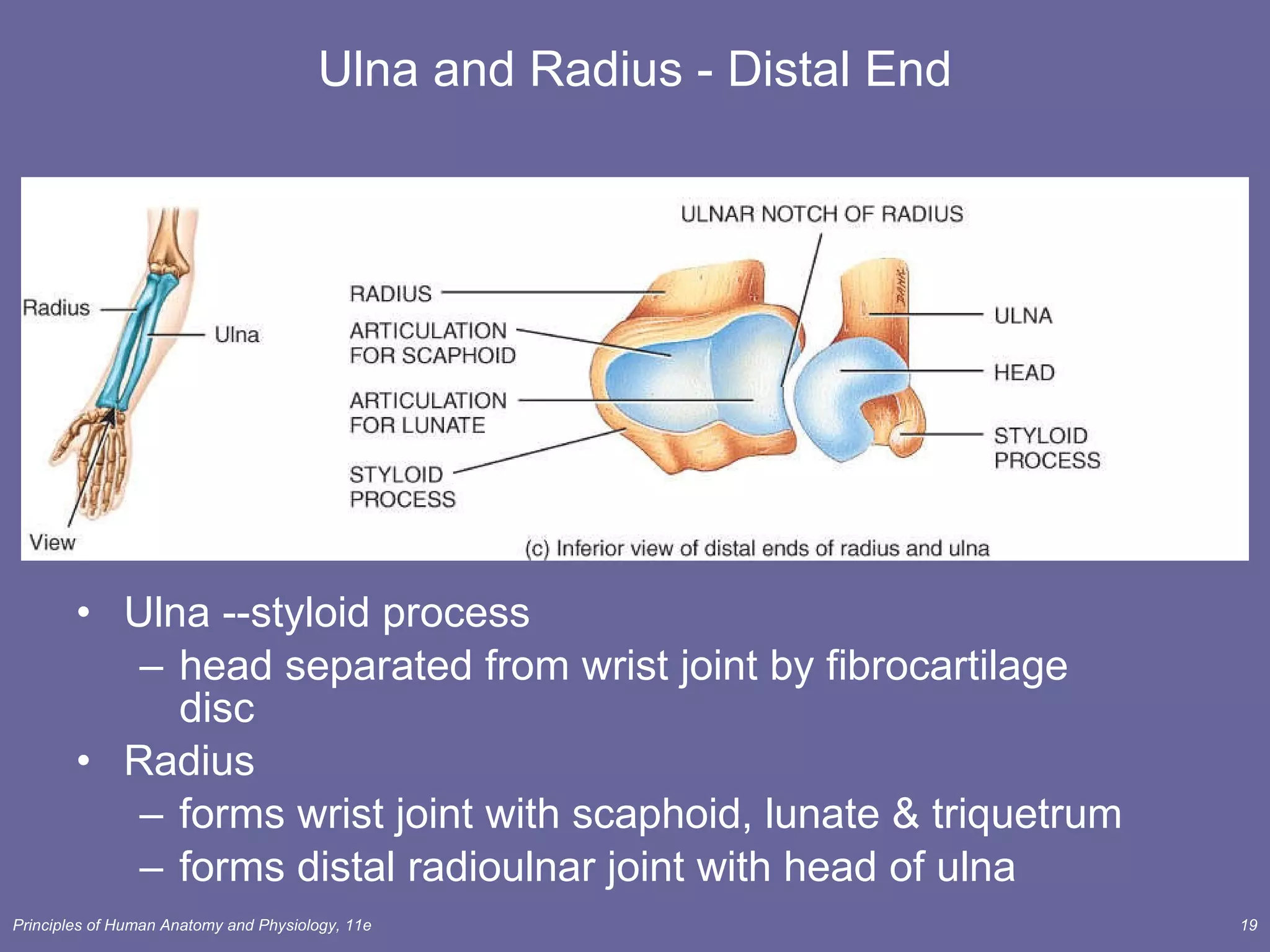
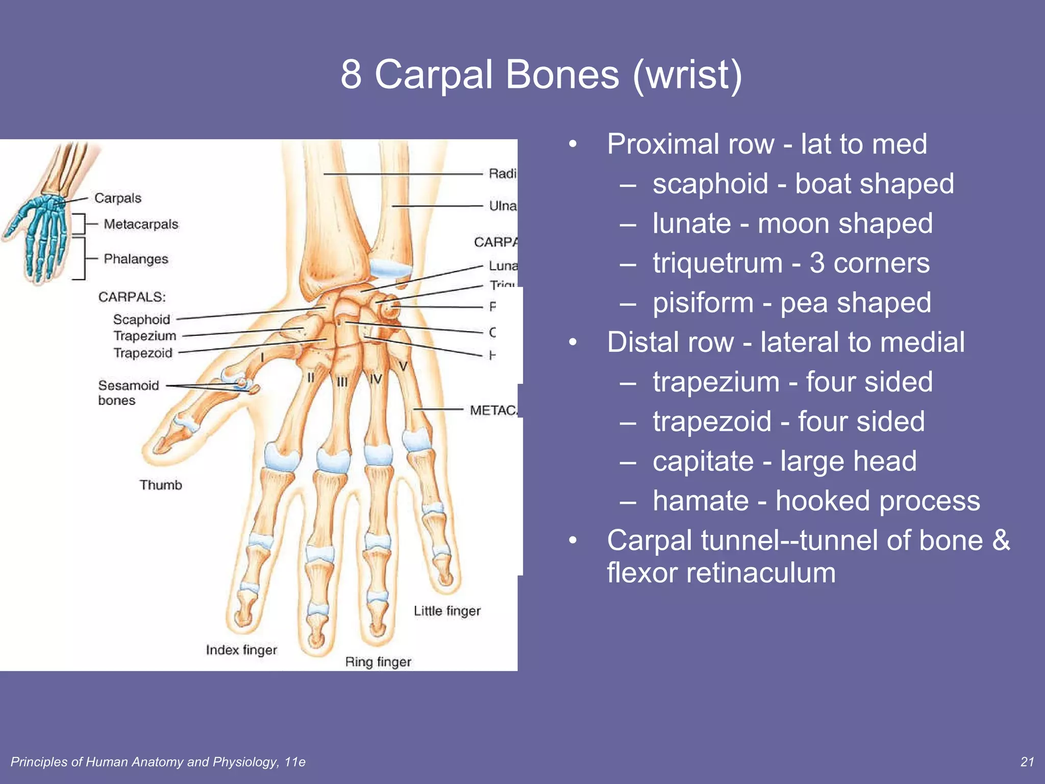
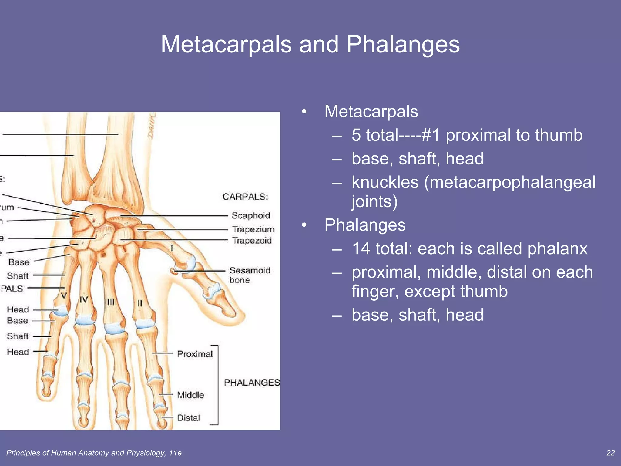
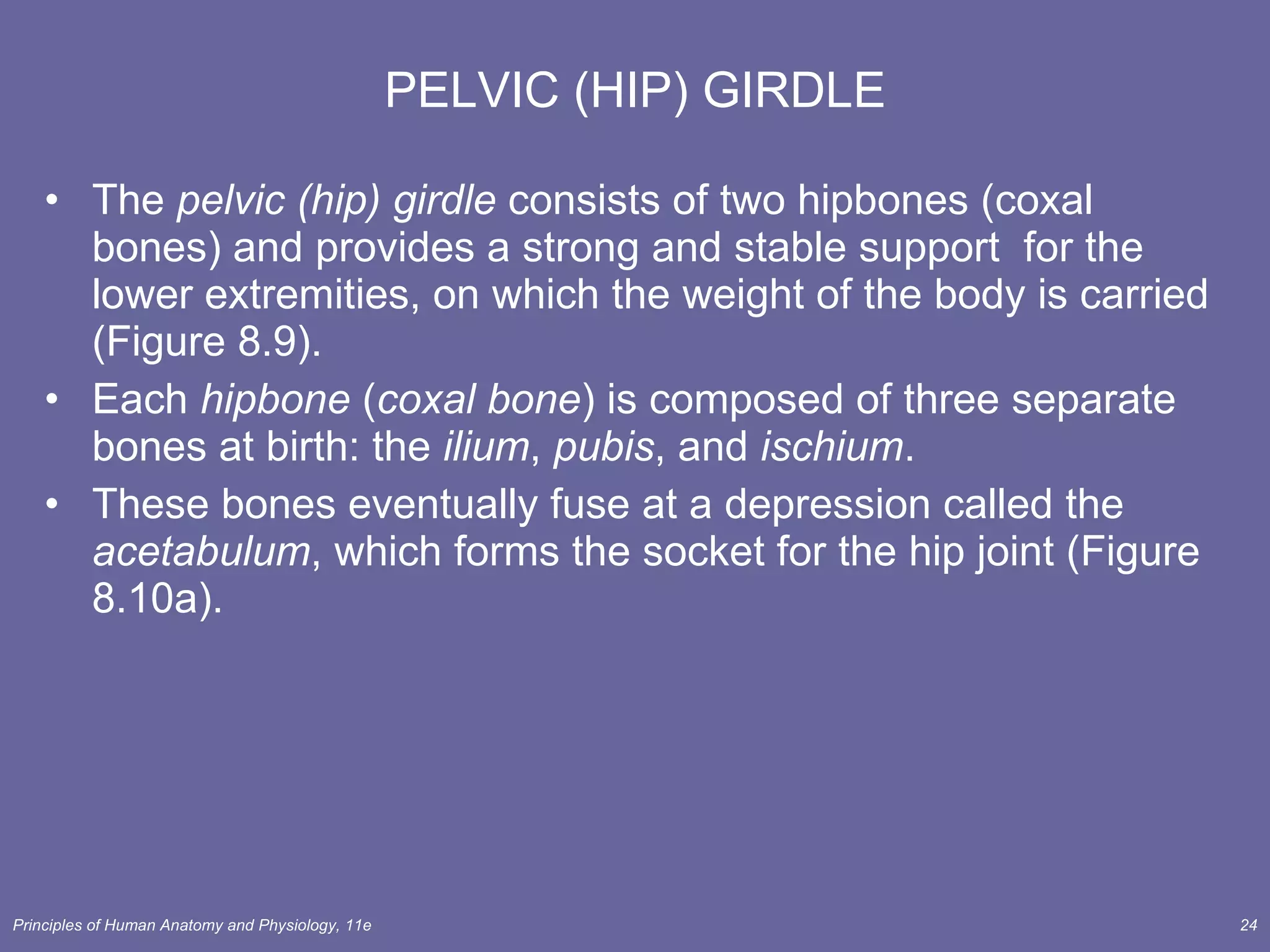
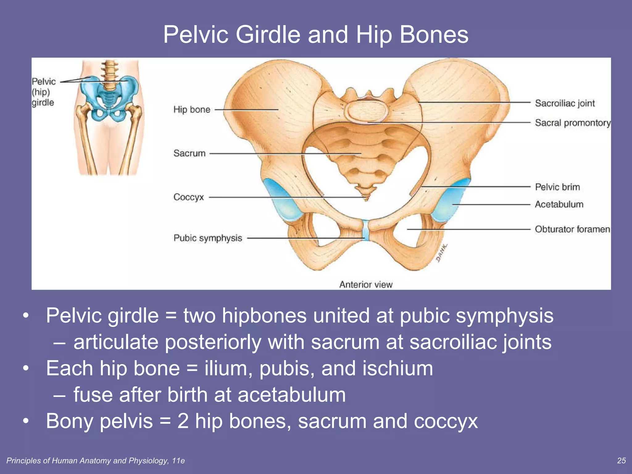
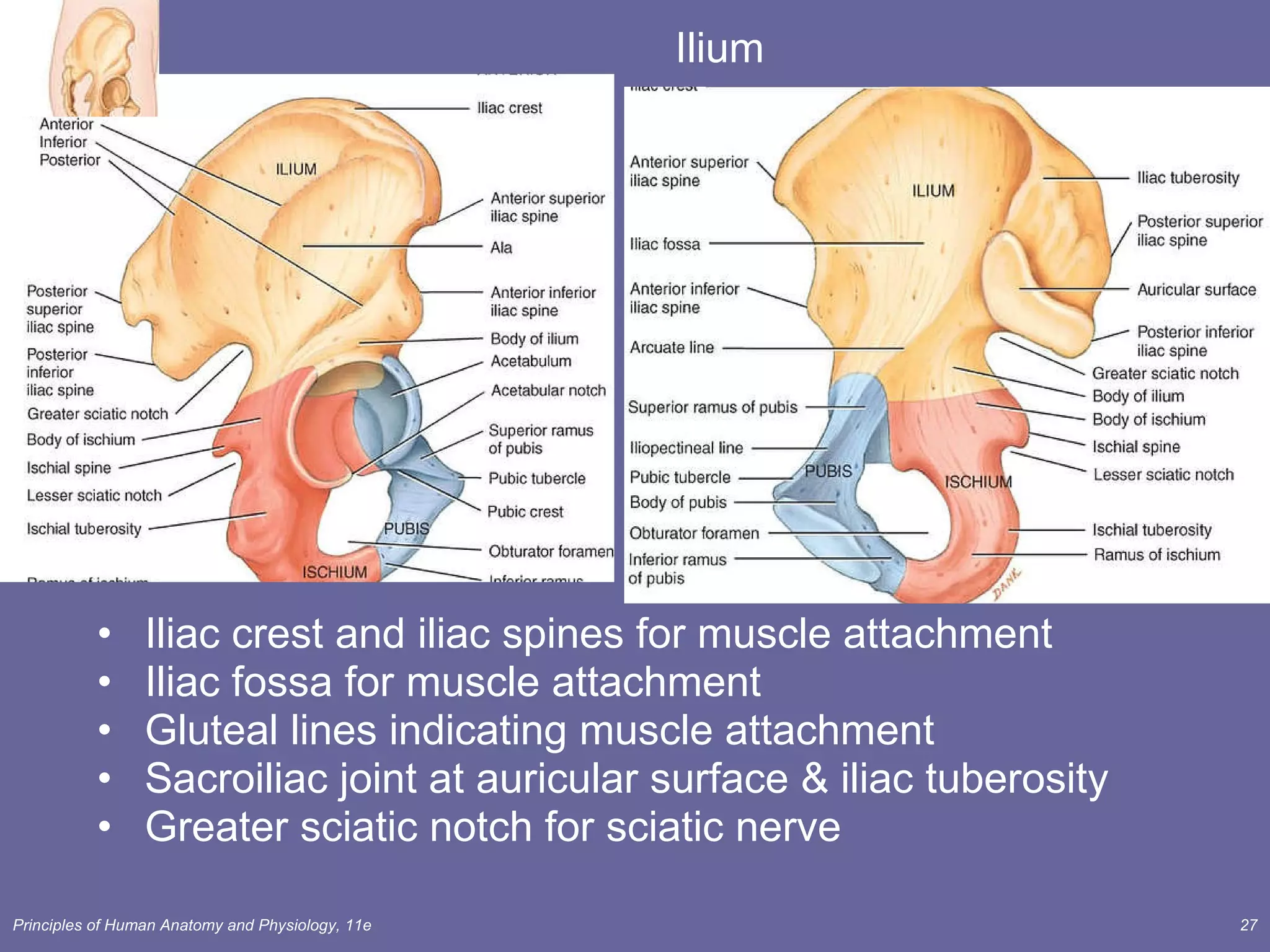
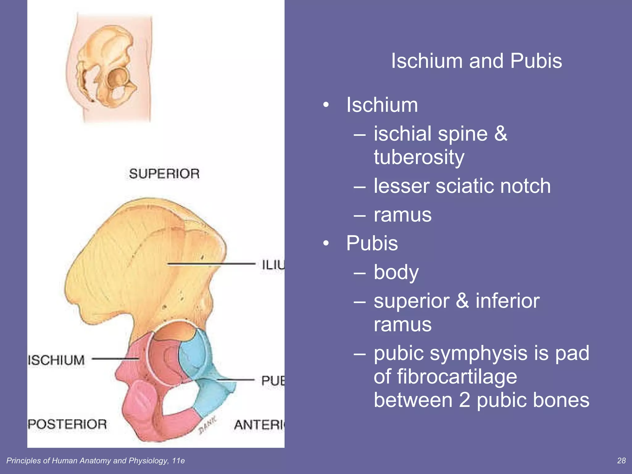
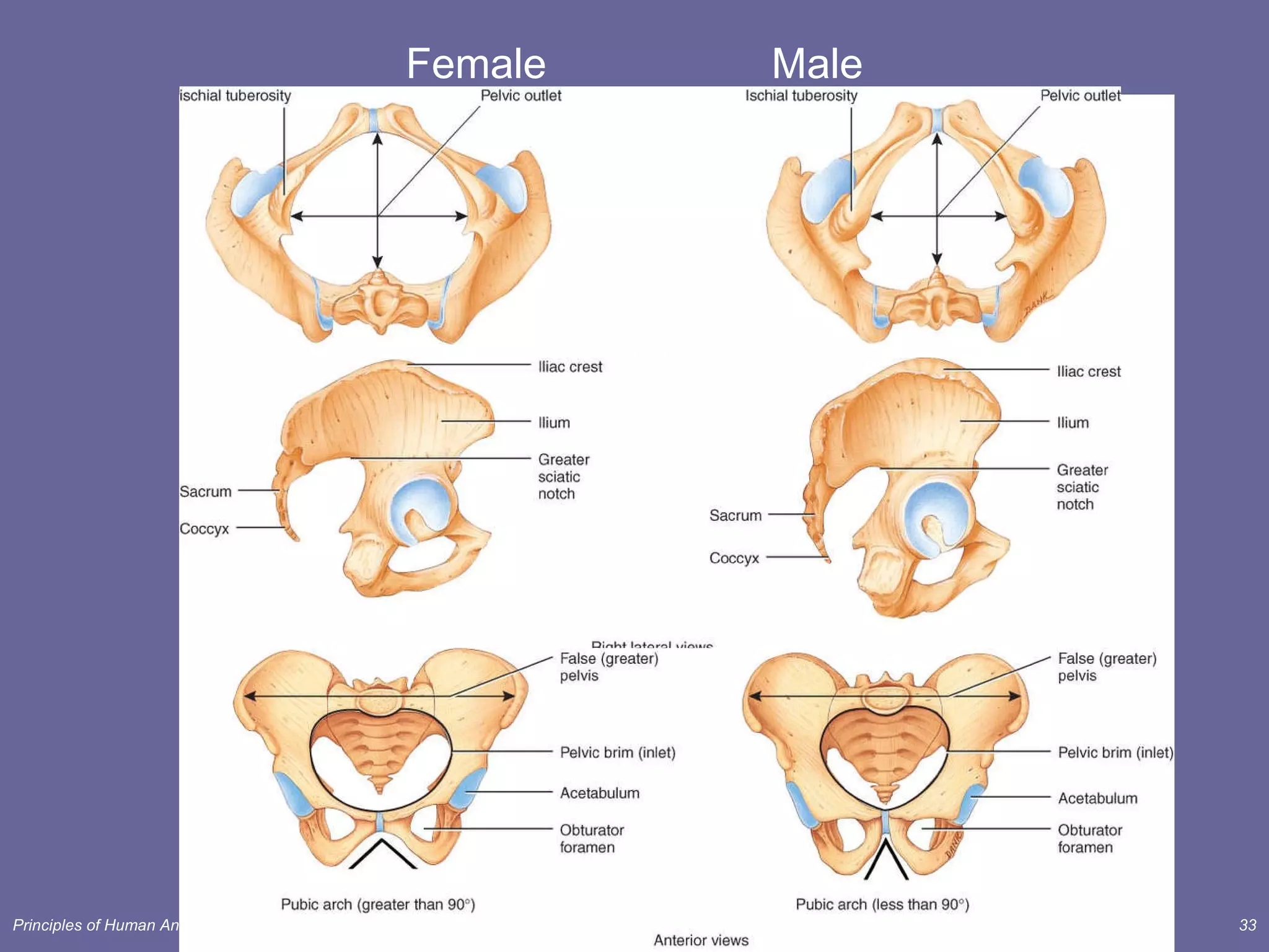
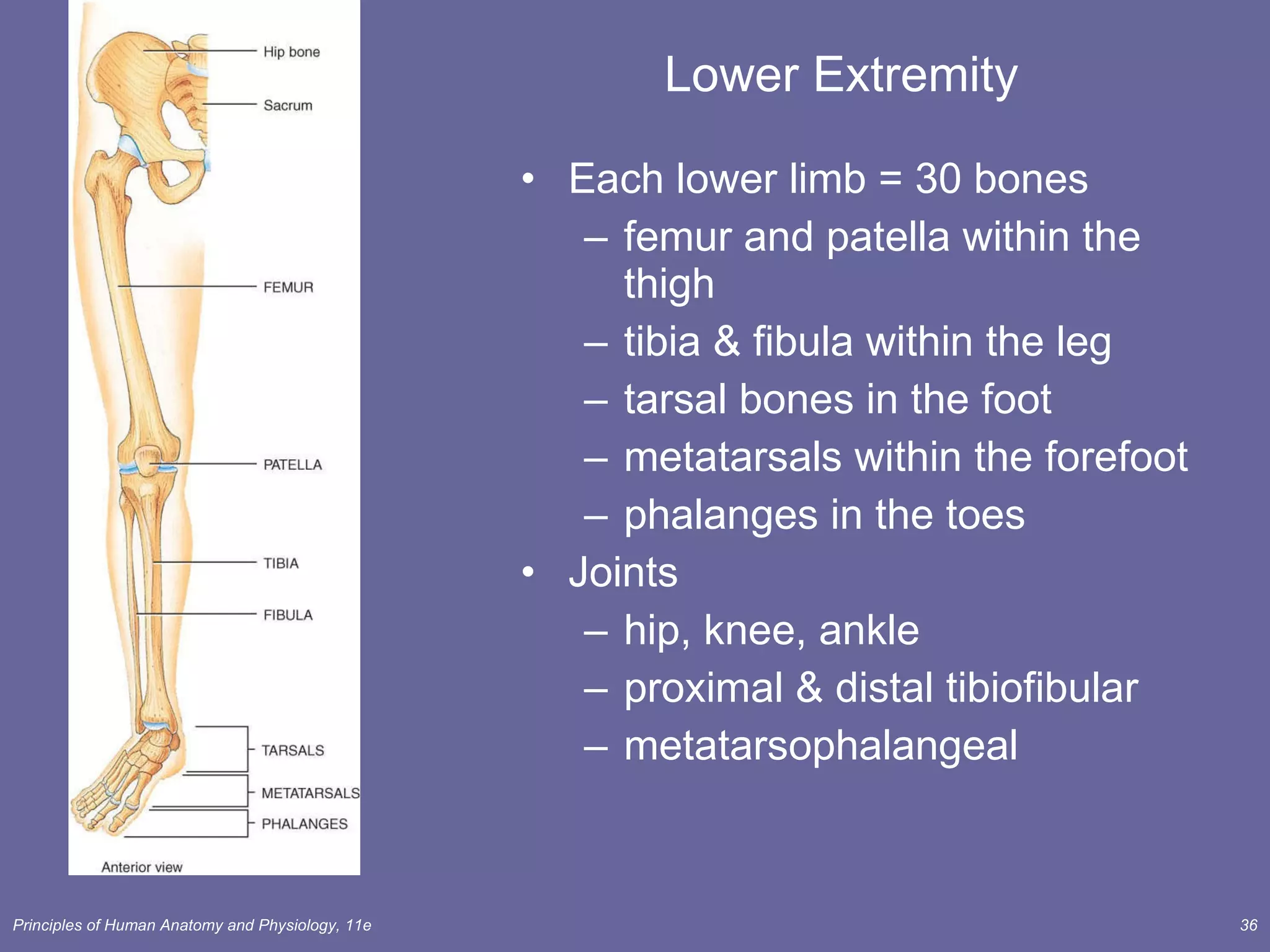
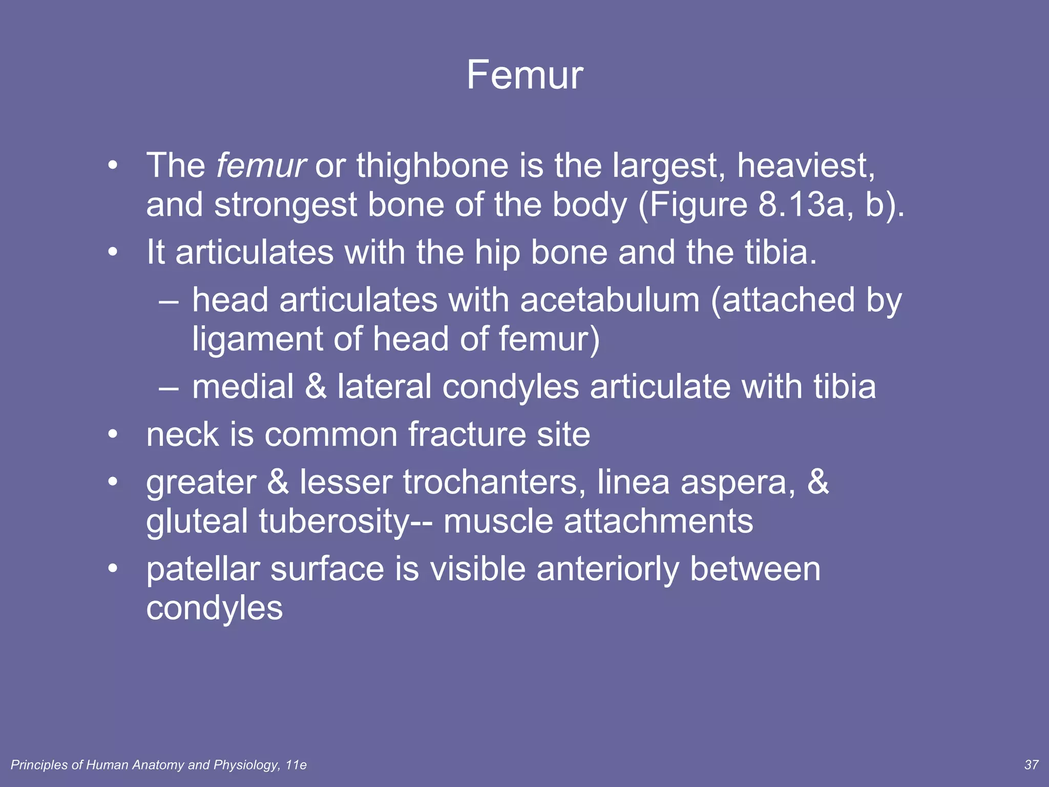
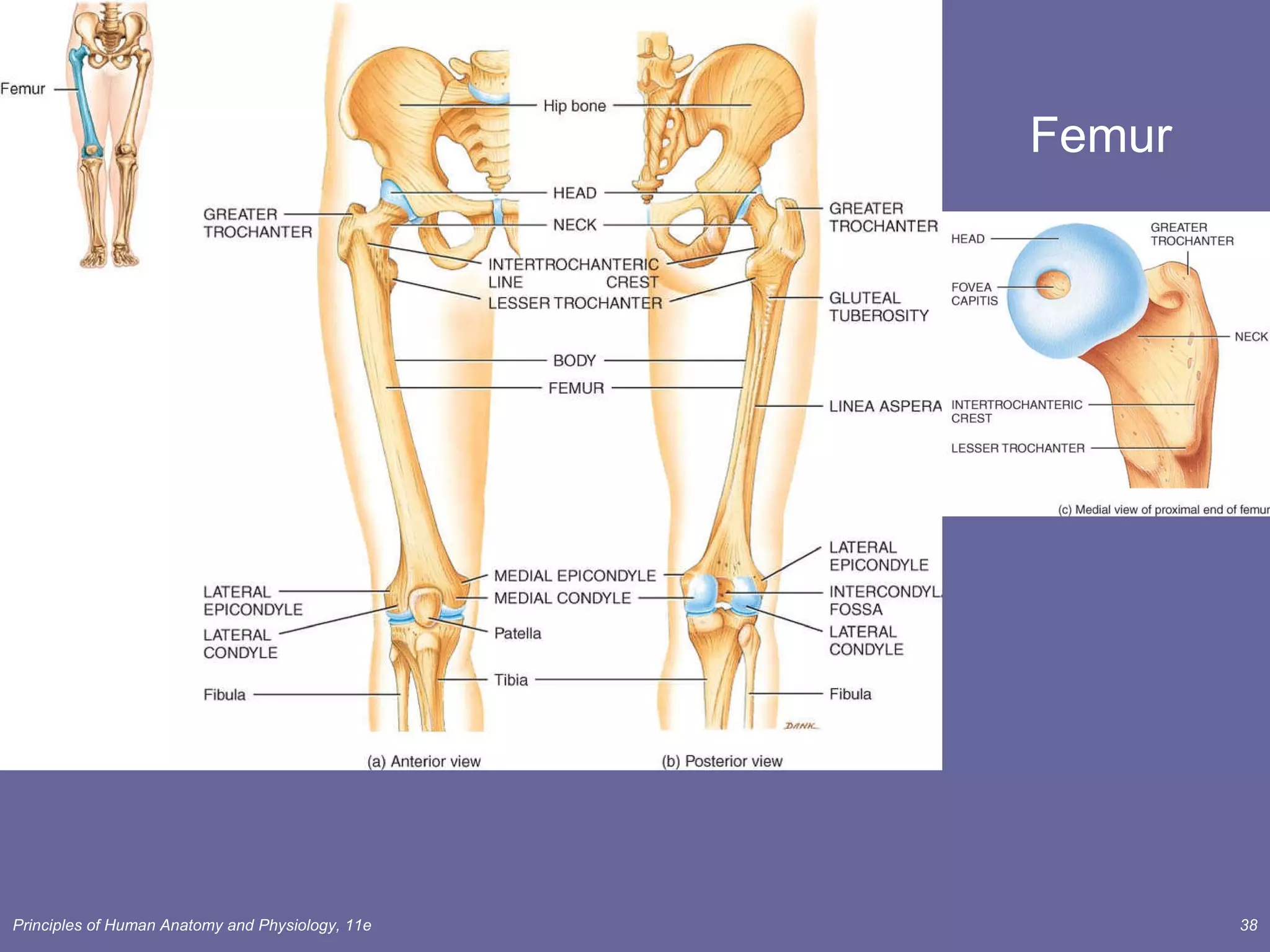
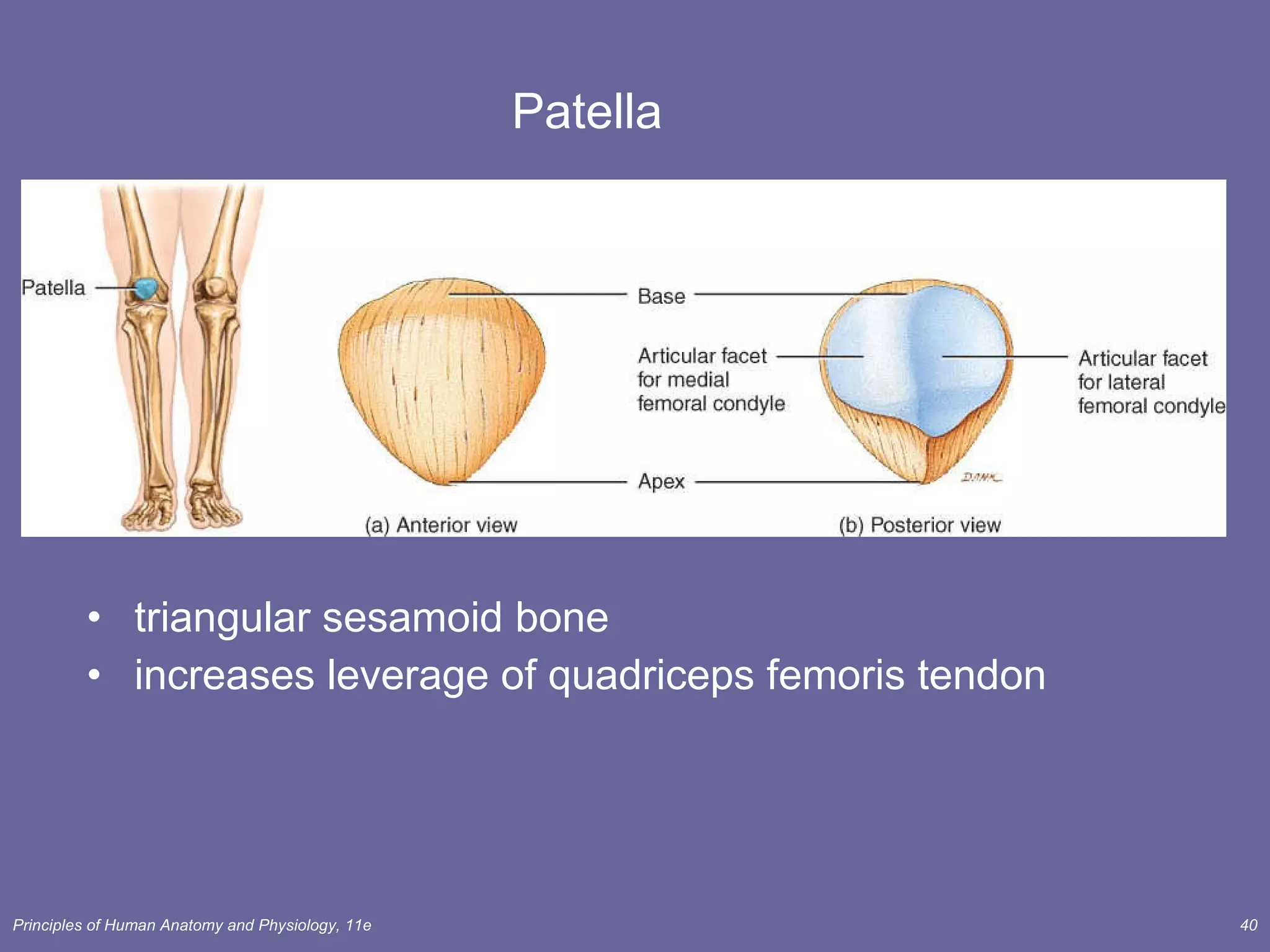
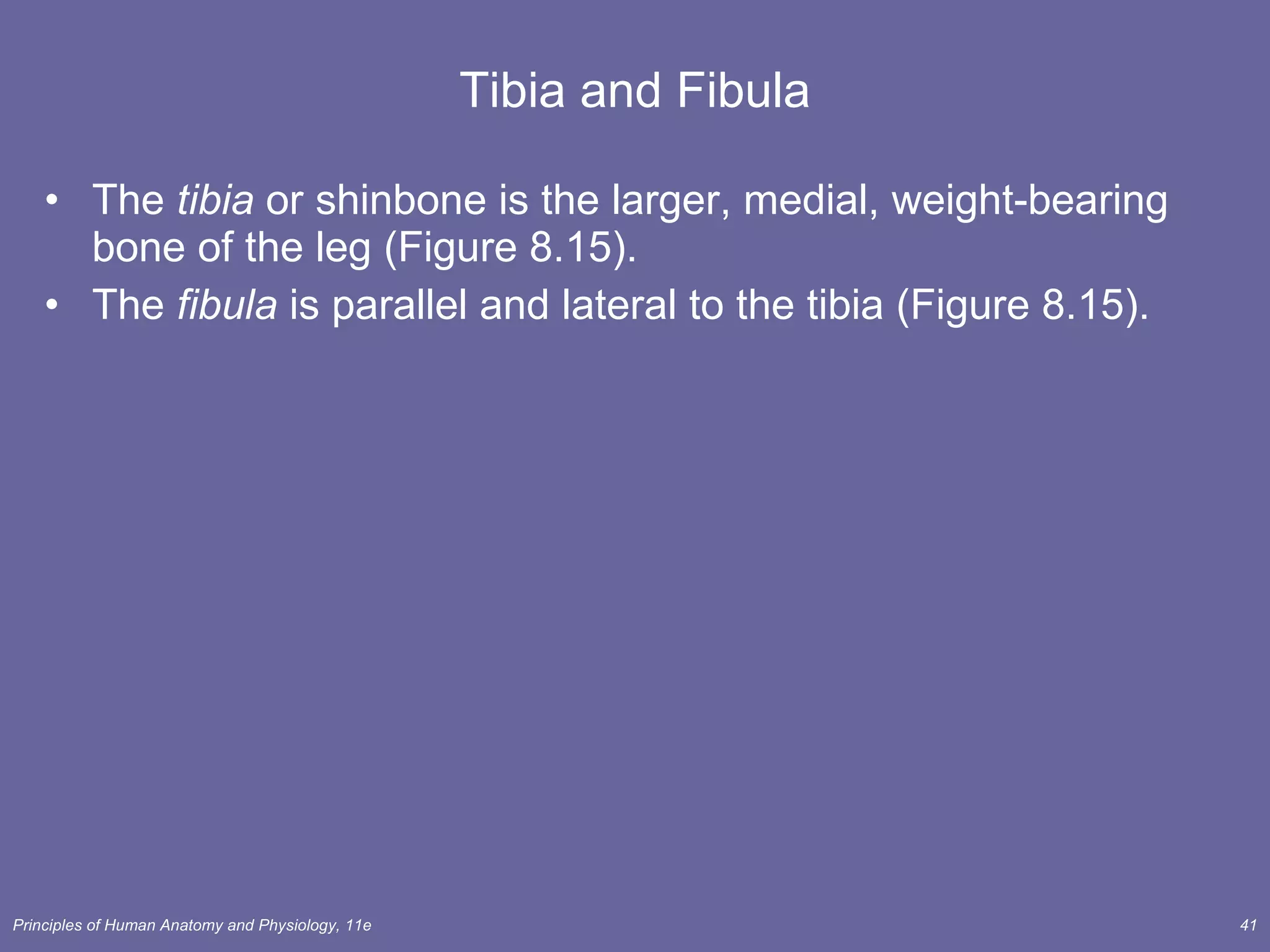
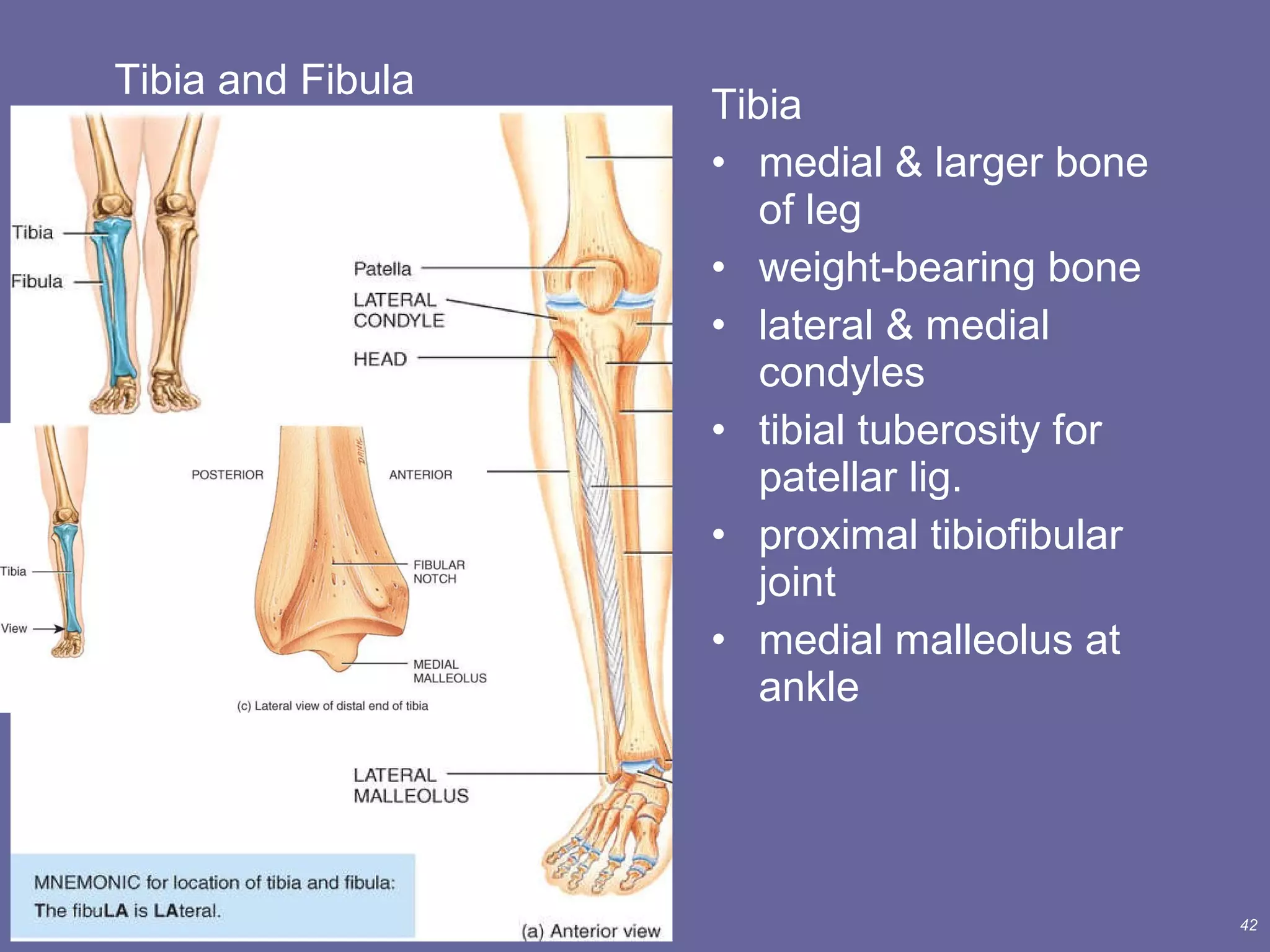
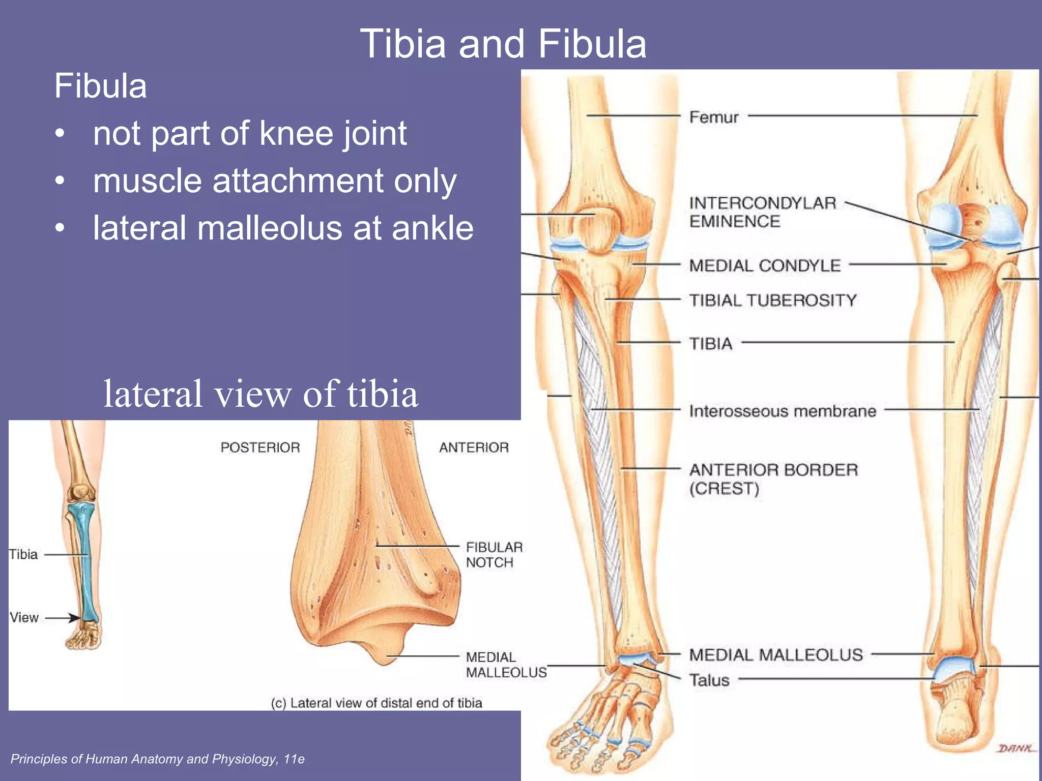
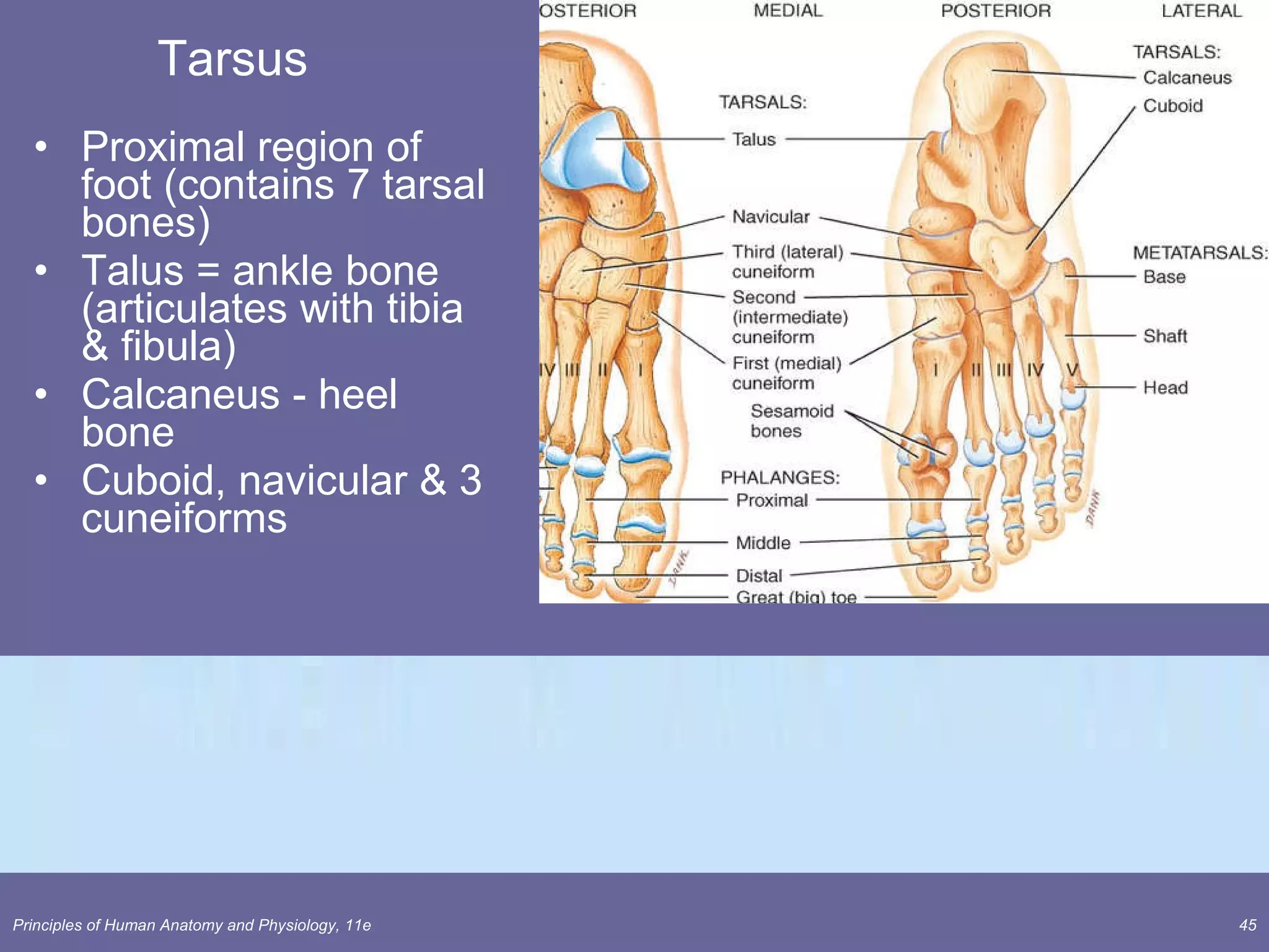
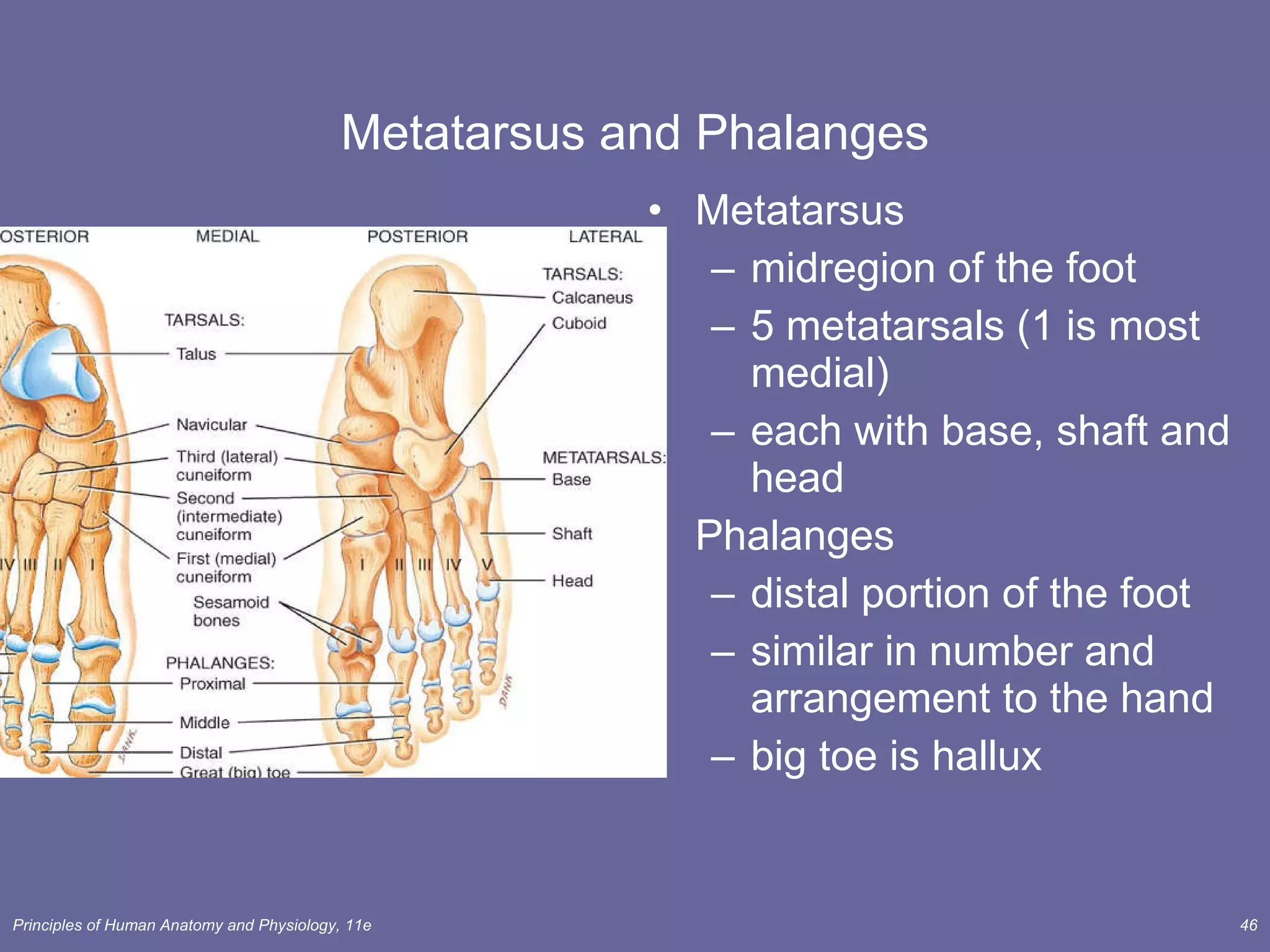
Chapter 8 The Skeletal System: Appendicular Skeleton Lecture Outline
INTRODUCTION The  appendicular skeleton  includes the bones of the upper and lower extremities and the shoulder and hip girdles. The appendicular skeleton functions primarily to facilitate movement.
Chapter 8 The Skeletal System: Appendicular Skeleton Pectoral girdle Pelvic girdle  Upper limbs Lower limbs
Pectoral (Shoulder) Girdle The  pectoral  or  shoulder girdle  attaches the bones of the upper limbs to the axial skeleton (Figure 8.1). Consists of scapula and clavicle  Clavicle articulates with sternum (sternoclavicular joint) Clavicle articulates with scapula (acromioclavicular joint) Scapula held in place by muscle only Upper limb attached to pectoral girdle at shoulder (glenohumeral joint)
Clavicle The  clavicle  or  collar bone  lies horizontally in the superior and anterior part of thorax superior to the first rib and articulates with the sternum and the clavicle (Figure 8.2). The clavicle, one of the most frequently broken bones in the body, transmits mechanical force from the upper limb to the trunk.
Clavicle (collarbone) S-shaped bone with two curves medial curve convex anteriorly/lateral one concave anteriorly Extends from sternum to scapula above 1st rib Fracture site is junction of curves  Ligaments attached to clavicle stabilize its position.
Scapula The  scapula  or  shoulder blade  articulates with the clavicle and the humerus (Figure 8.3). The scapulae articulate with other bones anteriorly, but are held in place posteriorly only by complex shoulder and back musculature.
Anterior Surface of Scapula Subscapular fossa filled with muscle  Coracoid process for muscle attachment
Posterior Surface of Scapula Triangular flat bone found in upper back region Scapular spine ends as acromion process a sharp ridge widening to a flat process Glenoid cavity forms shoulder joint with head of humerus Supraspinous & infraspinous fossa for muscular attachments
UPPER LIMB (EXTREMITY) Each upper limb consists of 30 bones including the humerus, ulna, radius, carpals, metacarpals, and phalanges (Figure 8.4).
Upper Extremity Each upper limb = 30 bones humerus within the arm ulna & radius within the forearm carpal bones within the wrist metacarpal bones within the palm phalanges in the fingers Joints  shoulder (glenohumeral), elbow, wrist, metacarpophalangeal, interphalangeal
Humerus The  humerus  is the longest and largest bone of the upper limb (Figure 8.5). It articulates proximally with the scapula and distally at the elbow with both the radius and ulna.
Humerus --- Proximal End Part of shoulder joint Head & anatomical neck  Greater & lesser tubercles for muscle attachments Intertubercular  sulcus or bicipital groove Surgical neck is fracture site Deltoid tuberosity Shaft
Humerus --- Distal End anterior and posterior Forms elbow joint with ulna and radius Capitulum  articulates with head of radius Trochlea articulation with ulna Olecranon fossa posterior depression for olecranon process of ulna  Medial & lateral epicondyles attachment of forearm muscles
Ulna and Radius The  ulna  is located on the medial aspect of the  forearm  (Figure 8.6). The  radius  is located on the lateral aspect (thumb side) of the forearm (Figure 8.6) The radius and ulna articulate with the humerus at the elbow joint (Figure 8.7a), with each other (Figure 8.7b, c), and with three carpal bones. (Figure 8.8)
Ulna & Radius --- Proximal End Ulna (on little finger side) trochlear notch articulates with humerus & radial notch with radius olecranon process forms point of elbow  Radius (on thumb side) head articulates with capitulum of  humerus & radial notch of ulna  tuberosity for muscle attachment
Elbow Joint Articulation of humerus with ulna and radius Ulna articulates with trochlea of humerus Radius articulates with capitulum of humerus Interosseous membrane between ulna & radius provides site for muscle attachment
Ulna and Radius - Distal End Ulna --styloid process head separated from wrist joint by fibrocartilage disc Radius  forms wrist joint with scaphoid, lunate & triquetrum forms distal radioulnar joint with head of ulna
Carpals, Metacarpal, and Phalanges The eight  carpal  bones, bound together by ligaments, comprise the  wrist  (Figure. 8.8). Five  metacarpal  bones are contained in the palm of each hand (Figure 8.8). Each hand contains 14  phalanges,  three in each finger and two in each thumb (Figure 8.8).
8 Carpal Bones (wrist) Proximal row - lat to med scaphoid - boat shaped lunate - moon shaped triquetrum - 3 corners pisiform - pea shaped Distal row - lateral to medial trapezium - four sided trapezoid - four sided capitate - large head hamate - hooked process Carpal tunnel--tunnel of bone & flexor retinaculum
Metacarpals and Phalanges Metacarpals 5 total----#1 proximal to thumb base, shaft, head knuckles (metacarpophalangeal joints) Phalanges 14 total: each is called phalanx proximal, middle, distal on each finger, except thumb base, shaft, head
Hand
PELVIC (HIP) GIRDLE The  pelvic (hip) girdle  consists of two hipbones (coxal bones) and provides a strong and stable support  for the lower extremities, on which the weight of the body is carried (Figure 8.9). Each  hipbone  ( coxal bone ) is composed of three separate bones at birth: the  ilium ,  pubis , and  ischium . These bones eventually fuse at a depression called the  acetabulum , which forms the socket for the hip joint (Figure 8.10a).
Pelvic Girdle and Hip Bones Pelvic girdle = two hipbones united at pubic symphysis articulate posteriorly with sacrum at sacroiliac joints Each hip bone = ilium, pubis, and ischium fuse after birth at acetabulum Bony pelvis = 2 hip bones, sacrum and coccyx
The Ilium  The larger of the three components of the hip bone and articulates (fuses) with the ischium and pubis (Figure 8.10b,c). Bone marrow aspiration or bone marrow biopsy are frequently performed on the iliac crest in adults. The  ischium  is the inferior, posterior portion of the hip bone (Figure 8.10b,c). The  pubis  is the anterior and inferior part of the hip bone (Figure 8.10b,c).
Ilium Iliac crest and iliac spines for muscle attachment Iliac fossa for muscle attachment Gluteal lines indicating muscle attachment Sacroiliac joint at auricular surface & iliac tuberosity Greater sciatic notch for sciatic nerve
Ischium and Pubis Ischium ischial spine & tuberosity lesser sciatic notch ramus Pubis body superior & inferior ramus pubic symphysis is pad of fibrocartilage between 2 pubic bones
Pelvis Pelvis = sacrum, coccyx & 2 hip bones Pelvic brim sacral promontory to symphysis pubis separates false from true pelvis false pelvis holds only abdominal organs Inlet & outlet Pelvic axis = path of babies head
True and False Pelves Together with the sacrum and coccyx, the two hipbones (coxal bones) form the  pelvis . The  greater  ( false ) and  lesser   ( true )  pelvis  are anatomical subdivisions of this basin-like structure (Figure 8.11a). Pelvimetry, the measurement of the size of the inlet and the outlet of the birth canal, is important during pregnancy
Female and Male Skeletons Male skeleton larger and heavier larger articular surfaces larger muscle attachments Female pelvis wider & shallower larger pelvic inlet & outlet more space in true pelvis pubic arch >90 degrees
COMPARISON OF FEMALE AND MALE PELVES Male bones are generally larger and heavier than those of the female; the male’s joint surfaces also tend to be larger. Muscle attachment points are more well-defined in the bones of a male than of a female due to the larger size of the muscles in males. A number of anatomical differences exist between the pelvic girdles of females and those of males, primarily related to the need for a larger pelvic outlet in females to facilitate childbirth (Table 8.1).
Female  Male
COMPARISON OF PECTORAL AND PELVIC GIRDLES The pectoral girdle does not directly articulate with the vertebral column; the pelvic girdle does. The pectoral girdle sockets are shallow and maximize movement; those of the pelvic girdle are deeper and allow less movement. The structure of the pectoral girdle offers more movement than strength; the pelvic girdle, more strength than movement.
LOWER LIMB (EXTREMITY) Each lower extremity is composed of 30 bones, including the  femur ,  tibia ,  fibula ,  tarsals ,  metatarsals , and  phalanges  (Figure 8.12).
Lower Extremity Each lower limb = 30 bones femur and patella within the thigh tibia & fibula within the leg tarsal bones in the foot metatarsals within the forefoot phalanges in the toes Joints  hip, knee, ankle  proximal & distal tibiofibular metatarsophalangeal
Femur The  femur  or thighbone is the largest, heaviest, and strongest bone of the body (Figure 8.13a, b). It articulates with the hip bone and the tibia. head articulates with acetabulum (attached by ligament of head of femur) medial & lateral condyles articulate with tibia neck is common fracture site  greater & lesser trochanters, linea aspera, & gluteal tuberosity-- muscle attachments patellar surface is visible anteriorly between condyles
Femur
Patella The  patella  or kneecap is a sesamoid bone located anterior to the knee joint (Figure 8.14). It functions to increase the leverage of the tendon of the quadriceps femoris muscle, to maintain the position of the tendon when the knee is bent, and to protect the knee joint. Patellofemoral stress syndrome  is a common knee problem in runners.
Patella triangular sesamoid bone  increases leverage of quadriceps femoris tendon
Tibia and Fibula The  tibia  or shinbone is the larger, medial, weight-bearing bone of the leg (Figure 8.15). The  fibula  is parallel and lateral to the tibia (Figure 8.15).
Tibia and Fibula Tibia medial & larger bone of leg weight-bearing bone lateral & medial condyles tibial tuberosity for patellar lig. proximal tibiofibular joint medial malleolus at ankle
Tibia and Fibula Fibula  not part of knee joint muscle attachment only lateral malleolus at ankle lateral view of tibia
Tarsals, Metatarsals, and Phalanges Seven  tarsal  bones constitute the  ankle  and share the weight associated with walking (Figure 8.16). Five  metatarsal  bones are contained in the  foot  (Figure 8.16). Fractures of the metatarsals are common among dancers, especially ballet dancers. The arrangement of  phalanges  in the  toes  is the same as that described for the fingers and thumb above - fourteen bones in each foot (Figure 8.16).
Tarsus Proximal region of foot (contains 7 tarsal bones) Talus = ankle bone (articulates with tibia & fibula) Calcaneus - heel bone Cuboid, navicular & 3 cuneiforms
Metatarsus and Phalanges Metatarsus midregion of the foot 5 metatarsals (1 is most medial) each with base, shaft and head Phalanges distal portion of the foot similar in number and arrangement to the hand big toe is hallux
Arches of the Foot The bones of the foot are arranged in two non-rigid arches that enable the foot to support the weight of the body; provide an ideal distribution of body weight over the hard and soft tissues, and provide leverage while walking (Figure 8.17). Flatfoot ,  clawfoot , and  clubfoot  are caused by decline, elevation, or rotation of the medial longitudinal arches.
Arches of the Foot Function distribute body weight over foot yield & spring back when weight is lifted Longitudinal arches along each side of foot Transverse arch across midfoot region navicular, cuneiforms & bases of metatarsals
Clinical Problems Flatfoot weakened ligaments allow bones of medial arch to drop Clawfoot medial arch is too elevated Hip fracture  1/2 million/year in US osteoporosis arthroplasty
DEVELOPMENTAL ANATOMY OF THE SKELETAL SYSTEM Bone forms from mesoderm by  intramembranous  or  endochondrial ossification . (Figure 6.6) The skull begins development during the fourth week after fertilization (Figure 8.18a) Vertebrae are derived from portions of cube-shaped masses of mesoderm called somites (Figure 10.10) Around the fifth week of embryonic life, extremities develop from  limb buds , which consist of mesoderm and ectoderm (Figure8.18b). By the sixth week, a constriction around the middle portion of the limb buds produces  hand plates  and  foot plates , which will become hands and feet. (Figure8.18c) By the seventh week, the arm, forearm and hand are evident in the upper limb bud and the thigh, leg, and foot appear in the lower limb bud. (Figure8.18d) By the eighth week the limb buds have developed into limbs. (Figure8.18e)
 
 
end

Lect 8 skeletal cont..

  • 1.
    Chapter 8 TheSkeletal System: Appendicular Skeleton Lecture Outline
  • 2.
    INTRODUCTION The appendicular skeleton includes the bones of the upper and lower extremities and the shoulder and hip girdles. The appendicular skeleton functions primarily to facilitate movement.
  • 3.
    Chapter 8 TheSkeletal System: Appendicular Skeleton Pectoral girdle Pelvic girdle Upper limbs Lower limbs
  • 4.
    Pectoral (Shoulder) GirdleThe pectoral or shoulder girdle attaches the bones of the upper limbs to the axial skeleton (Figure 8.1). Consists of scapula and clavicle Clavicle articulates with sternum (sternoclavicular joint) Clavicle articulates with scapula (acromioclavicular joint) Scapula held in place by muscle only Upper limb attached to pectoral girdle at shoulder (glenohumeral joint)
  • 5.
    Clavicle The clavicle or collar bone lies horizontally in the superior and anterior part of thorax superior to the first rib and articulates with the sternum and the clavicle (Figure 8.2). The clavicle, one of the most frequently broken bones in the body, transmits mechanical force from the upper limb to the trunk.
  • 6.
    Clavicle (collarbone) S-shapedbone with two curves medial curve convex anteriorly/lateral one concave anteriorly Extends from sternum to scapula above 1st rib Fracture site is junction of curves Ligaments attached to clavicle stabilize its position.
  • 7.
    Scapula The scapula or shoulder blade articulates with the clavicle and the humerus (Figure 8.3). The scapulae articulate with other bones anteriorly, but are held in place posteriorly only by complex shoulder and back musculature.
  • 8.
    Anterior Surface ofScapula Subscapular fossa filled with muscle Coracoid process for muscle attachment
  • 9.
    Posterior Surface ofScapula Triangular flat bone found in upper back region Scapular spine ends as acromion process a sharp ridge widening to a flat process Glenoid cavity forms shoulder joint with head of humerus Supraspinous & infraspinous fossa for muscular attachments
  • 10.
    UPPER LIMB (EXTREMITY)Each upper limb consists of 30 bones including the humerus, ulna, radius, carpals, metacarpals, and phalanges (Figure 8.4).
  • 11.
    Upper Extremity Eachupper limb = 30 bones humerus within the arm ulna & radius within the forearm carpal bones within the wrist metacarpal bones within the palm phalanges in the fingers Joints shoulder (glenohumeral), elbow, wrist, metacarpophalangeal, interphalangeal
  • 12.
    Humerus The humerus is the longest and largest bone of the upper limb (Figure 8.5). It articulates proximally with the scapula and distally at the elbow with both the radius and ulna.
  • 13.
    Humerus --- ProximalEnd Part of shoulder joint Head & anatomical neck Greater & lesser tubercles for muscle attachments Intertubercular sulcus or bicipital groove Surgical neck is fracture site Deltoid tuberosity Shaft
  • 14.
    Humerus --- DistalEnd anterior and posterior Forms elbow joint with ulna and radius Capitulum articulates with head of radius Trochlea articulation with ulna Olecranon fossa posterior depression for olecranon process of ulna Medial & lateral epicondyles attachment of forearm muscles
  • 15.
    Ulna and RadiusThe ulna is located on the medial aspect of the forearm (Figure 8.6). The radius is located on the lateral aspect (thumb side) of the forearm (Figure 8.6) The radius and ulna articulate with the humerus at the elbow joint (Figure 8.7a), with each other (Figure 8.7b, c), and with three carpal bones. (Figure 8.8)
  • 16.
    Ulna & Radius--- Proximal End Ulna (on little finger side) trochlear notch articulates with humerus & radial notch with radius olecranon process forms point of elbow Radius (on thumb side) head articulates with capitulum of humerus & radial notch of ulna tuberosity for muscle attachment
  • 17.
    Ulna & Radius--- Proximal End Ulna (on little finger side) trochlear notch articulates with humerus & radial notch with radius olecranon process forms point of elbow Radius (on thumb side) head articulates with capitulum of humerus & radial notch of ulna tuberosity for muscle attachment
  • 18.
    Elbow Joint Articulationof humerus with ulna and radius Ulna articulates with trochlea of humerus Radius articulates with capitulum of humerus Interosseous membrane between ulna & radius provides site for muscle attachment
  • 19.
    Ulna and Radius- Distal End Ulna --styloid process head separated from wrist joint by fibrocartilage disc Radius forms wrist joint with scaphoid, lunate & triquetrum forms distal radioulnar joint with head of ulna
  • 20.
    Carpals, Metacarpal, andPhalanges The eight carpal bones, bound together by ligaments, comprise the wrist (Figure. 8.8). Five metacarpal bones are contained in the palm of each hand (Figure 8.8). Each hand contains 14 phalanges, three in each finger and two in each thumb (Figure 8.8).
  • 21.
    8 Carpal Bones(wrist) Proximal row - lat to med scaphoid - boat shaped lunate - moon shaped triquetrum - 3 corners pisiform - pea shaped Distal row - lateral to medial trapezium - four sided trapezoid - four sided capitate - large head hamate - hooked process Carpal tunnel--tunnel of bone & flexor retinaculum
  • 22.
    Metacarpals and PhalangesMetacarpals 5 total----#1 proximal to thumb base, shaft, head knuckles (metacarpophalangeal joints) Phalanges 14 total: each is called phalanx proximal, middle, distal on each finger, except thumb base, shaft, head
  • 23.
  • 24.
    PELVIC (HIP) GIRDLEThe pelvic (hip) girdle consists of two hipbones (coxal bones) and provides a strong and stable support for the lower extremities, on which the weight of the body is carried (Figure 8.9). Each hipbone ( coxal bone ) is composed of three separate bones at birth: the ilium , pubis , and ischium . These bones eventually fuse at a depression called the acetabulum , which forms the socket for the hip joint (Figure 8.10a).
  • 25.
    Pelvic Girdle andHip Bones Pelvic girdle = two hipbones united at pubic symphysis articulate posteriorly with sacrum at sacroiliac joints Each hip bone = ilium, pubis, and ischium fuse after birth at acetabulum Bony pelvis = 2 hip bones, sacrum and coccyx
  • 26.
    The Ilium The larger of the three components of the hip bone and articulates (fuses) with the ischium and pubis (Figure 8.10b,c). Bone marrow aspiration or bone marrow biopsy are frequently performed on the iliac crest in adults. The ischium is the inferior, posterior portion of the hip bone (Figure 8.10b,c). The pubis is the anterior and inferior part of the hip bone (Figure 8.10b,c).
  • 27.
    Ilium Iliac crestand iliac spines for muscle attachment Iliac fossa for muscle attachment Gluteal lines indicating muscle attachment Sacroiliac joint at auricular surface & iliac tuberosity Greater sciatic notch for sciatic nerve
  • 28.
    Ischium and PubisIschium ischial spine & tuberosity lesser sciatic notch ramus Pubis body superior & inferior ramus pubic symphysis is pad of fibrocartilage between 2 pubic bones
  • 29.
    Pelvis Pelvis =sacrum, coccyx & 2 hip bones Pelvic brim sacral promontory to symphysis pubis separates false from true pelvis false pelvis holds only abdominal organs Inlet & outlet Pelvic axis = path of babies head
  • 30.
    True and FalsePelves Together with the sacrum and coccyx, the two hipbones (coxal bones) form the pelvis . The greater ( false ) and lesser ( true ) pelvis are anatomical subdivisions of this basin-like structure (Figure 8.11a). Pelvimetry, the measurement of the size of the inlet and the outlet of the birth canal, is important during pregnancy
  • 31.
    Female and MaleSkeletons Male skeleton larger and heavier larger articular surfaces larger muscle attachments Female pelvis wider & shallower larger pelvic inlet & outlet more space in true pelvis pubic arch >90 degrees
  • 32.
    COMPARISON OF FEMALEAND MALE PELVES Male bones are generally larger and heavier than those of the female; the male’s joint surfaces also tend to be larger. Muscle attachment points are more well-defined in the bones of a male than of a female due to the larger size of the muscles in males. A number of anatomical differences exist between the pelvic girdles of females and those of males, primarily related to the need for a larger pelvic outlet in females to facilitate childbirth (Table 8.1).
  • 33.
  • 34.
    COMPARISON OF PECTORALAND PELVIC GIRDLES The pectoral girdle does not directly articulate with the vertebral column; the pelvic girdle does. The pectoral girdle sockets are shallow and maximize movement; those of the pelvic girdle are deeper and allow less movement. The structure of the pectoral girdle offers more movement than strength; the pelvic girdle, more strength than movement.
  • 35.
    LOWER LIMB (EXTREMITY)Each lower extremity is composed of 30 bones, including the femur , tibia , fibula , tarsals , metatarsals , and phalanges (Figure 8.12).
  • 36.
    Lower Extremity Eachlower limb = 30 bones femur and patella within the thigh tibia & fibula within the leg tarsal bones in the foot metatarsals within the forefoot phalanges in the toes Joints hip, knee, ankle proximal & distal tibiofibular metatarsophalangeal
  • 37.
    Femur The femur or thighbone is the largest, heaviest, and strongest bone of the body (Figure 8.13a, b). It articulates with the hip bone and the tibia. head articulates with acetabulum (attached by ligament of head of femur) medial & lateral condyles articulate with tibia neck is common fracture site greater & lesser trochanters, linea aspera, & gluteal tuberosity-- muscle attachments patellar surface is visible anteriorly between condyles
  • 38.
  • 39.
    Patella The patella or kneecap is a sesamoid bone located anterior to the knee joint (Figure 8.14). It functions to increase the leverage of the tendon of the quadriceps femoris muscle, to maintain the position of the tendon when the knee is bent, and to protect the knee joint. Patellofemoral stress syndrome is a common knee problem in runners.
  • 40.
    Patella triangular sesamoidbone increases leverage of quadriceps femoris tendon
  • 41.
    Tibia and FibulaThe tibia or shinbone is the larger, medial, weight-bearing bone of the leg (Figure 8.15). The fibula is parallel and lateral to the tibia (Figure 8.15).
  • 42.
    Tibia and FibulaTibia medial & larger bone of leg weight-bearing bone lateral & medial condyles tibial tuberosity for patellar lig. proximal tibiofibular joint medial malleolus at ankle
  • 43.
    Tibia and FibulaFibula not part of knee joint muscle attachment only lateral malleolus at ankle lateral view of tibia
  • 44.
    Tarsals, Metatarsals, andPhalanges Seven tarsal bones constitute the ankle and share the weight associated with walking (Figure 8.16). Five metatarsal bones are contained in the foot (Figure 8.16). Fractures of the metatarsals are common among dancers, especially ballet dancers. The arrangement of phalanges in the toes is the same as that described for the fingers and thumb above - fourteen bones in each foot (Figure 8.16).
  • 45.
    Tarsus Proximal regionof foot (contains 7 tarsal bones) Talus = ankle bone (articulates with tibia & fibula) Calcaneus - heel bone Cuboid, navicular & 3 cuneiforms
  • 46.
    Metatarsus and PhalangesMetatarsus midregion of the foot 5 metatarsals (1 is most medial) each with base, shaft and head Phalanges distal portion of the foot similar in number and arrangement to the hand big toe is hallux
  • 47.
    Arches of theFoot The bones of the foot are arranged in two non-rigid arches that enable the foot to support the weight of the body; provide an ideal distribution of body weight over the hard and soft tissues, and provide leverage while walking (Figure 8.17). Flatfoot , clawfoot , and clubfoot are caused by decline, elevation, or rotation of the medial longitudinal arches.
  • 48.
    Arches of theFoot Function distribute body weight over foot yield & spring back when weight is lifted Longitudinal arches along each side of foot Transverse arch across midfoot region navicular, cuneiforms & bases of metatarsals
  • 49.
    Clinical Problems Flatfootweakened ligaments allow bones of medial arch to drop Clawfoot medial arch is too elevated Hip fracture 1/2 million/year in US osteoporosis arthroplasty
  • 50.
    DEVELOPMENTAL ANATOMY OFTHE SKELETAL SYSTEM Bone forms from mesoderm by intramembranous or endochondrial ossification . (Figure 6.6) The skull begins development during the fourth week after fertilization (Figure 8.18a) Vertebrae are derived from portions of cube-shaped masses of mesoderm called somites (Figure 10.10) Around the fifth week of embryonic life, extremities develop from limb buds , which consist of mesoderm and ectoderm (Figure8.18b). By the sixth week, a constriction around the middle portion of the limb buds produces hand plates and foot plates , which will become hands and feet. (Figure8.18c) By the seventh week, the arm, forearm and hand are evident in the upper limb bud and the thigh, leg, and foot appear in the lower limb bud. (Figure8.18d) By the eighth week the limb buds have developed into limbs. (Figure8.18e)
  • 51.
  • 52.
  • 53.