Cell & Tissues document provides an overview of cell structure and function including:
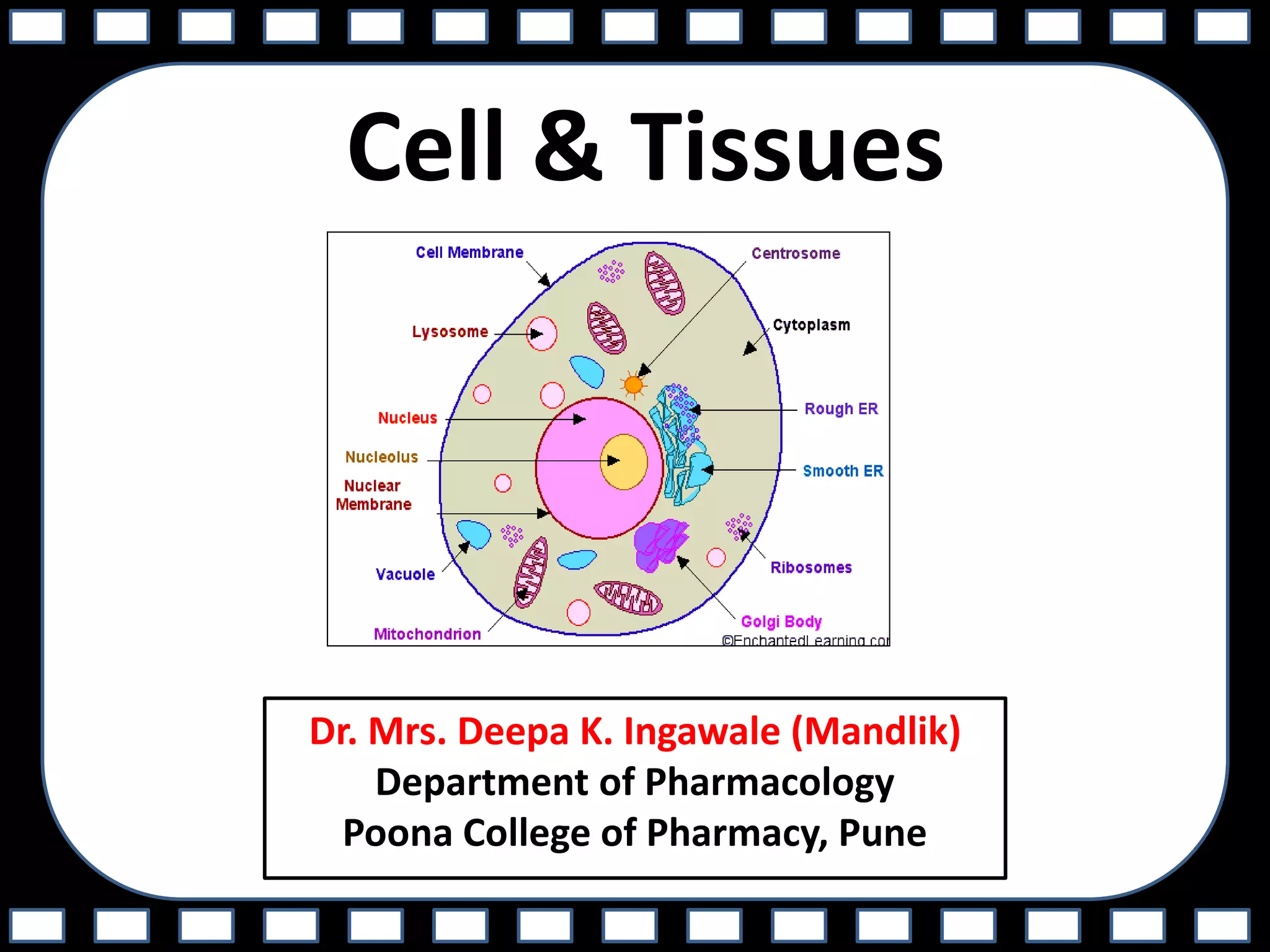
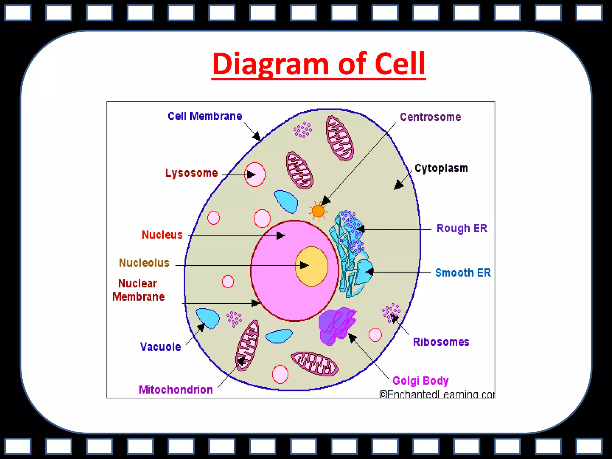
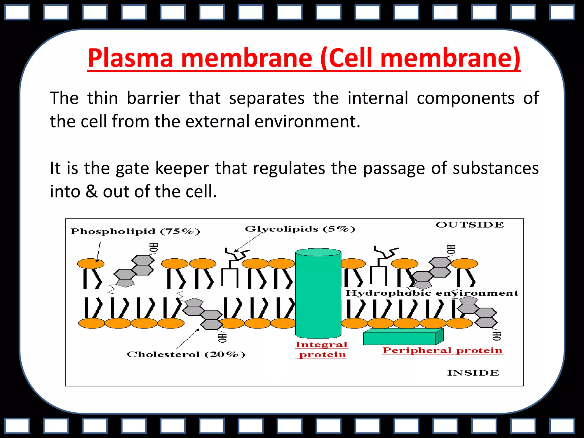
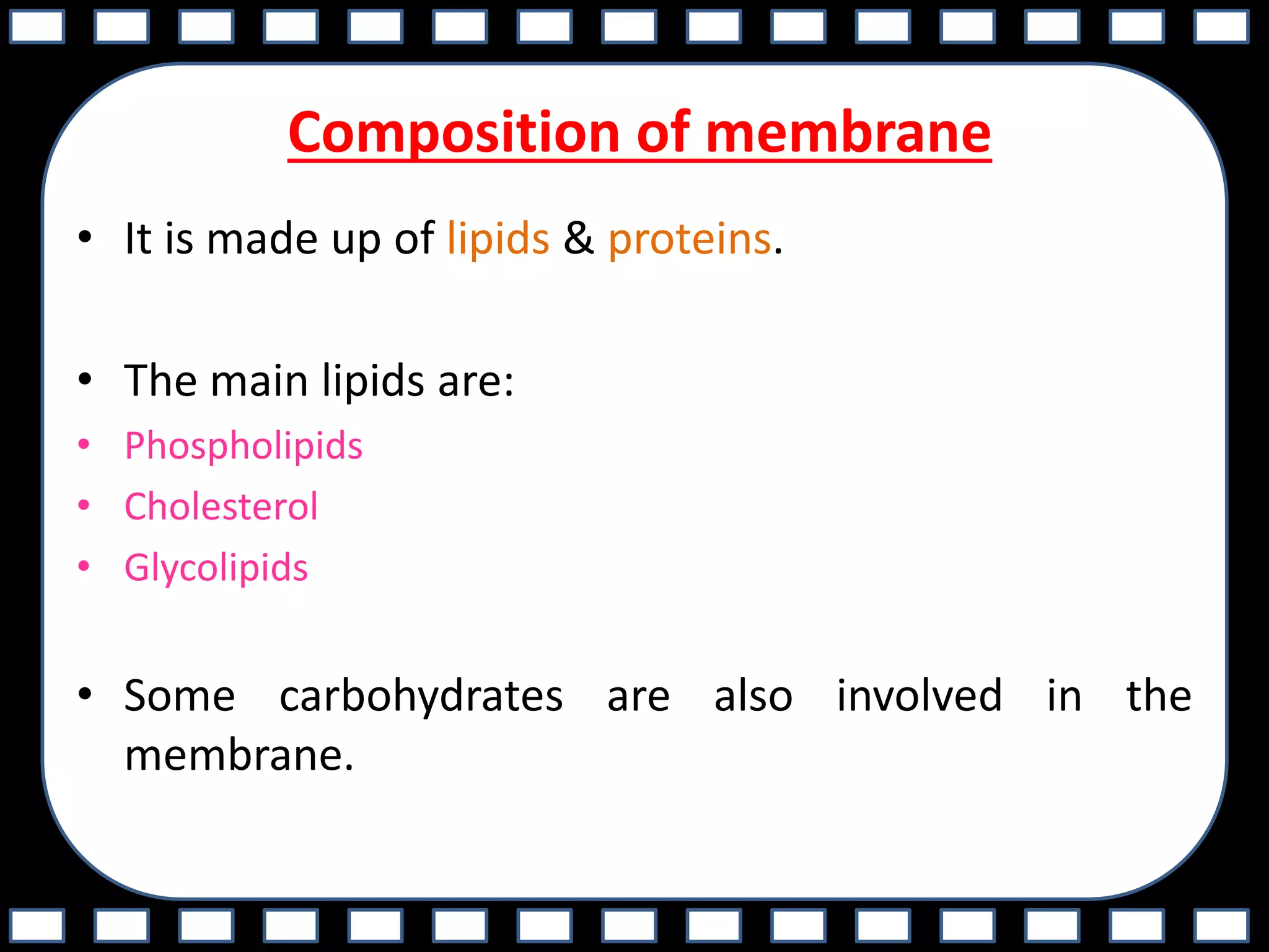
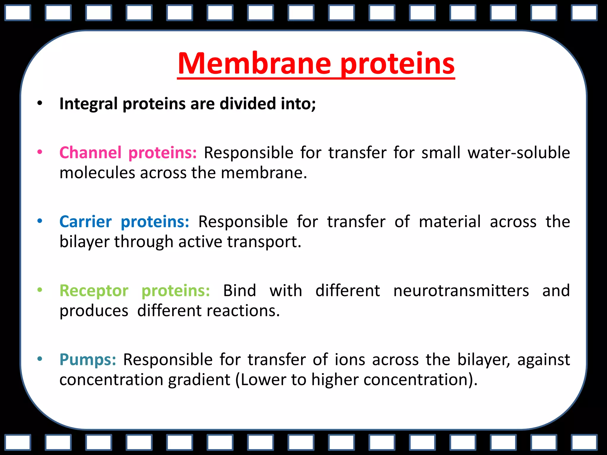
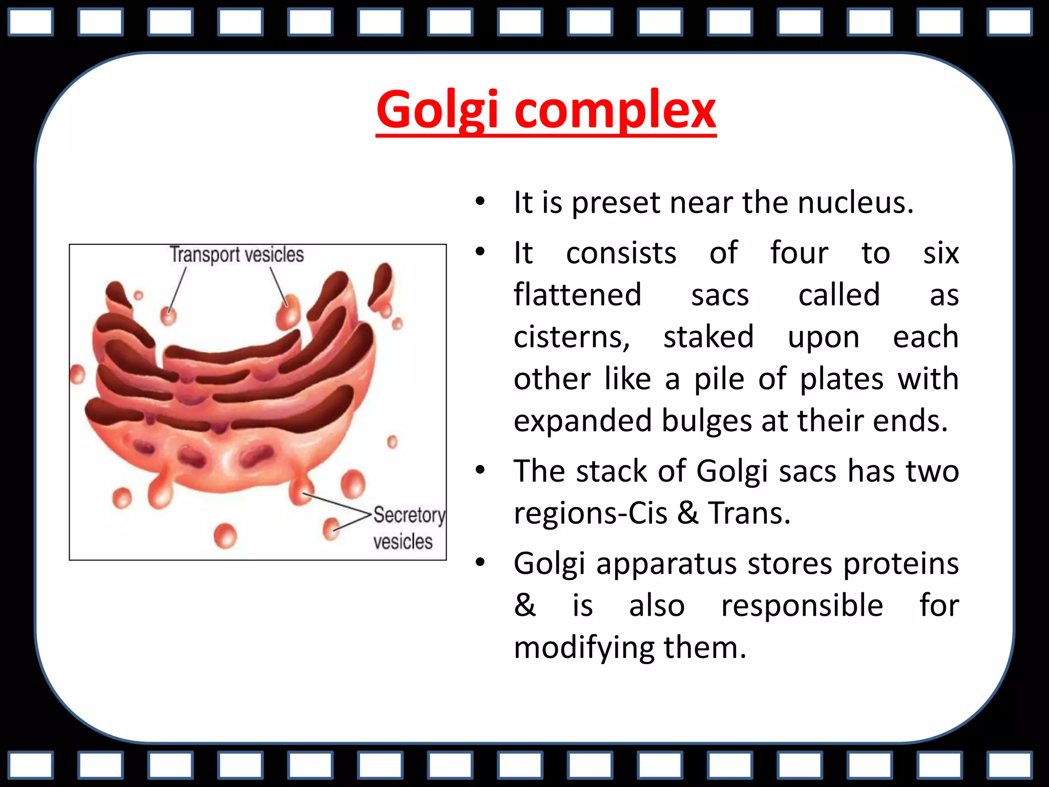
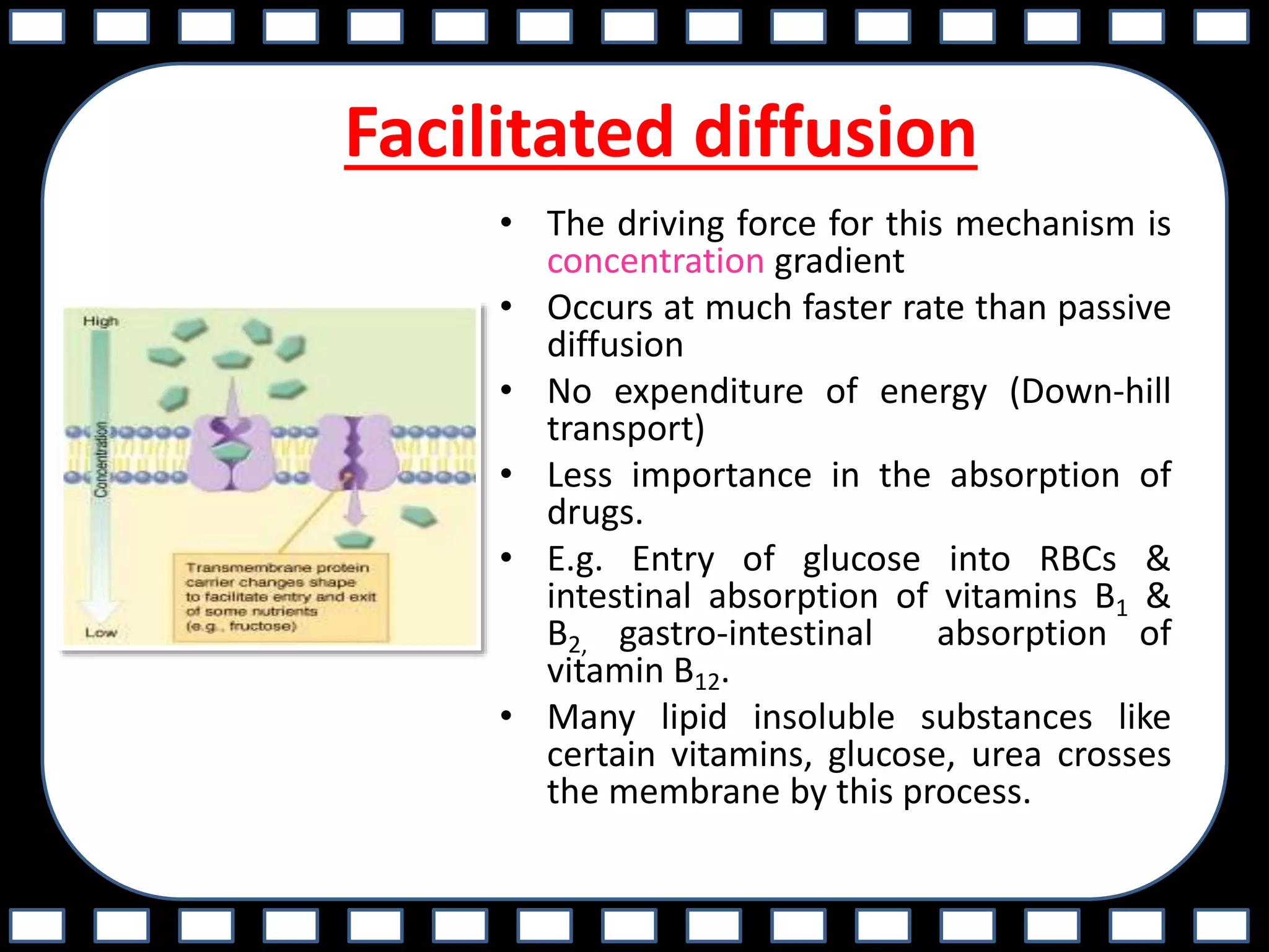
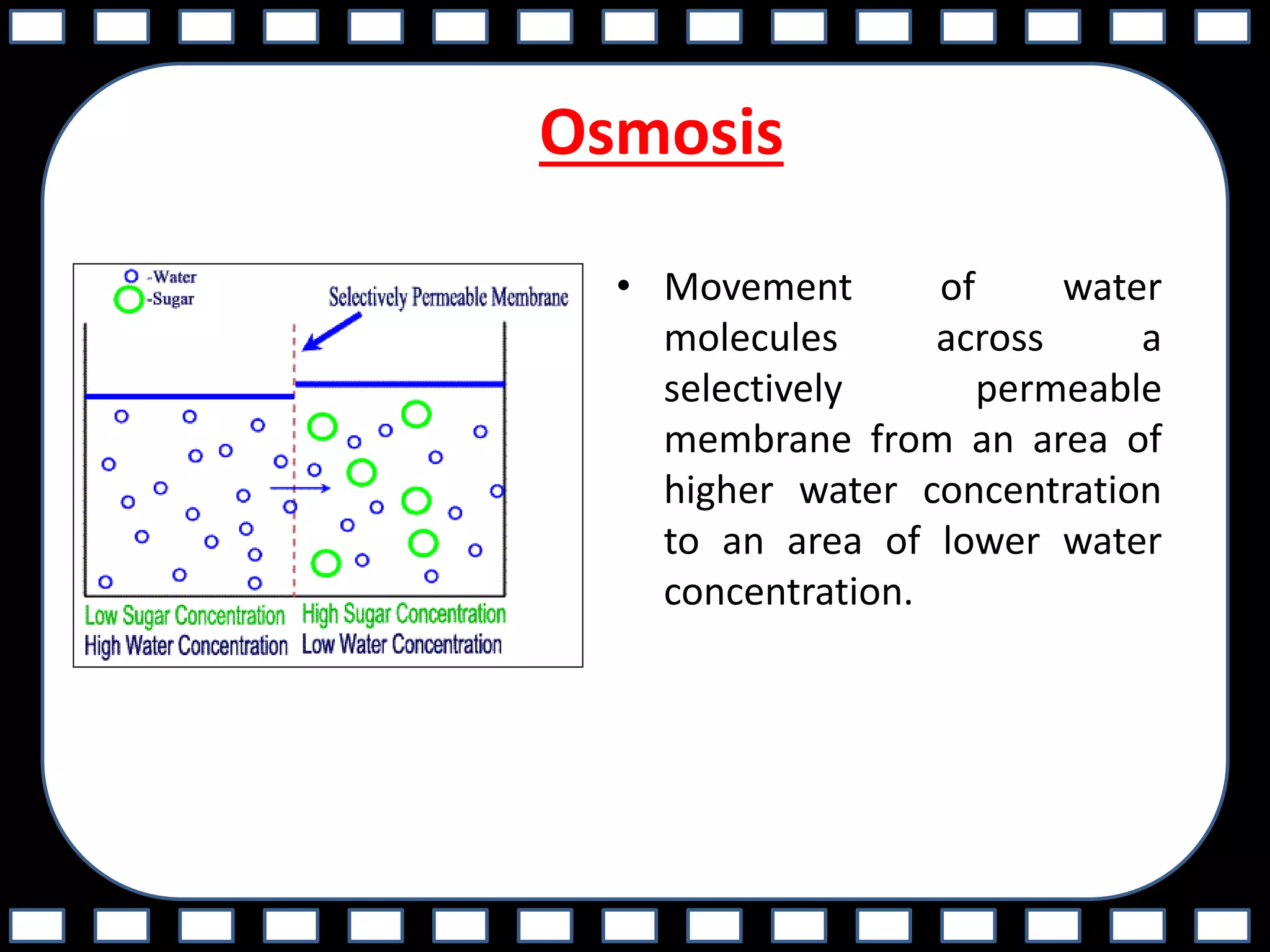
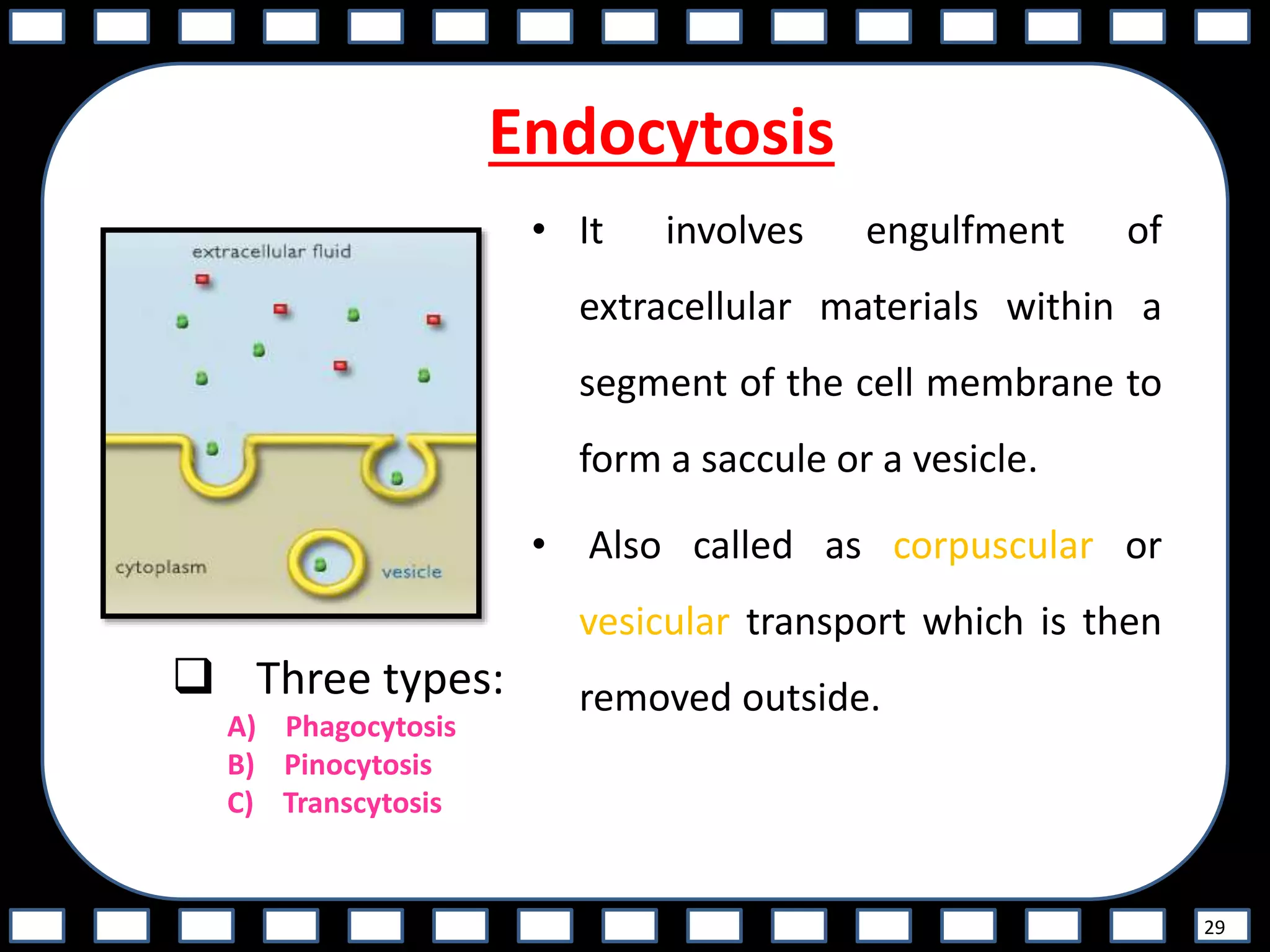
1. It defines cells and their basic parts like the plasma membrane, cytoplasm, nucleus, and various organelles. The plasma membrane regulates passage of substances into and out of the cell via proteins and lipids.
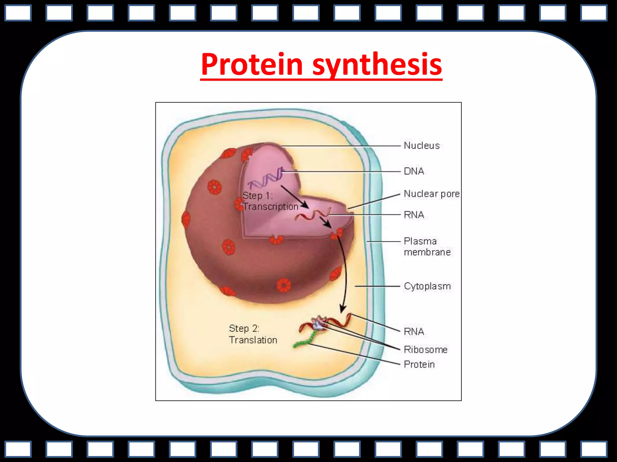
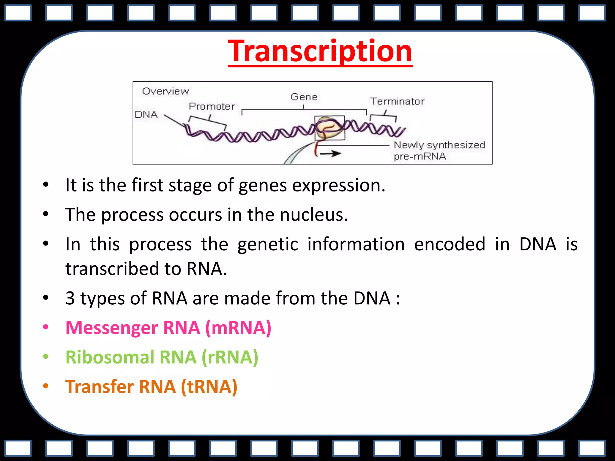
2. Protein synthesis is summarized as transcription of DNA to mRNA in the nucleus, followed by translation of mRNA to proteins by ribosomes.
3. Cell division and the stages of cell cycle are also briefly discussed. The document aims to describe cellular structures, functions, transport mechanisms, and protein synthesis at the basic level.